Copyright © 2017 The Korean Society for Bone and Mineral Research
This is an Open Access article distributed under the terms of the Creative Commons Attribution Non-Commercial Li- cense (http://creativecommons.org/licenses/by-nc/4.0/) which permits unrestricted non-commercial use, distribu- tion, and reproduction in any medium, provided the original work is properly cited.
Association of Serum 25-hydroxy-vitamin D
Concentration and Arterial Stiffness among Korean Adults in Single Center
Jung Hae Lee, Heuy Sun Suh
Department of Family Medicine, Gachon University Gil Medical Center, Gachon University School of Medicine, Incheon, Korea
Background: There are growing concerns about the role of vitamin D deficiency in car- diovascular diseases. Therefore, we investigated the correlation between serum 25-hy- droxy-vitamin D (25[OH]D) and arterial stiffness among Korean adults. Methods: We ret- rospectively reviewed the medical charts of 302 people (115 women and 187 men) who visited a tertiary hospital from January 2015 to December 2016. Serum 25(OH)D was measured using the radioimmunoassay technique, and brachial-ankle pulse wave veloc- ity (baPWV) was measured using an automatic wave analyzer. We obtained the doctor’s report on the medical history of the participants, their alcohol consumption and smok- ing habits, and their exercise status. Metabolic syndrome was diagnosed based on guide- lines from the National Cholesterol Education Program (NCEP)-Adult Treatment Panel (ATP III) and the International Diabetes Federation (IDF). Results of basic blood tests and physical assessment were also collected. Results: In the Pearson correlation analysis, se- rum 25(OH)D and baPWV showed a statistically significant inverse relationship (r=-0.279, P<0.001). Using multiple regression analysis, and after adjusting for possible confound- ers, serum 25(OH)D concentration was found to be significantly associated with baPWV (β=-0.121, P=0.011). Conclusions: We observed an association between serum 25(OH) D concentration and arterial stiffness. Further studies involving larger sample sizes will be needed to confirm this associations.
Key Words: Adult, 25-hydroxyvitamin D3, Vascular stiffness
INTRODUCTION
Vitamin D is synthesized in the body, and can be considered a hormone that has endocrine, autocrine, and paracrine functions. Vitamin D regulates calcium homeostasis and mineral metabolism by influencing the activities of the intes- tines, bones and kidneys. In addition, vitamin D is essential for the induction of cellular differentiation as well as for the inhibition of angiogenesis.[1,2] Vitamin D also has a number of nonskeletal effects that may favorably affect the cardiovas- cular system, for example, downregulation of the renin-angiotensin system,[3]
stimulation of insulin sensitivity in pancreatic beta (β)-cells,[4] and modulation of inflammation.[5]
Presently, vitamin D deficiency is recognized as a worldwide concern,[6] and in Koreans especially, this deficiency is very common. The fourth Korean National Corresponding author
Heuy Sun Suh
Department of Family Medicine, Gachon University Gil Hospital, 21 Namdong-daero, 774 beon-gil, Namdong-gu, Incheon 21565, Korea
Tel: +82 32-460-2735 Fax: +82 32-460-3354 E-mail: [email protected] Received: January 31, 2017 Revised: February 11, 2017 Accepted: February 12, 2017
No potential conflict of interest relevant to this article was reported.
Health and Nutrition Examination Survey (KNHANES V-1 and 2) reported a particularly high prevalence of vitamin D deficiency (25-hydroxy-vitamin D [25(OH)D] <20 ng/mL) in up to 60% of Koreans aged ≥10 years (65.9% of males and 77.7% of females).[7]
There are growing concerns about the role of vitamin D deficiency in cardiovascular diseases. Previous clinical stud- ies have confirmed that low vitamin D levels (defined as serum 25(OH)D concentration below 20 ng/mL according to guidelines from The Endocrine Society can impair vas- cular function. This may compromise vascular compliance (the elastic property of blood vessels), thereby manifesting as increased arterial stiffness.[8]
Arterial stiffness is an early marker of atherosclerotic dis- eases. Increased arterial stiffness has been shown to be an independent risk factor for both cardiovascular disease and overall mortality in elderly patients.[9-12] Pulse wave velocity (PWV) is a simple, economical, and the most wide- ly used non-invasive examination method for measuring arterial stiffness.[13] Several studies have investigated the association between vitamin D status and arterial stiffness using PWV. However, most of these studies were performed in Western countries,[14-19] with only a few Asian studies.
[20-22] Moreover, only a few studies have involved Kore- ans,[23,24] and a recent study has observed no significant relationship between vitamin D status and arterial stiffness.
[24] Even a meta-analysis of various studies has produced the same result.[25] However, there are many studies that have shown vitamin D to have a positive effect against ar- terial stiffness.[26-30] We hypothesized that a higher con- centration of serum 25(OH)D leads to reduced arterial stiff- ness. Therefore, we evaluated the relationship between 25(OH)D concentration and brachial-ankle PWV (baPWV) in Korean adults.
METHODS
1. Study subjectsA total of 302 participants (age≥20 years) who had un- dergone medical checkups at a tertiary healthcare facility, between January 2015 and December 2016 were included in the study. Information about lifestyle, medical history, drug history, and alcohol consumption, smoking habits and exercise status were obtained from the doctor’s notes.
Body weight and height were measured in light clothing
without shoes, and body mass index (BMI) was calculated as the weight divided by the height squared (kg/m2). Waist circumference (WC) was measured at the midpoint between the iliac crest and the lower rib margin. All participants had already undergone a basic regular blood examination, as well as examinations for baPWV and 25(OH)D. Blood sam- ples were collected from all participants after at least 8 hr of fasting. Participants were made to rest for at least 5 min before the blood pressure (BP) was measured on the right arm using and electronic device.
After reviewing the health screening charts, patients with a history of dyslipidemia (n=34), cardiovascular diseases (n=8), cerebrovascular diseases (n=2), any type of cancer (n=13) were excluded from the study.
2. Study methods
Vitamin D status is commonly assessed by measuring 25(OH)D level. This is because unlike 1,25 dihydroxy-vita- min D (1,25[OH]2D), 25(OH)D has a longer half-life and its activity is not affected by the parathyroid hormone.[31]
Blood samples were analyzed and 25(OH)D measured, us- ing a radioimmunoassay (ADVIA Centaur XPT Immunoas- say System; Siemens, Erlangen, Germany). The lowest mea- surable concentration of 25(OH)D was 4.2 ng/mL. Vitamin D deficiency was defined as a serum concentration of 25(OH) D, <20 ng/mL, while vitamin D insufficiency was defined as a serum concentration of 25(OH)D ≥20 ng/mL, but <30 ng/mL.[32,33]
Participants were made to rest for 5 min in the supine position before PWV was measured using an automatic waveform analyzer (VP-1000 plus; Omron Co., Ltd., Kyoto, Japan). The PWV between the brachial arteries and the an- kle was measured by placing both arms and the ankle in a cuff, to which an oscillometric sensor was implanted. In this study, the average value of both the right and left baPWV was calculated and used in the analyses.[34]
Regular alcohol drinkers were defined as those whose records showed a consumption of more than one bottle per week, while all other participants were defined as non- drinkers. Smokers were defined as those who had smoked more than five packs of cigarettes during their lifetimes and were smoking currently. The remaining participants were defined as nonsmokers. Regular exercise was defined as performing exercise for more than 3 days in a week for 30 min per session.
Information about any previous diagnoses of hyperten- sion (HTN) and diabetes mellitus (DM) were obtained throu- gh health interviews. Guidelines from the National Choles- terol Education Program (NCEP)-Adult Treatment Panel (ATP III) and the International Diabetes Federation (IDF) were employed in diagnosing metabolic syndrome.
The presence of metabolic syndrome was defined as when a participants met ≥3 of any of the following diagnostic criteria. 1) WC ≥90 cm (male), ≥85 cm (female), 2) triglyc- erides (TG) ≥150 mg/dL, 3) high-density lipoprotein cho- lesterol (HDL-C) ≤40 mg/dL (male), ≤50 mg/dL (female), 4) BP ≥130/85 mmHg or previously diagnosed HTN, 5) fast- ing blood glucose (FBG) ≥110 mg/dL or previously diag- nosed type 2 DM.
3. Statistical analysis
Statistical analyses were performed using the SPSS version 12.0 (SPSS Inc., Chicago, IL, USA), and the variables are pre- sented as mean±standard deviation. Results were comput- ed as two-tailed, and the level of statistical significance was set at P<0.05. We compared baseline characteristics be- tween the male and female participants using the chi-square
test and independent t-test. The difference in PWV was ana- lyzed according to history of HTN, DM, presence of metabolic syndrome, smoking, alcohol consumption, and exercise habit with the independent t-test; the Pearson correlation coeffi- cients were used to assess the relationships among biochem- ical factors, clinical factors, and PWV.
Using independent t-test and Pearson correlation analy- sis, a multiple regression analysis was conducted on statis- tically significant variables as covariates and PWV as the dependent variable. Some variables (alcohol consumption, smoking, exercise) that were not statistically significant (P>0.05), but could be confounding factors were also in- cluded as covariates. In the multiple regression analysis that included systolic BP (sBP), diastolic BP (dBP), and FBG as covariates, although a history of HTN and DM were sta- tistically significant factors according to the independent t-test, they were excluded in the multiple regression analy- sis owing to multicollinearity.
RESULTS
The baseline characteristics of the patients are reported Table 1. Baseline characteristics of the patients
Female (n=115) Male (n=187) Total (n=302) P-value
Age (yr) 55.6±9.6 54.7±8.7 55.1±9.1 0.402
BMI (kg/m2) 23.8±3.6 25.0±2.8 24.5±3.2 0.004
WC (cm) 77.4±8.8 84.9±7.4 82.0±8.7 <0.001
sBP (mmHg) 123.4±16.7 124.1±13.3 123.9±14.7 0.712
dBP (mmHg) 72.6±10.9 78.4±9.2 76.2±10.3 <0.001
25(OH)D (ng/mL) 19.9±7.9 20.5±6.4 20.3±7.0 0.524
baPWV (cm/s) 1,406.8±253.5 1,421.6±237.7 1,416.0±243.6 0.609
Total cholesterol (mg/dL) 193.1±35.9 198.6±34.2 196.5±34.9 0.182
LDL-C (mg/dL) 116.6±32.6 125.5±33.5 122.1±33.4 0.025
TG (mg/dL) 105.5±57.5 151.5±98.0 134.0±87.7 <0.001
HDL-C (mg/dL) 55.1±14.4 45.9±10.4 49.4±12.9 <0.001
FBG (mg/dL) 87.9±22.7 94.1±25.8 91.7±24.8 0.028
Alcohol (%) 41 (35.7) 158 (84.5) 199 (65.9) <0.001a)
Smoking (%) 7 (6.1) 164 (87.7) 171 (56.6) <0.001a)
Exercise (%) 35 (30.4) 53 (28.3) 88 (29.1) 0.698a)
HTN (%) 26 (22.6) 42 (22.5) 68 (22.5) 1.000a)
DM (%) 9 (7.8) 17 (9.1) 26 (8.6) 0.834a)
Metabolic syndrome (%) 23 (20.0) 50 (26.7) 73 (24.2) 0.214a)
The data is presented as mean±standard deviation.
a)Differences were assessed by the Chi-square test.
BMI, body mass index; WC, waist circumference; sBP, systolic blood pressure; dBP, diastolic blood pressure; 25(OH)D, 25-hydroxy-vitamin D; baPWV, brachial-ankle pulse wave velocity; LDL-C, low-density lipoprotein cholesterol; TG, triglycerides; HDL-C, high-density lipoprotein cholesterol; FBG, fast- ing blood glucose; HTN, hypertension; DM, diabetes mellitus.
in Table 1. A total of 302 participants were included in the study. This consisted of 115 women and 187 men with mean age of 55.6 years and 54.7 years, respectively. The differ- ences in BMI, WC, dBP, low-density lipoprotein cholesterol (LDL-C), TG, HDL-C, FBG, alcohol consumption status, and smoking status were statistically different between men and women.
Mean serum concentration of 25(OH)D was lower in wom- en (19.9 ng/mL) than in men (20.5 ng/mL), although the difference was statistically insignificant (P=0.524). Howev- er, the prevalence of vitamin D deficiency (i.e. 25(OH)D <20 ng/mL) was significantly higher in women than in men (55.7%
and 42.2% respectively, P=0.025) (Data is not shown).
Mean baPWV was higher in men, although the difference was not significant. (1,406.8 cm/s vs. 1,421.6 cm/s, P=0.609) (Table 1). An independent t-test conducted to analyze the relationship between baPWV and various underlying con- ditions yielded statistically significant differences for HTN (1,557.7 cm/s vs. 1,374.8 cm/s, P<0.001), DM (1,521.9 cm/s vs. 1,406.0 cm/s, P=0.020) and presence of metabolic syn-
drome (1,540.5 cm/s vs. 1,376.3 cm/s, P<0.001) (Table 2).
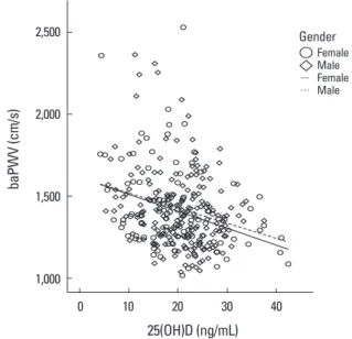
Univariate relationships between baPWV and parame- ters are shown in Table 3. baPWV positively correlated with age, WC, sBP, dBP, TG, and FBG and baPWV negatively cor- related with 25(OH)D (r=-0.279, P<0.001) and HDL-C.
In a fully adjusted multiple regression analysis, age, sBP, FBG, WC, and 25(OH)D (β=-0.121, P=0.011) were indepen- dently associated with baPWV, whereas sex, BMI, dBP, alco- hol consumption, smoking, exercise status, presence of metabolic syndrome, and all lipid profiles (total cholester- ol, LDL-C, TG, and HDL-C) were not. This regression model accounted for 51.7% (R2=0.517) of the total variance of baPWV (Table 4). The Durbin-Watson coefficient was 1.911, the fit test for multiple linear regression model was suit-
Table 2. Independent t-test between brachial-ankle pulse wave ve- locity and multiple confounders
Category
P-value Exist Not exist
Alcohol 1413.2±222.2 1421.4±281.5 0.797
Smoking 1426.9±244.0 1401.7±243.2 0.373
Exercise 1398.8±230.6 1423.0±248.9 0.434
HTN 1557.7±238.6 1374.8±229.6 <0.001
DM 1521.9±206.1 1406.0±244.8 0.020
Metabolic syndrome 1540.5±286.9 1376.3±214.0 <0.001 HTN, hypertension; DM, diabetes mellitus.
Table 3. Pearson’s correlation coefficients between brachial-ankle pulse wave velocity and parameters
Pearson’s correlation coefficient P-value
Age 0.432 <0.001
WC 0.130 0.024
sBP 0.455 <0.001
dBP 0.347 <0.001
25(OH)D -0.279 <0.001
TG 0.228 <0.001
HDL-C -0.128 0.026
FBG 0.384 <0.001
WC, waist circumference; sBP, systolic blood pressure; dBP, diastolic blood pressure; 25(OH)D, 25-hydroxyvitamin D; TG, triglycerides; HDL-C, high density lipoprotein cholesterol; FBG, fasting blood glucose.
Table 4. Multiple regression analysis for brachial-ankle pulse wave velocity
β coefficient t P-value
Age 0.448 10.105 <0.001
sBP 0.390 5.122 <0.001
FBG 0.268 5.563 <0.001
WC -0.147 -2.005 0.046
25(OH)D -0.121 -2.550 0.011
In this model, gender, body mass index, diastolic blood pressure, lipid profiles (total cholesterol, low density lipoprotein, triglycerides, high density lipoprotein), alcohol, smoking, exercise status, and presence of metabolic syndrome were also included as covariates, but they were not independently associated with brachial-ankle pulse wave velocity.
sBP, systolic blood pressure; FBG, fasting blood glucose; WC, waist cir- cumference; 25(OH)D, 25-hydroxy-vitamin D.
Fig. 1. Correlation between 25-hydroxy-vitamin D (25[OH]D) and bra- chial-ankle pulse wave velocity (baPWV), scatter diagram.
Fig. 1 Correlation between 25-hydroxyvitamin D(25(OH)D) and brachial-ankle pulse wave velocity(baPWV), Scatter diagram.
0.00 10.00 20.00 30.00 40.00
25(OH)D(ng/mL) 1000
1500 2000 2500
baPWV(cm/s)
Gender Female Male ㅡ Female
… Male
2,500
2,000
1,500
1,000
baPWV (cm/s)
0 10 20 30 40 25(OH)D (ng/mL)
Fig. 1 Correlation between 25-hydroxyvitamin D(25(OH)D) and brachial-ankle pulse wave velocity(baPWV), Scatter diagram.
0.00 10.00 20.00 30.00 40.00
25(OH)D(ng/mL) 1000
1500 2000 2500
baPWV(cm/s)
Gender Female Male ㅡ Female
… Male Female MaleFemale Male Gender
able (F=19.049, P<0.001) and all variance inflation factors of independent variables were under 10. The results of the correlation between 25(OH)D and baPWV, are represented in a scatter diagram (Fig. 1).
DISCUSSION
Our study demonstrated that arterial stiffness is signifi- cantly associated with low serum concentration of 25(OH) D. The Korean Vascular Research Working Group (KVRWG) suggested age and BP as major factors that influence arte- rial function. The present study, however, demonstrated a statistically significant relationship between 25(OH)D and arterial stiffness even after adjustment for age and BP. In addition, we found relatively higher mean serum concen- trations of 25(OH)D in participants (19.9 ng/mL in women, 20.5 ng/mL in men, aged 29-84 years) from this study than in other Korean adults (aged 20-87 years) from KNHANES database between 2008 and 2010. (16.9 ng/mL in women, 19.5 ng/mL in men).[24] We also found arterial stiffness to statistically correlate with FBG and WC.
The effects of vitamin D on the cardiovascular system are very important. Vitamin D receptors are found in many cells of the cardiovascular system.[31] Several adequate mechanisms explain how vitamin D may influence the car- diovascular system. An in vitro study has demonstrated that adequate vitamin D is associated with increased vas- cular endothelial cell (EC) expression of nuclear factor-kap- pa beta and interleukin-6, two important inflammatory mediators.[35] Moreover, vitamin D may attenuate the ad- verse effects of advanced glycation end products on EC ex- pression, which would have otherwise provoked dysfunc- tion.[36] Another study suggested that exogenous supple- mentation of vitamin D resulted in decreased EC prolifera- tion and that EC stress upregulates vitamin D receptor ex- pression on EC creating an autocrine or paracrine role for vitamin D with the potential to modulate EC adhesion and vascular smooth muscle cell migration and proliferation.
[37] These results only confirm that vitamin D is important for proper functioning of the cardiovascular system.
Although several previous studies have presented incon- sistent finding,[15,38-40] two other cross-sectional studies observed an inverse relationship between serum 25(OH)D level and arterial stiffness.[15,32] In the Framingham off- spring study as well, vitamin D deficiency was found to be
associated with an increased incidence of cardiovascular diseases.[41] Several studies have observed a successful decrease in baPWV prior to supplementation with vitamin D.[26-30] However, a European cross-sectional study [40]
as well as a cross-sectional analysis of the Korean Longitu- dinal Study on Health and Aging (KLoSHA) [39] showed no association between serum concentration of 25(OH)D and baPWV.
A recent cross-sectional study among Korean adults con- firmed that the positive association between 25(OH)D and baPWV was reversed when BP was controlled.[24] As men- tioned earlier, BP plays a major role in influencing arterial function. Arterial stiffness results from structural and func- tional changes to the vascular tree,[34] and high BP may affect those changes.[42] One study among Koreans dem- onstrated that a statistically significant association between 25(OH)D and baPWV was only existent in patients with type 2 DM.[23] They concluded that such a relationship between 25(OH)D and type 2 DM is believed to be caused by insulin resistance and impaired β-cell function among vitamin D deficient patients.[43-45]
Many studies have had inconsistent result in this area until quite recently, our study has a few strengths. To the best of our knowledge, our study may represent the first evidence of significant linear association between serum 25(OH)D concentration and arterial stiffness among Kore- an adults. Our study also included data on sBP, dBP, FBG, history of HTN and history of DM as possible confounding variables. Through multiple linear regression analysis, we demonstrated that sBP and FBG significantly correlated with baPWV. Nonetheless, we found serum 25(OH)D con- centration to be an independent risk factor of arterial stiff- ness (β=-0.121, P=0.011). These results are in contrast with those observed in most recent cross-sectional studies among Korean adults,[24] although their study was conducted with a rural population, whereas our study was conducted with an urban population. Also, in our study, we obtained information from the screening medical chart of those who went for health medical examinations at the healthcare fa- cility. Thus these individuals are more concerned about their health. In addition, there may be some differences in terms of physical activity, sun exposure, their sunscreen use, and eating habits.
Most of the vitamin D in the body is synthesized in the skin, through absorption of ultraviolet rays. Owing to the
decreasing amount of time spent outdoors, the number of vitamin D deficient people is increasing. A study conduct- ed in obese older adults demonstrated that physical activi- ty may be required to enhance the effect of vitamin D.[46]
Therefore, it is important to spend more time outdoors and increase physical activity level.
Our study has a few limitations. Firstly, the study was per- formed on individuals only in a single center in Korea, with a sample size of only 302 participants. Therefore, the find- ings may not be representative of the total adult popula- tion in Korea. Second, this was a cross-sectional study that employed assessment of screening charts, thereby render- ing it difficult to determine causal relationships. Addition- ally, data collection errors may have occurred, as informa- tion regarding tobacco and alcohol use, exercise status, and medical history were obtained from the doctor’s con- sultation notes and may not be a true reflection of the ac- tual situation. Furthermore, we used single measurements of serum 25(OH)D concentration as indicators of vitamin D status; thus this may not accurately reflect long-term vita- min D status. Lastly, we did not consider other factors that could directly affect vitamin D status such as seasonal vari- ation, duration of sun exposure, sunscreen use, skin pig- mentation, vitamin D supplementation or medication, eat- ing habits, and parathyroid hormone level.
In conclusion, an adequate serum concentration of 25(OH) D has a beneficial effect on arterial stiffness after adjusting for cardiometabolic factors. However, owing to certain lim- itations such as small sample size and confounders such as seasonal variations and the unknown parathyroid hormone levels, larger prospective studies are necessary to confirm these associations.
REFERENCES
1. Banerjee P, Chatterjee M. Antiproliferative role of vitamin D and its analogs--a brief overview. Mol Cell Biochem 2003;
253:247-54.
2. Ordonez-Moran P, Larriba MJ, Pendas-Franco N, et al. Vita- min D and cancer: an update of in vitro and in vivo data.
Front Biosci 2005;10:2723-49.
3. Li YC, Kong J, Wei M, et al. 1,25-Dihydroxyvitamin D(3) is a negative endocrine regulator of the renin-angiotensin sys- tem. J Clin Invest 2002;110:229-38.
4. Norman AW, Frankel JB, Heldt AM, et al. Vitamin D deficien-
cy inhibits pancreatic secretion of insulin. Science 1980;
209:823-5.
5. Hewison M. Vitamin D and immune function: an overview.
Proc Nutr Soc 2012;71:50-61.
6. Holick MF. Vitamin D deficiency. N Engl J Med 2007;357:
266-81.
7. Jung IK. Prevalence of vitamin D deficiency in Korea: Re- sults from KNHANES 2010 to 2011. J Nutr Health 2013;46:
540-51.
8. Melamed ML, Muntner P, Michos ED, et al. Serum 25-hy- droxyvitamin D levels and the prevalence of peripheral arterial disease: results from NHANES 2001 to 2004. Arte- rioscler Thromb Vasc Biol 2008;28:1179-85.
9. Meaume S, Rudnichi A, Lynch A, et al. Aortic pulse wave velocity as a marker of cardiovascular disease in subjects over 70 years old. J Hypertens 2001;19:871-7.
10. Laurent S, Boutouyrie P, Asmar R, et al. Aortic stiffness is an independent predictor of all-cause and cardiovascular mortality in hypertensive patients. Hypertension 2001;37:
1236-41.
11. Cruickshank K, Riste L, Anderson SG, et al. Aortic pulse-wave velocity and its relationship to mortality in diabetes and glucose intolerance: an integrated index of vascular func- tion? Circulation 2002;106:2085-90.
12. Blacher J, Safar ME, Guerin AP, et al. Aortic pulse wave ve- locity index and mortality in end-stage renal disease. Kid- ney Int 2003;63:1852-60.
13. Hametner B, Wassertheurer S, Kropf J, et al. Oscillometric estimation of aortic pulse wave velocity: comparison with intra-aortic catheter measurements. Blood Press Monit 2013;18:173-6.
14. Ponda MP, Huang X, Odeh MA, et al. Vitamin D may not improve lipid levels: a serial clinical laboratory data study.
Circulation 2012;126:270-7.
15. Giallauria F, Milaneschi Y, Tanaka T, et al. Arterial stiffness and vitamin D levels: the Baltimore longitudinal study of aging. J Clin Endocrinol Metab 2012;97:3717-23.
16. Gepner AD, Colangelo LA, Blondon M, et al. 25-hydroxyvi- tamin D and parathyroid hormone levels do not predict changes in carotid arterial stiffness: the Multi-Ethnic Study of Atherosclerosis. Arterioscler Thromb Vasc Biol 2014;34:
1102-9.
17. Martins D, Meng YX, Tareen N, et al. The effect of short term vitamin D supplementation on the inflammatory and oxi- dative mediators of arterial stiffness. Health (Irvine Calif)
2014;6:1503-11.
18. Witham MD, Adams F, Kabir G, et al. Effect of short-term vitamin D supplementation on markers of vascular health in South Asian women living in the UK--a randomised con- trolled trial. Atherosclerosis 2013;230:293-9.
19. Wang L, Manson JE, Buring JE, et al. Dietary intake of dairy products, calcium, and vitamin D and the risk of hyperten- sion in middle-aged and older women. Hypertension 2008;
51:1073-9.
20. Schmitz KJ, Skinner HG, Bautista LE, et al. Association of 25-hydroxyvitamin D with blood pressure in predominant- ly 25-hydroxyvitamin D deficient Hispanic and African Amer- icans. Am J Hypertens 2009;22:867-70.
21. Sun X, Cao ZB, Tanisawa K, et al. Associations between the Serum 25(OH)D concentration and lipid profiles in Japa- nese men. J Atheroscler Thromb 2015;22:355-62.
22. Uemura H, Katsuura-Kamano S, Yamaguchi M, et al. Asso- ciation between dietary calcium intake and arterial stiff- ness according to dietary vitamin D intake in men. Br J Nutr 2014;112:1333-40.
23. Lee JI, Oh SJ, Ha WC, et al. Serum 25-hydroxyvitamin D con- centration and arterial stiffness among type 2 diabetes.
Diabetes Res Clin Pract 2012;95:42-7.
24. Kang JY, Kim MK, Jung S, et al. The cross-sectional relation- ships of dietary and serum vitamin D with cardiometabol- ic risk factors: Metabolic components, subclinical athero- sclerosis, and arterial stiffness. Nutrition 2016;32:1048-56.
e1.
25. Rodríguez AJ, Scott D, Srikanth V, et al. Effect of vitamin D supplementation on measures of arterial stiffness: a sys- tematic review and meta-analysis of randomized controlled trials. Clin Endocrinol (Oxf) 2016;84:645-57.
26. Dong Y, Stallmann-Jorgensen IS, Pollock NK, et al. A 16- week randomized clinical trial of 2000 international units daily vitamin D3 supplementation in black youth: 25-hy- droxyvitamin D, adiposity, and arterial stiffness. J Clin En- docrinol Metab 2010;95:4584-91.
27. Mcgreevy C, Barry M, Bennett K, et al. The effect of vita- min D replacement on arterial stiffness in an elderly com- munity based population. Ir J Med Sci 2013;182:S234-5.
28. Dreyer G, Tucker AT, Harwood SM, et al. Ergocalciferol and microcirculatory function in chronic kidney disease and concomitant vitamin d deficiency: an exploratory, double blind, randomised controlled trial. PLoS One 2014;9:e99461.
29. Pilz S, Gaksch M, Kienreich K, et al. Effects of vitamin D on
blood pressure and cardiovascular risk factors: a random- ized controlled trial. Hypertension 2015;65:1195-201.
30. Witham MD, Adams F, McSwiggan S, et al. Effect of inter- mittent vitamin D3 on vascular function and symptoms in chronic fatigue syndrome--a randomised controlled trial.
Nutr Metab Cardiovasc Dis 2015;25:287-94.
31. Park HA, Kim SY. Recent advance on vitamin D. J Korean Med Assoc 2013;56:310-8.
32. Holick MF, Siris ES, Binkley N, et al. Prevalence of vitamin D inadequacy among postmenopausal North American wom- en receiving osteoporosis therapy. J Clin Endocrinol Metab 2005;90:3215-24.
33. Jacobs ET, Alberts DS, Foote JA, et al. Vitamin D insufficien- cy in southern Arizona. Am J Clin Nutr 2008;87:608-13.
34. Tomiyama H, Matsumoto C, Shiina K, et al. Brachial-ankle PWV: current status and future directions as a useful mark- er in the management of cardiovascular disease and/or cardiovascular risk factors. J Atheroscler Thromb 2016;23:
128-46.
35. Zhang Y, Leung DY, Richers BN, et al. Vitamin D inhibits mo- nocyte/macrophage proinflammatory cytokine produc- tion by targeting MAPK phosphatase-1. J Immunol 2012;
188:2127-35.
36. Talmor Y, Golan E, Benchetrit S, et al. Calcitriol blunts the deleterious impact of advanced glycation end products on endothelial cells. Am J Physiol Renal Physiol 2008;294:
F1059-64.
37. Raymond MA, Désormeaux A, Labelle A, et al. Endothelial stress induces the release of vitamin D-binding protein, a novel growth factor. Biochem Biophys Res Commun 2005;
338:1374-82.
38. Şeker T, Gür M, Kuloğlu O, et al. Serum 25-hydroxyvitamin D is associated with both arterial and ventricular stiffness in healthy subjects. J Cardiol 2013;62:361-5.
39. Lim S, Shin H, Kim MJ, et al. Vitamin D inadequacy is asso- ciated with significant coronary artery stenosis in a com- munity-based elderly cohort: the Korean Longitudinal Study on Health and Aging. J Clin Endocrinol Metab 2012;97:169- 78.
40. Deleskog A, Piksasova O, Silveira A, et al. Serum 25-hydroxy- vitamin D concentration in subclinical carotid atheroscle- rosis. Arterioscler Thromb Vasc Biol 2013;33:2633-8.
41. Wang TJ, Pencina MJ, Booth SL, et al. Vitamin D deficiency and risk of cardiovascular disease. Circulation 2008;117:
503-11.
42. Lim J, Pearman ME, Park W, et al. Impact of blood pressure perturbations on arterial stiffness. Am J Physiol Regul In- tegr Comp Physiol 2015;309:R1540-5.
43. Mathieu C, Gysemans C, Giulietti A, et al. Vitamin D and diabetes. Diabetologia 2005;48:1247-57.
44. Chiu KC, Chu A, Go VL, et al. Hypovitaminosis D is associ- ated with insulin resistance and beta cell dysfunction. Am J Clin Nutr 2004;79:820-5.
45. Pittas AG, Lau J, Hu FB, et al. The role of vitamin D and cal- cium in type 2 diabetes. A systematic review and meta- analysis. J Clin Endocrinol Metab 2007;92:2017-29.
46. Scott D, Ebeling PR, Sanders KM, et al. Vitamin d and physi- cal activity status: associations with five-year changes in body composition and muscle function in community- dwelling older adults. J Clin Endocrinol Metab 2015;100:
670-8.