255
INTRODUCTION
Breast cancer is the most common malignancy in female worldwide. It also became the leading incidence of female malignancy in Korea since 2001.(1,2) A significant feature of Korean women with breast cancer is that the median age of onset of breast cancer is around 45 years, approximately 15 years younger when compared with Western women in the Unites States.(1,2) Young women with breast cancer have more aggressive lesions than old women with breast cancer manifested by more elevated S-fraction, higher hormone receptor negative tumors, and poorer outcomes.(3,4)
It is well known that in approximately 20∼30% of breast carcinoma in Western women, tumor cells show an ampli- fication and/or over-expression of the tyrosine kinase receptor HER-2/neu (HER-2).(5,6) HER-2 encodes a transmembrane protein (p185) with an extracellular growth factor receptor do- main and an intracellular tyrosine kinase domain, and it is a member of the EGFR (HER-1), HER-3, and HER-4. HER-2 poised to interact with other receptors in the absence of direct ligand binding.(7) Over-expression of HER-2 has been shown test may be useful for selecting systemic chemotherapy in Korean patients with early onset breast cancer. And the specific anti-HER-2 therapy will be helpful to a large proportion of Korean patients who have more tumors with HER-2 overexpression than White patients. (Journal of Korean Breast Cancer Society 2003;6:255-262)
ꠏꠏꠏꠏꠏꠏꠏꠏꠏꠏꠏꠏꠏꠏꠏꠏꠏꠏꠏꠏꠏꠏꠏꠏꠏꠏꠏꠏꠏꠏꠏꠏꠏꠏꠏꠏꠏꠏꠏꠏꠏꠏꠏꠏꠏ Key Words: HER-2/neu, Breast cancer, FISH
Purpose: The purpose of this work was to investigate the prognostic significance of of HER-2/neu (HER-2) oncogene protein overexpression in Korean women with early-onset breast cancer by immunohistochemistry. Furthermore, the results of the test were correlated with HER-2 oncogene gene amplification assessed by fluorescence in situ hybridi- zation (FISH).
Methods: HER-2 status in 60 cases of breast cancer diagnosed at the age of 45 years or younger was investi- gated by a rabbit polyclonal antibody (Dako) and by ap- plying the Hercep Test, and the results were compared with FISH analysis in all Hercep Test 2+ and 3+ speci- mens (28 cases) and five HER-2/neu negative specimens.
Results: FISH revealed that HER-2 was exclusively am- plified in cases with Hercep Test 3+ (20/21). Cases with Hercep Test 3+ or FISH positive tumors were significantly associated with estrogen and progesterone receptor nega- tive tumors. No association was found between HER-2 sta- tus and axillary node status. In univariate analysis, FISH status was significantly associated with poor prognosis, but Hercep Test status was not a significant prognostic factor.
Conclusion: The finding of higher positive tumors and poor prognostic factor of Her-2 in Korean women with early- onset breast cancer may have potential implication for local and systemic management of breast cancer. HER-2
Characteristics of HER-2/neu Oncogene in Korean Women with Early-onset Breast Cancer by Immunohistochemistry
and Fluorescence In Situ Hybridization
Departments of Radiation Oncology, 1Surgery and 2Clinical Pathology, Soonchunhyang University, Seoul, Korea,
3Department of Internal Medicine, Gacheon University, Incheon, Korea, 4Departments of Pathology and
5Therapeutic Radiology, Yale University, New Haven, CT, USA
Doo Ho Choi, Min Hyuk Lee1, Yong Hee Ahn1, Dong Wha Lee2, Dong Bok Shin3, Darryl Carter4, Bonnie L. King and Bruce G. Haffty5
Correspondence: Doo Ho Choi, Department of Radiation Oncology, Soonchunhyang University Hospital, 657 Hanam-dong, Yongsan-gu, Seoul 140-743, Korea. Tel: 82-2-709-9412, Fax: 82-2-709-9414, E-mail: [email protected] Received:2 September, 2003 Accepted: 5 December, 2003 This work was partly supported by the Ethel F. Donaghue Women's Health Investigator Program at Yale, the Patterson Trust.
This work was presented at the 7th annual meeting of the Korean Breast Cancer Society 2003.
to be associated with a poor prognosis in patients with node-positive breast cancer and possibly also in patients with node-negative breast cancer.(5,6,8) Accurate determination of the HER-2 alteration has now become important because therapeutic decisions for patients are influenced by this infor- mation in both the adjuvant and advanced stage settings.
HER-2 alteration is shown to signify resistant to tamoxifen therapy(9-11) and CMF chemotherapy.(12) On the other hand, an increase sensitivity to anthracycline-containing regimen was observed.(13-15) More importantly, the monoclonal antiboby Trastuzumab (Herceptin, Genentech, Inc, CA, USA) directed against the HER-2 protein has resulted in inhibition of tumor growth in both preclinical and clinical studies.(16,17) This effect can be increased when used in combination with chemotherapeutic agents.(18,19) This effect was particularly apparent in patients with a strong (3+) expression of the HER-2 antigen or gene amplification on tumor cells or gene amplification. In the previous study, we reported higher pro- portion of young Korean patients than White patients had breast tumors with HER-2 overexpression or amplification.(20) In this study, we analyzed the HER-2 status by immuno- histochemistry (IHC) and compared the results with the clinical parameters including survival rates and failure pat- terns. Also, the different grades of protein over-expression were compared with results obtained by fluorescence in situ hybridization (FISH).
METHODS
The study population consisted of 60 cases of breast cancer that was diagnosed with the age of 45 years or younger who had been treated at the Soonchunhyang University Hospital, Seoul, Korea from 1995 to 2000. These cases were retrieved from hospital breast cancer registry. The median and range for the age at onset were 37 and 22 to 45 years, respectively (Table 1). The median follow-up period for the cohort at the time of analysis was 4.5 years. Thirty-eight patients under- went modified radical mastectomy, and 22 patients underwent quadrantectomy and radiotherapy. Patients who underwent modified radical mastectomy received adjuvant high dose che- motherapy and postoperative radiotherapy, if axillary lymph node was positive in 4 or more than 4 lymph nodes. All the other patients received 6 cycles of CMF chemotherapy and/or tamoxifen. Systemic chemotherapy was not delivered accor- ding to HER-2 or FISH status. Paraffin-embedded tissues from the primary tumors were obtained from the hospital archives.
The staining was performed on 5μm-thick tissue sections prepared from paraffin-embedded archival tissues. The sections were dried in 60oC heated oven for one hour. The sections were dewaxed in xylene and rehydrated through graded alcohols to distilled water. The antigen retrieval method utilized a steam bath at 97oC for 20 minutes for determination of ER, PR, cyclin D1 and HER-2. The Dako autostainer was used to perform the immunohistochemical staining. Tris-buffered saline was used for all the wash steps.
Sections were treated with hydrogen peroxide for 5 minutes.
Since the Dako LSAB plus and Envision detection kits were used, no blocking steps were required. The sections were incubated in antibody for 30 minutes at room temperature.
Secondary antigen retrieval was performed by using detection kits Envision for ER and PR, Dako Lsab+ for cyclin D1 and Hercep Test fo HER-2). Labelling was performed with DAB. All sections were counterstained with hematoxylin, Table 1. Percentage distribution of characteristics for Korean
women with early-onset breast cancer (N=60) ꠚꠚꠚꠚꠚꠚꠚꠚꠚꠚꠚꠚꠚꠚꠚꠚꠚꠚꠚꠚꠚꠚꠚꠚꠚꠚꠚꠚꠚꠚꠚꠚꠚꠚꠚꠚꠚꠚꠚꠚꠚꠚꠚꠚꠚꠚꠚꠚꠚꠚꠚ
Characteristics No. of patients (%) ꠏꠏꠏꠏꠏꠏꠏꠏꠏꠏꠏꠏꠏꠏꠏꠏꠏꠏꠏꠏꠏꠏꠏꠏꠏꠏꠏꠏꠏꠏꠏꠏꠏꠏꠏꠏꠏꠏꠏꠏꠏꠏꠏꠏꠏꠏꠏꠏꠏꠏꠏ
Age (years)
Mean 36.9
Median 37
Range 22∼45
Tumor size (cm)
Mean 2.8
Median 2
Range 1.0∼8.0
T-stage
Tis 2 (3.3)
T1 27 (45.0)
T2 28 (46.7)
T3 3 (5.0)
Histology
IDC 57 (95.0)
DCIS 2 (3.4)
Medullary ca 1 (1.7)
Nuclear Grade
Grade 1 0 (0)
Grade 2 43 (71.7)
Grade 3 17 (28.3)
Axillary node status
Positive 29 (48.3)
Negative 31 (51.7)
ꠏꠏꠏꠏꠏꠏꠏꠏꠏꠏꠏꠏꠏꠏꠏꠏꠏꠏꠏꠏꠏꠏꠏꠏꠏꠏꠏꠏꠏꠏꠏꠏꠏꠏꠏꠏꠏꠏꠏꠏꠏꠏꠏꠏꠏꠏꠏꠏꠏꠏꠏ IDC= infiltrating ductal carcinoma; DCIS = ductal carcinoma in situ.
dehydrated with ethanol and permanently coverslipped.
Tumors known to express the markers served as positive control. For HER-2, primary antibody was omitted for negative control and DAKO K5204 was used for reference grade. A cut-point of ≥10% tumor cells stained without considering intensity was used to categorize positive results for ER and PR. For cyclin D1, H-score (defined as the pro- duct of intensity and distribution) was determined and higher than H-score 75 was considered positive. For HER-2, only membrane staining was scored as positive, and cytoplasmic staining was ignored. A numeric score ranging from 0 to 3 that reflects the staining intensity and patterns in 10% or more of tumor cell is employed. Numeric score of 2+ or 3+
was considered positive by Hercep Test guidelines. All im- munostained sections were examined and scored by two of the authors. Any discrepancies were resolved by subsequent consultation.
All tumors with 2+ or 3+ Hercep Test scores and 5 samples with HER-2/neu 0 or 1+ cases were evaluated for gene amplification by interphase FISH. FISH analysis was performed with the PathVysionTM HER-2 DNA Probe Kit (Vysis, Downers Grove, IL. USA) using two directly labeled fluorescent DNA probes complementary to the HER-2/neu gene locus (LSI HER-2/neu SpectrumRedTM) and to chromo- some 17 peri-centromeric satellite DNA (CEP17 Spectrum- GreenTM) as previously described.(20) In brief, slides with 5 m tissue sections were baked at 60oC overnight, deparaffin pretreated in sodium thiocyanate (Vysis), digested with pepsin (4 mg/ml in 0.01 M HCl; Vysis), and rinsed in graded etha- nols. Ten l of probe/hybridization mixture including blocking
DNA were applied to each tissue section, sealed with rubber cement. The probes and tissues were codenatured and hy- bridized overnight by using the Vysis Hybrite hybridization system at 37oC. After air drying the slides were counter stained with 4',6-diamindino-2-phenylindole (DAPI) and cover-slip- ped. HER-2 gene amplification was quantified by comparing the ratio of LSI HER-2 to CEP 17 probe signals in accor- dance with the PathVysion HER-2 DNA Probe kit criteria.
All cases with more than a mean number of four fluore- scence signals per two signals of the centromere of chro- mosome 17 were considered amplified. Scoring was done in a blinded fashion by a single observer (Bonnie L. King) inde- pendent of IHC results.
All patient's data including clinical, pathologic, and out- come measures were entered into a computerized database employing SPSS (Statistical Package for the Social Science).
For analysis of extent of bivariate correlations, Person's Cor- relation Coefficients were used. We used Kaplan-Meier to calculate disease free survival probabilities, and the log-rank test was used to compare survival distributions of cases. For multivariate analysis, Cox proportional hazard model was used.
RESULTS
A total of three cases were disqualified from the study of prognosis either on the basis of absence of tumor cells in the tissue section, DCIS or unsatisfactory FISH analysis. IHC and FISH assays were conducted as blinded from each other - as well as to clinical information. No patients were excluded on
Fig. 1. Estimated relapse free survival probabilities invasive breast cancer stratified by conventional immunohistochemistry criteria (Hercep Test 0/+1 versus 2+ and 3+) by Kaplan- Meier method.
Fig. 2. Relapse free survival rates of patients stratified by fluore- scence in situ hybridization.
the basis of age or the length of available follow-up.
Mean number of axillary sampling was 16.9 (range 6∼39) and mean number of positive axillary node metastasis was 2.47. Korean women in this study cohort over-expressed HER-2 in 47.5% of the cases (HER-2 2+ and HER-2 3+ by Hercep Test), including 55% in node negative patients.
Among the twenty-nine patients with positive axillary lymph node, thirteen and twelve patients had positive tumors for Hercep Test and FISH study, respectively. Twelve patients out of twenty FISH positive tumor also had axillary lymph node metastasis. The positive percentages of the ER, PR, and cyclin D1 were 47.5%, 42.4%, and 40.9%, respectively. FISH analysis was successful on 27/28 (96%) of the tumors, and gene amplification was detected in 20/27 (74%) of the evaluative tumors. Of the cases, 19/20 (95%) of the HER-2 3+ and 1/7 (16%) of the HER-2 2+ cases were FISH positive. All the evaluative tumors with HER-2 0/1+ were
FISH negative (score range, 1.05∼1.15), and we assumed and analyzed that all the cases with Hercep Test 0/1+ not tested by FISH were FISH negative.
Probability of survival of patients according to Hercep Test demonstrated that patients with HER-2 over-expression had tendency but did not have decreased survival (Fig. 1; P=
0.0883). However the decreased probability of survival was observed in HER-2 3+ group (data not shown). HER-2/neu over-expression had no impact on estrogen receptor status, but there was a significant proportion of estrogen receptor negativity in patients with a 3+ staining compared with those with both 2+ and negative cases (P=0.009). Patients with FISH positive tumors showed decreased survival (Fig. 2;
P=0.0439), Survival difference was observed rather early in the cases with axillary node metastasis (Fig. 3). Moreover, the range of HER-2/CEP ratio in the treatment related failure group was higher than those without recurrence (4.55∼12.5 Fig. 3. Relapse free survival rates of patients according to FISH status, subdivided by lymph node status.
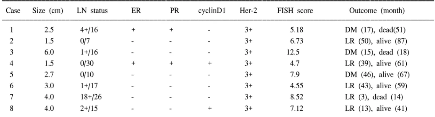
Table 2. Clinical courses and outcome of FISH amplified cases with relapse
ꠚꠚꠚꠚꠚꠚꠚꠚꠚꠚꠚꠚꠚꠚꠚꠚꠚꠚꠚꠚꠚꠚꠚꠚꠚꠚꠚꠚꠚꠚꠚꠚꠚꠚꠚꠚꠚꠚꠚꠚꠚꠚꠚꠚꠚꠚꠚꠚꠚꠚꠚꠚꠚꠚꠚꠚꠚꠚꠚꠚꠚꠚꠚꠚꠚꠚꠚꠚꠚꠚꠚꠚꠚꠚꠚꠚꠚꠚꠚꠚꠚꠚꠚꠚꠚꠚꠚꠚꠚꠚꠚꠚꠚꠚꠚꠚꠚꠚꠚꠚꠚꠚꠚꠚꠚꠚꠚꠚ
Case Size (cm) LN status ER PR cyclinD1 Her-2 FISH score Outcome (month)
ꠏꠏꠏꠏꠏꠏꠏꠏꠏꠏꠏꠏꠏꠏꠏꠏꠏꠏꠏꠏꠏꠏꠏꠏꠏꠏꠏꠏꠏꠏꠏꠏꠏꠏꠏꠏꠏꠏꠏꠏꠏꠏꠏꠏꠏꠏꠏꠏꠏꠏꠏꠏꠏꠏꠏꠏꠏꠏꠏꠏꠏꠏꠏꠏꠏꠏꠏꠏꠏꠏꠏꠏꠏꠏꠏꠏꠏꠏꠏꠏꠏꠏꠏꠏꠏꠏꠏꠏꠏꠏꠏꠏꠏꠏꠏꠏꠏꠏꠏꠏꠏꠏꠏꠏꠏꠏꠏꠏ
1 2.5 4+/16 + + - 3+ 5.18 DM (17), dead(51)
2 1.5 0/7 - - - 3+ 6.73 LR (50), alive (87)
3 6.0 1+/16 - - - 3+ 12.5 DM (15), dead (18)
4 1.5 0/30 + + + 3+ 4.7 LR (39), alive (61)
5 2.7 0/10 - - - 3+ 7.9 DM (46), alive (67)
6 3.0 1+/17 - - - 3+ 4.55 LR (43), alive (59)
7 4.0 18+/26 - - - 3+ 8.52 LR (3), dead (14)
8 4.0 2+/15 - - + 3+ 7.12 LR (13), alive (41)
ꠏꠏꠏꠏꠏꠏꠏꠏꠏꠏꠏꠏꠏꠏꠏꠏꠏꠏꠏꠏꠏꠏꠏꠏꠏꠏꠏꠏꠏꠏꠏꠏꠏꠏꠏꠏꠏꠏꠏꠏꠏꠏꠏꠏꠏꠏꠏꠏꠏꠏꠏꠏꠏꠏꠏꠏꠏꠏꠏꠏꠏꠏꠏꠏꠏꠏꠏꠏꠏꠏꠏꠏꠏꠏꠏꠏꠏꠏꠏꠏꠏꠏꠏꠏꠏꠏꠏꠏꠏꠏꠏꠏꠏꠏꠏꠏꠏꠏꠏꠏꠏꠏꠏꠏꠏꠏꠏꠏ DM = distant metastasis; LR = locoregional recurrence.
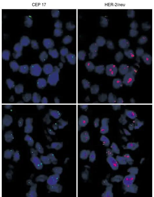
versus 0.9∼7.9). A total of eight cases in FISH positive patients had locoregional or distant failure (Table 2). Fig. 4 shows 2 cases with FISH+ who experienced relapse after treatment. Proportional hazard regression analysis was con- ducted to compare FISH-determined HER-2 amplification with the most commonly used clinical prognostic markers including tumor size, histologic grade, hormone receptor, cyclin D1 and lymph node status. Only lymph node status was found to be a predictor of disease-free survival, inde- pendent of other variables (P=0.040). FISH (P=0.057) and T-stage (P=0.099) were marginally significant prognostic factors.
DISCUSSION
The HER-2 gene was first identified as a transforming oncogene in the DNA of chemically induced neuroblastomas in the rat.(21) The HER-2 oncogene is overexpressed in 20∼
30% of breast carcinomas from Western women. And in approximately 90∼95% of these cases, overexpression is a direct result of gene amplification. The HER-2 gene product is composed of a cytoplasmic domain with tyrosine kinase activity, a transmembrane domain, and an extracellular do- Fig. 4. FISH analysis demonstrating HER-2/neu gene amplification cases with treatment related failure (The bottom panel is case 1 in
Table 2 and the top panel is case 2 in the Table 2).
main that may be shed from the surface of breast cancer cells. Her-2 may be involved in the pathogenesis and clinical aggressiveness of HER-2 overexpressing tumors.
Tissue-based detection of the HER-2 alteration by IHC and FISH offers clear advantage over other approaches such as Southern, Northern, and Western blot and PCR-based analy- sis, which suffer from dilution artifacts resulting from mixture of normal and abnormal cell proliferations within tissue. And the difficulties of dealing with frozen tissue in the clinical medicine have made testing of HER-2 on formalin-fixed tissues a necessity. And Hercep Test and FISH have been approved by the US Food and Drug Administration for use in the selection of patients for therapy with the Herceptin.
Both IHC and FISH permit for the specific detection of the alteration in individual cells maintaining original architecture, which permits analysis in its histologic context. With this aspect and availability of reagents and low cost, IHC was popularly used. However, it has significant defects including antigen alterations by fixation procedures, sensitivities of anti-HER-2 antibody, and inconsistent interpretation of the results. An advantage of FISH is that it circumvents antigenic changes that occur in formalin-fixed/paraffin-embedded tissues, a major limitation inherent to IHC. The polyclonal antiserum used in the FDA approved Hercep Test is currently recommended scoring method. Samples scoring at 3+ are regarded as unequivocally positive and 0/1+ as negative.
Borderline 1+/2+ and 2+ require confirmation with alter- native methods, preferably FISH.
Concordance between FISH and IHC in this study was consistent with what has been found in other studies. This issue has become of paramount importance in the clinical setting because the HER-2 gene product represents a specific target for treatment with trastuzumab, and optimal use of this therapy requires accurate and reliable determination of HER-2 status. Strongly positive (3+) HER-2 tumors were highly associated with gene amplification in this study, whereas weakly positive (2+) HER-2 tumors showed only a minor association. Tsuda et al. reported the concordance rate of the high-level amplification with an HER-2 score of 3+ was 97%, while only 25% of HER-2 2+ showed high level amplification (22). Jacobs et al. also found a high level of concordance (91%) between FISH and IHC by Hercep Test in the evaluation of HER-2 status in 100 cases.(23) Lebeau et al. also reported HER-2 3+ were always associated with gene amplification, with 75% of the HER-2 2+ lacked gene amplification. HER-2 overexpression was demonstrated in 42% of the tumors with Hercep Test and FISH identified
gene amplification in 28% of the tumors.(24) For HER-2 2+
cases, it is unclear whether they represent highly sensitive staining or some cases show overexpression without ampli- fication. Tubbs et al. reported that most Hercep Test-positive cases with a score of 2+ and a normal gene copy should be regarded as true false-positive, which means unresponsiveness to Herceptin.(25) Perez et al. recommended that all specimens with a HER-2 2+ result should be evaluated by FISH for HER-2 gene amplification before making a decision to recommend anti-HER-2/neu therapy,(19) and recent data sug- gested the necessity of central laboratory testing to confirm HER-2 status.(26,27)
In the previous study, 47.5% of the tumors have HER-2 overexpressing tumors and the majority of the IHC 3+ cases have HER-2 gene amplification in Korean women with early-onset breast cancers.(20) These results are somewhat higher than those of previous studies in Western countries.
Recently, Jeffrey et al. at Stanford University also observed higher HER-2/neu gene amplification in Korean patients with breast cancer than Caucasian breast cancer by cDNA microarray method (personal communication). These results suggest that there may be biologic and molecular alterations not identified yet and supports the necessity for further large scale research and identification of etiology between the two racial groups.
The current study shows that the FISH data provide better prognostic information and segregate higher risk breast cancer patients more accurately than IHC. But, the high level of correlation between IHC 3+ staining and amplification detected by FISH from this study and other reports makes it difficulty to justify the routine use of FISH for determination of HER-2 status. Although FISH has many advantages, the major disadvantages of FISH versus IHC are the high cost, the time-consuming tissue preparation and evaluation of slides and requirement of a fluorescence microscope. Recently, a new chromogenic in situ hybridization (CISH) method allows detection of gene amplification, using peroxidase reaction with direct evaluation of the histopathologic features; and it provides comparable results to FISH, with significant cost saving.(28-30)
Analyses with HercepTest revealed that 3+ HER-2 tumors revealed a significantly worse course of the disease. This finding is corroborated by the fact that in this study, almost exclusively HER-2 3+ cases were found to be amplified in the FISH assay. In comparison, HER-2 2+ status was of no negative prognostic value. Previous studies have demonstrated that subjects whose tumors have 3+ HER-2 overexpression
have a marked improvement in clinical response when com- pared with subjects whose tumors have 2+ overexpression.(31) Interestingly enough, HER-2 2+ cases had a trend toward a longer disease-free survival when compared with HER-2 negative cases in this study. This finding was observed in other reports.(8)
Association between HER-2 receptor positivity (2+ and 3+) and hormone receptor status was not found, but HER-2 3+ or FISH positivity was strongly associated with hormone receptor negativity in these patients. Many studies also reported that IHC 3+ cases were significantly associated with estrogen receptor negativity or lower estrogen receptor level.(8,32) Preclinical studies have suggested that the estrogen- dependent cultured human breast cancer cell lines are ren- dered hormone-independent(33) and levels of ER and PR significantly decreased after transfection with multiple copies of the HER-2 gene.(34) Further investigations are necessary to define the responsiveness of HER-2-positive tumors to hormone therapy with selective estrogen-receptor modulators and aromatase inhibitors.
Patients with axillary lymph node positive and HER-2 amplification revealed worse survival compared with those without axillary lymph node metastasis and HER-2 am- plification. And patients with node negative with gene am- plification also revealed a clear trend toward poorer survival in this study. Larger numbers of patients and a longer follow-up period would be required for the definitive role of HER-2 in Korean patients with node positive and negative cancers. Few studies reported outcome of Korean women with breast cancer by HER-2 status. Han et al. reported erb-B protein expression as a significant factor to determine re- currence, but they did not confirm the results by HER-2 gene amplification.(35) Paik et al. reported the outcome by IHC and FISH and they could not find prognostic significance of HER-2 status.(36)
CONCLUSION
In conclusion, the finding of higher HER-2 overexpressing tumors and their impact on poor prognostic outcome in Korean women with early-onset breast cancer may have potential implication for local and systemic management of breast cancer. HER-2 test may be useful for selecting systemic chemotherapy in many Korean patients with early onset breast cancer. And the specific anti-HER-2 therapy will be helpful to a large proportion of Korean patients who have more tumors with HER-2 overexpression than those of
Caucasian patients.
REFERENCES
1) Hunter CP. Epidemiology, stage at diagnosis, and tumor biology of breast carcinoma in multiracial and multiethnic populations. Cancer 2000;88:1193-202.
2) Ministry of Health and Welfare, Annual Report of the Korea Central Cancer Registry 2001 (published in 2003).
3) Chung M, Chang HR, Bland KI, Wanebo HJ. Younger women with breast carcinoma have a poor prognosis than older women. Cancer 1996;77:97-103.
4) Shavers VL, Harlan LC, Stevens JL. Racial/ethnic variation in clinical presentation, treatment, and survival among breast cancer patients under age 35. Cancer 2003;97:134-47.
5) Slamon DJ, Clark GM, Wong SG, Levin WJ, Ullrich A, McGuire WL. Human breast cancer: correlation of relapse and survival with amplification of the HER-2/neu oncogene.
Science 1987;235:177-82.
6) Slamon DJ, Godolphin W, Jones LA, Holt JA, Wong SG, Keith DE, et al. Studies of the HER-2/neu proto-oncogene in human breast and ovarian cancer. Science 1989;244:707-12.
7) Cho HS, Mason K, Ramyar KX, Stanley AM, Gabelli SB, Denney Jr DW, et al. Structure of the extracellular region of HER2 alone and in complex with the Herceptin Fab. Nature 2003;421:756-60.
8) Birner P, Oberhuber G, Stani J, Reithofer C, Samonigg H, Hausmaninger H, et al. Evaluation of the United States Food and Drug Administration-approved scoring and test system of HER-2 protein expression in breast cancer. Clin Cancer Res 2001;7:1669-75.
9) Elledge RM, Green S, Ciocca D, Pugh R, Alled DC, Clark GM, et al. HER-2 expression and response to tamoxifen in estrogen receptor positive breast cancer: a Southwest Oncology Group Study. Clin Cancer Res 1998;4:7-12.
10) Berry DA, Muss HB, Thor AD, Dressler L, Liu ET, Broad- water G, et al. HER-2/neu and p53 expression versus tamo- xifen resistance in estrogen receptor positive breast cancer. J Clin Oncol 2000;18:3471-9.
11) Osborne CK, Bardou V, Hopp TA, Chamness GC, Hilsenbeck SG, Fuqua SAW, et al. Role of the estrogen receptor coacti- vator AIBI (SRC-3) and HER-2/neu in tamoxifen resistance in breast cancer. J Natl Cancer Inst 2003;95:353-61.
12) Stal O, Sullivan S, Wingren S, Skoog L, Rutqvist LE, Carsten- sen JM, et al. C-erb-2 expression and benefit from adjuvant chemotherapy and radiotherapy of breast cancer. Eur J Cancer 1995;31A:2185-90.
13) Muss HB, Thor AD, Berry DA, Kute T, Liu ET, Koerner F, et al. C-erb-2 expression and response to adjuvant chemo- therapy in women with node-positive early breast cancer. N Engl J Med 1994;330:1260-6.
14) Thor AD, Berry DA, Budman DR, Muss HB, Kute T,
Henderson IC, et al. ErbB-2, p53, and efficacy of adjuvant therapy in lymph node positive breast cancer. J Natl Cancer Inst 1998;90:1346-60.
15) Paik S, Bryant J, Tan-Chiu E, Yothers G, Park C, Wickerham L, et al. HER2 and choice of adjuvant chemotherapy for invasive breast cancer: National Surgical Adjuvant Breast and Bowel Project Protocol B-15. J Natl Cancer Inst 2000;92:
1991-8.
16) Pietras RJ, Fendly BM, Chazin VR, Pegram MD, Howel SB, Slamon DJ. Antibody to HER-2/neu receptor blocks DNA repair after cisplatin in human breast and ovarian cancer cells.
Oncogene 1994;9:1829-38.
17) Slamon DJ, Leyland-Jones B, Shak S, Fuchs H, Patron V, Bajamonde A, et al. Use of chemotherapy plus a monoclonal antibody against HER2 for metastatic breast cancer that overexpresses HER2. N Engl J Med 2001;344:783-92.
18) Cobleigh MA, Vogel CL, Tripathy D, Robert NJ, Scholl S, Fehrenbacher L, et al. Multinational study of the efficacy and safety of humanized anti-HER2 monoclonal antibody in women who have HER-2-overexpressing metastatic breast cancer that has progressed after chemotherapy for metastatic disease. J Clin Oncol 1999;17:2639-48.
19) Perez EA, Roche PC, Jenkins RB, Reynolds CA, Halling KC, Ingle JN, et al. HER2 testing in patients with breast cancer:
poor correlation between weak positivity by immunohistoche- mistry and gene amplification by fluorescence in situ hybridi- zation. Mayo Clin Proc 2002;77:148-54.
20) Choi DH, Shin DB, Lee MH, Lee DH, Dhandapani D, King BL, et al. A comparison of five immunohistochemical biomar- kers and HER-2/neu gene amplification by fluorescence in situ hybridization in White and Korean patients with early onset breast carcinoma. Cancer 2003;98:1587-95.
21) Padhy LC, Shih C, Cowing D, Finkelstein R, Weinberg KA.
Identification of a phosphoprotein specifically induced by the transforming DNA of rat neuroblastomas. Cell 1982;28:865- 71.
22) Tsuda H, Akiyama F, Terasaki H, Hasegawa T, Kurosumi M, Shimadzu M, et al. Detection of HER-2/neu (c-erb B-2) DNA amplification in primary breast carcinoma. Interobserver repro- ducibility and correlation with immunohistochemical HER-2 overexpression. Cancer 2001;92:2965-74.
23) Jacobs TW, Gown AM, Yaziji H, Barnes MJ, Schnitt SJ. Com- parison of fluorescence in situ hybridization and immunohisto- chemistry for the evaluation of HER-2/neu in breast cancer.
J Clin Oncol 1999;17:1974-82.
24) Lebeau A. Deimling D, Kaltz C, Sendelhofert A, Iff A, Luthart B, et al. Her-2/neu analysis in archival tissue samples of hu- man breast cancer: comparison of immunohistochemistry and fluorescence in situ hybridization. J Clin Oncol 2001;19:353- 63.
25) Tubbs RR, Pettay JD, Roche PC, Stoler MH, Jenkins RB,
Grogan TM. Discrepancies in clinical laboratory testing of eligibility for trastuzumab therapy: apparent immunohistoche- mical false-positives do not get the message. J Clin Oncol 2001;19:2714-21.
26) Paik S, Bryant J, Tan-Chiu E, Romond E, Hiller W, Park K, et al. Real-world performance of HER2 testing-National Surgical Adjuvant Breast and Bowel Project experience. J Natl Cancer Inst 2002;94:852-4.
27) Roche PC, Suman VJ, Jenkins RB, Davidson NE, Martino S, Kaufman PA, et al. Concordance between local and central laboratory HER2 testing in the Breast Intergroup Trial N9831.
J Natl Cancer Inst 2002;94:855-7.
28) Tanner M, Gancberg D, Leo AD, Larsimont D, Rous G, Piccart MJ, et al. Chromogenic in situ hybridization: A practical alternative for fluoresce in situ hybridization to detect HER-2/neu oncogene amplification in archival breast cancer samples. Am J Pathol 2000;157:1467-72.
29) Kumamoto H, Sasano H, Taniguchi T, Suzuchi T, Morya T, Ichinohasama R. Chromogenic in situ hybridization analysis of HER-2/neu status in breast carcinoma. Application in scree- ning of patients for trastuzumab (Herceptin) therapy. Pathol Int 2001;51:579-84.
30) Zhao J, Wu R, Au A, Marquez A, Yu Y, Shi Z. Determination of HER2 gene amplification by Chromogenic in situ hybridi- zation (CISH) in archival breast carcinoma. Mod Pathol 2002;15:657-65.
31) Vogel CL, Cobleigh MA, Tripathy, Gutheil JC, Harris LN, Fehrenhacher L, et al. Efficacy and safety of trastuzumab as a single agent in first line treatment of HER2-overexpressing metastatic breast cancer. J Clin Oncol 2002;20:719-26.
32) Ferrero-Pous M, Hacene K, Bouchet C, Le Doussal V, Tu- biana-Hulin M, Spyratos F. Relationship between c-erbB-2 and other tumor characteristics in breast cancer prognosis. Clin Cancer Res 2000;6:4745-54.
33) Benz CC, Scott GK, Sarup JC, Johnson RM, Tripathy D, Coro- nado E, et al. Estrogen-dependent, tamoxifen-resistant tumori- genic growth of MCF-7 cells transfected with HER2/neu.
Breast Cancer Res Treat 1993;24:85-95.
34) Konecny G, Pauletti G, Pegram M, Untch M, Dandekar S, Aguilar Z, et al. Quantitative association between HER-2/neu and steroid hormone receptors in hormone receptor-positive primary breast cancer. J Natl Cancer Inst 2003;95:142-53.
35) Han SW, Yun IJ, Noh DY, Choe KJ, Song SY, Chi JG.
Abnormal expression of four novel molecular markers repre- sents a highly aggressive phenotype in breast cancer. Immuno- histochemical assay of p53, nm23, erbB-2, and Cathepsin D protein. J Surg Oncol 1997;65:22-7.
36) Park KM, Kim JY, Lim SJ. Comparing fluorescence in situ hybridization and immunohistochemistry to detect HER-2/neu status in breast carcinoma. Korean J Pathol 2002;36:243-8.