ABSTRACT
Objective: The aim of this study was to compare survival outcomes in two groups of patients with recurrent epithelial ovarian cancer (EOC) with initial recurrence detection by cancer antigen 125 (CA-125) elevation or imaging, and underwent secondary cytoreductive surgery (SCS).
Methods: A retrospective review of the medical records was performed on 99 recurrent EOC patients who underwent SCS at the Samsung Medical Center between January 2002 and December 2013. For follow-up after primary treatment, patients were routinely assessed by CA-125 levels every 3 months and computed tomography (CT) scan (or magnetic resonance imaging [MRI]) every 6 months for first 3 years, and by CA-125 every 6 months and CT scan (or MRI) every 12 months thereafter.
Results: The first recurrence was initially identified by either CA-125 elevation (n=41, 41.4%) or by imaging study (n=58, 58.6%). None of the patients showed the symptoms as initial sign of recurrence. There were higher percentages of extra-pelvic recurrence (87.8%) and multiple recurrences (78.0%) in the group diagnosed by CA-125 elevation. The proportion of no residual disease after SCS was comparably lower in the CA-125 group (22.0% vs. 72.4%).
There were 19 cancer-associated deaths (19.2%) within a median follow-up period of 67 months. The group diagnosed by imaging had better overall survival from initial diagnosis (OS1), overall survival after SCS (OS2), progression-free survival after the initial treatment (PFS1) and progression-free survival after SCS compared to those of the CA-125 group (PFS2).
Conclusion: EOC patients with recurrence initially detected by imaging study showed better survival outcomes than patients diagnosed by CA-125 elevation.
Keywords: Diagnostic Techniques and Procedures; Ovarian Neoplasms; Recurrence
INTRODUCTION
Epithelial ovarian cancer (EOC) is known to be strongly associated with malignancy-related death in women [1,2]. In most cases, EOC is asymptomatic until late in the disease course.
This delay in diagnosis is related to poor prognosis. Debulking surgery and chemotherapy are the primary therapies for EOC. Most patients respond to primary treatment; 75% of patients have a complete response. However, 40% to 60% of all EOC patients and 75% of patients with advanced stages eventually experience recurrence [3-7]. Diagnosing and effectively
Original Article
Received: Feb 14, 2016 Revised: Apr 14, 2016 Accepted: Apr 28, 2016 Correspondence to Tae-Joong Kim
Department of Obstetrics and Gynecology, Samsung Medical Center, Sungkyunkwan University School of Medicine, 81 Irwon-ro, Gangnam-gu, Seoul 06351, Korea.
E-mail: [email protected] Copyright © 2016. Asian Society of Gynecologic Oncology, Korean Society of Gynecologic Oncology
This is an Open Access article distributed under the terms of the Creative Commons Attribution Non-Commercial License (http://
creativecommons.org/licenses/by-nc/4.0/) which permits unrestricted non-commercial use, distribution, and reproduction in any medium, provided the original work is properly cited.
ORCID E Sun Paik
http://orcid.org/0000-0002-6172-5860 Tae-Joong Kim
http://orcid.org/0000-0002-9693-9164 Yoo-Young Lee
http://orcid.org/0000-0002-6951-4558 Chel Hun Choi
http://orcid.org/0000-0002-0199-6669 Jeong-Won Lee
http://orcid.org/0000-0002-6945-0398 Byoung-Gie Kim
http://orcid.org/0000-0002-0572-8450 Duk-Soo Bae
http://orcid.org/0000-0003-0016-1704 Conflict of Interest
E Sun Paik, Tae-Joong Kim, Yoo-Young Lee, Chel Hun Choi, Jeong-Won Lee, Byoung-Gie Kim, Duk-Soo Bae
Department of Obstetrics and Gynecology, Samsung Medical Center, Sungkyunkwan University School of Medicine, Seoul, Korea
Comparison of survival outcomes
after recurrence detected by cancer
antigen 125 elevation versus imaging
study in epithelial ovarian cancer
After primary treatment for EOC, the current National Comprehensive Cancer Network (NCCN) guideline recommends that patients followed up every 2 to 4 months for 2 years, then 3 to 6 months for 3 years, then annually after 5 years. During the follow-up period, patients are screened for recurrence based upon their clinical signs, serum tumor marker (cancer antigen 125 [CA-125]) levels, or radiologic studies [8,9]. Monitoring CA-125 alone is insufficient to detect recurrent disease [10-13]. Similarly, it is not currently advised to treat asymptomatic patients who have elevated tumor markers, but no imaging evidence of recurrence [10-13].
There is no defined standard treatment for recurrent EOC at this time. Currently, the NCCN guideline suggests that physicians consider secondary cytoreductive surgery (SCS) for platinum sensitive patients with radiologic and/or clinical relapse. Although the results of randomized trials addressing the role of SCS (GOG213, DESKTOP III, SOCceR) are not yet published, a Cochrane review based on the results of previous retrospective studies reported that complete reduction in SCS is associated with significant improvement in overall survival (OS) [14].
There has been number of criteria for selecting appropriate candidates for SCS [15,16].
However, negative predictive value of scores for surgical SCS continues to be poor [17] and additional information is needed for selecting candidates for SCS. In this study, we sought to understand which patients with recurrent EOC might be appropriate for SCS in addition to prior selection criteria. In order to do so, we compared survival outcomes in two groups of patients that differed by how their recurrence was initially diagnosed.
MATERIALS AND METHODS
We performed a retrospective review of all EOC patients who underwent surgery at Samsung Medical Center between January 2002 and December 2013. Data were obtained from the electric medical records. This study was approved by the Institutional Review Board.
1. Patients
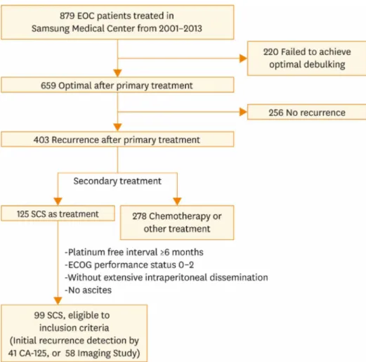
Among patients who had primary treatment for EOC in Samsung Medical Center, 99 EOC patients were identified for having SCS after recurrence. Patients who had undergone SCS were included according to the following inclusion criteria: (1) optimal debulking after primary treatment, (2) platinum-free interval (PFI) ≥6 months, (3) good performance status before surgery as defined by Eastern Cooperative Oncology Group (performance status 0 to 2), (4) single or multiple recurrence sites without extensive intraperitoneal dissemination or ascites on imaging. These inclusion criteria were based on those used in other major retrospective studies [16,18,19]. None of the candidates had received chemotherapy prior to SCS. Flowchart of included patients is shown in Fig. 1.
Patients who underwent SCS were divided into two groups according to the initial diagnostic methods: CA-125 elevation or positive imaging findings. None of the patients showed the symptoms as initial sign of recurrence. The diagnostic criteria for recurrence or metastasis during the follow-up period included rising CA-125 levels or clinical signs of recurrence, which include positive conventional imaging findings.
2. Treatment and follow-up
The standard primary surgical treatment consisted of hysterectomy, bilateral salpingo- oophorectomy, omentectomy, retroperitoneal (pelvic and paraaortic) lymphadenectomy, and tumorectomy of any metastatic lesions. Peritoneal washing was routinely performed.
Peritoneal biopsies were performed if any abnormalities were identified. After primary debulking surgery (PDS), either abdominopelvic computed tomography (CT) or pelvic magnetic resonance imaging (MRI) was performed to confirm the disease status.
Within two weeks of surgery, patients started their first cycle of platinum based combination chemotherapy. Cycles were repeated every 3 weeks until 4 to 6 cycles were completed. The CA-125 level was routinely checked to determine whether CA-125 was in the normal range.
After primary treatment, patients were assessed by physical examination, complete blood count and chemistries, including CA-125 levels, every 3 months for the first 3 years, and every 6 months thereafter. Chest radiography and abdominopelvic CT scan (or alternatively abdominopelvic MRI) were performed every 6 months for the first 3 years, and every 12 months thereafter (although this may not be standard follow-up schedule, it had been proceeding in our institution as routine protocol). Additional diagnostic procedures were performed according to specific clinical suspicions. If recurrence was suspected with
Fig. 1. Flowchart of included patients. CA-125, cancer antigen 125; ECOG, Eastern Cooperative Oncology Group;
EOC, epithelial ovarian cancer; SCS, secondary cytoreductive surgery.
symptoms or CA-125 elevation, additional imaging studies were performed. Recurrence could have been detected by imaging studies with or without CA-125 elevation. After detection of recurrence in EOC patients, time and method of secondary treatment were decided by physicians, usually SCS or chemotherapy. After secondary treatment was performed, the same follow up schedules and treatment were conducted as were those after PDS.
For each patient, the method of recurrence detection was determined from a review of the electric medical record. Detection was attributed to an office visit if the patient presented with symptoms of recurrence or clinical signs on exam. In contrast, detection was attributed to imaging if this was the first or only evidence of disease recurrence. Lastly, detection was attributed to CA-125 level if elevations in this marker were the initial abnormal finding leading to the diagnosis. Elevated CA-125 levels were defined as >35.0 U/mL by the laboratory at our institution. Forty-one of the 99 patients who underwent SCS were initially diagnosed with recurrence based on a CA-125 elevation. In contrast, 58 patients were diagnosed by imaging findings, and did not have elevated CA-125 levels. Patients with both CA-125 elevation and imaging findings were categorized as CA-125 elevation group, and no patients were diagnosed by symptoms alone.
3. Statistical analysis
Summary statistics were used to describe the data. Continuous variables are presented as medians (range). Categorical variables are presented as frequencies (percentages).
The baseline clinical characteristics and study outcomes were compared between the two groups using the Mann-Whitney test for continuous variables, and the chi-square test or Fisher exact test for categorical variables, as appropriate. In order to define the recurrence pattern, the location of the recurrence site was divided into two categories. Recurrences in the pelvis occurring below the pelvic brim were considered pelvic recurrences. Extra-pelvic recurrences were those outside of this defined area. The spread patterns were classified as peritoneal, lymphatic, or hematogenous metastases. Peritoneal recurrences were defined as cases with a recurrence of metastatic lesions in the peritoneum of the pelvis and abdominal area. Hematogenous recurrences were defined as visceral metastases, such as in the liver and lung parenchyma. Lymphatic metastases were defined as metastases to lymph nodes, mostly pelvic and paraaortic, as well as distant lymph nodes. The Kaplan-Meier method was used for survival analysis. Multivariate analyses were performed with the Cox proportional hazards model to evaluate the importance of the recurrence type and other clinicopathological features as prognostic factors. Multivariate p-values were used to characterize the independence of these factors. A 95% CI was used to quantify the relationship between survival time and each independent factor. Statistical analyses were performed using SPSS ver. 21.0 (IBM Co., Armonk, NY, USA). A two-sided p≤0.05 was considered statistically significant. The PFI was defined as the time from the completion of primary treatment to recurrence, death or loss to follow-up. The overall survival from initial diagnosis (OS1) was described as the time between diagnosis and the patient's death or loss to follow-up.
The overall survival after SCS (OS2) was described as the time from SCS to the date of the patient's death or loss to follow-up. We defined the progression-free survival from the initial treatment (PFS1) as the time between the diagnosis and first relapse, or the final follow-up visit. We defined progression-free survival from SCS (PFS2) as the period between the time of SCS to relapse, or the final follow-up visit.
RESULTS
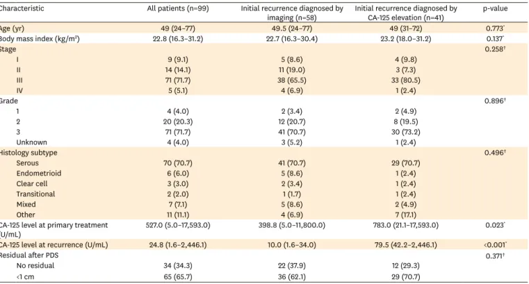
The baseline patient characteristics are shown in Table 1. The largest proportion of patients had stage 3 (n=71, 71.7%), grade 3 (n=71, 71.7%), and serous type (n=70, 70.7%) disease.
There were no statistically significant differences between the two groups with regard to age, BMI, stage, grade, histology or residual disease after PDS. In contrast, there were statistically significant differences in the median CA-125 levels at primary treatment as well as at recurrence between the two groups.
Table 2 shows the recurrence patterns of the first relapse and the surgical characteristics of SCS. There were statistically significant differences in the locations and number of recurrence lesions. In the group with CA-125 elevation, 87.8% of patients had extra-pelvic recurrence, compared to 58.6% of the group diagnosed by imaging. A higher percentage of multiple recurrence lesions were found in the CA-125 elevation group (78%) than in the imaging diagnosis group (39.7%). Differences in recurrence sites and spread patterns (peritoneal/
lymphatic/hematogenous) between two groups were not statistically significant.
With regard to surgical SCS characteristics, 72.4% of patients diagnosed with imaging had no residual disease after SCS; in contrast, only 22.0% of those diagnosed by CA-125 elevation had no residual disease (p<0.001). Also, the median PFI was significantly longer in patients with imaging diagnoses compared to that of the CA-125 group (26 months vs. 19 months, p=0.003). There were no significant differences for interval from initial recurrence detection to SCS between two groups (p=0.324).
Table 1. Baseline patient characteristics
Characteristic All patients (n=99) Initial recurrence diagnosed by
imaging (n=58) Initial recurrence diagnosed by
CA-125 elevation (n=41) p-value
Age (yr) 49 (24–77) 49.5 (24–77) 49 (31–72) 0.773*
Body mass index (kg/m2) 22.8 (16.3–31.2) 22.7 (16.3–30.4) 23.2 (18.0–31.2) 0.137*
Stage 0.258†
I 9 (9.1) 5 (8.6) 4 (9.8)
II 14 (14.1) 11 (19.0) 3 (7.3)
III 71 (71.7) 38 (65.5) 33 (80.5)
IV 5 (5.1) 4 (6.9) 1 (2.4)
Grade 0.896†
1 4 (4.0) 2 (3.4) 2 (4.9)
2 20 (20.3) 12 (20.7) 8 (19.5)
3 71 (71.7) 41 (70.7) 30 (73.2)
Unknown 4 (4.0) 3 (5.2) 1 (2.4)
Histology subtype 0.496†
Serous 70 (70.7) 41 (70.7) 29 (70.7)
Endometrioid 6 (6.0) 5 (8.6) 1 (2.4)
Clear cell 3 (3.0) 2 (3.4) 1 (2.4)
Transitional 2 (2.0) 1 (1.7) 1 (2.4)
Mixed 7 (7.1) 5 (8.6) 2 (4.9)
Other 11 (11.1) 4 (6.9) 7 (17.1)
CA-125 level at primary treatment
(U/mL) 527.0 (5.0–17,593.0) 398.8 (5.0–11,800.0) 783.0 (21.1–17,593.0) 0.023*
CA-125 level at recurrence (U/mL) 24.8 (1.6–2,446.1) 10.0 (1.6–34.0) 79.5 (42.2–2,446.1) <0.001*
Residual after PDS 0.371†
No residual 34 (34.3) 22 (37.9) 12 (29.3)
<1 cm 65 (65.7) 36 (62.1) 29 (70.7)
Values are presented as median (range) or number (%).
CA-125, cancer antigen 125; PDS, primary debulking surgery.
*Mann-Whitney test. †Fisher exact test.
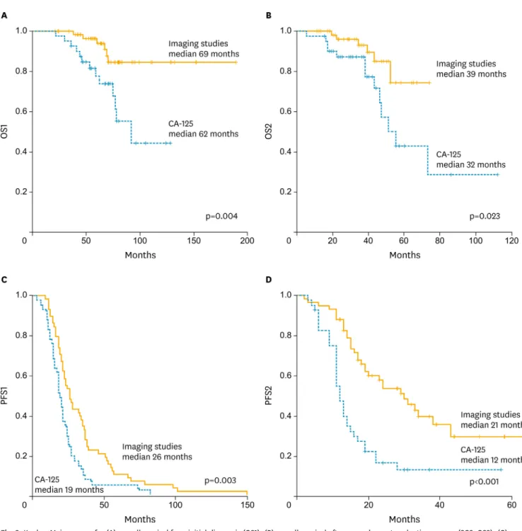
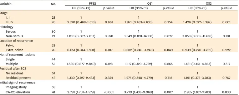
The survival analyses are shown in Fig. 2. The Kaplan-Meier curves for OS1, OS2, PFS1, and PFS2, revealed that patients in the imaging group had significantly better survival than did those with elevated CA-125. The median OS1, OS2, PFS1, and PFS2 were comparably longer in the imaging group than they were in the CA-125 group. In multivariate analysis with clinicopathological variables and recurrent patterns, CA-125 elevation at recurrence diagnosis was a significant factor for poorer survival in this patients group (Table 3).
DISCUSSION
There were differences in recurrence patterns and survival outcomes between two groups of patients who had secondary cytoreduction with distinct initial methods of recurrence diagnosis. It is difficult to directly derive the role of SCS in this study, because patients included in this study were limited to patients who underwent SCS for treatment, not all patients with recurrence. We compared the characteristics and survival of patients with different initial diagnostic methods of EOC recurrence to determine which group is appropriate for SCS.
The group diagnosed by imaging demonstrated a high percentage of pelvic recurrence and single site recurrence. This may be attributed to residual disease after SCS. The residual disease after SCS was relatively more frequent in the elevated CA-125 group than in the imaging group. Although it was not clearly demonstrated in our data, these differences may Table 2. Recurrence patterns and surgical characteristics by group
Characteristic All patients (n=99) Initial recurrence
diagnosed by imaging (n=58)
Initial recurrence diagnosed by CA-125
elevation (n=41)
p-value
Recurrence site 0.653*
Peritoneal surface 37 (37.4) 25 (43.1) 12 (29.3)
Mesentery and omentum 15 (15.2) 7 (12.1) 8 (19.5)
Lymph nodes 17 (17.2) 10 (17.2) 7 (17.1)
Spleen 13 (13.1) 8 (13.8) 5 (12.2)
Liver 12 (12.1) 6 (10.3) 6 (14.6)
Diaphragm 5 (5.1) 2 (3.4) 3 (7.3)
Location of recurrence 0.002*
Pelvic 29 (29.3) 24 (41.4) 5 (12.2)
Extra-pelvic 70 (70.7) 34 (58.6) 36 (87.8)
No. of recurrent lesions <0.001*
Single 44 (44.4) 35 (60.3) 9 (22.0)
Multiple 55 (55.6) 23 (39.7) 32 (78.0)
Spread pattern 0.852*
Peritoneal 78 (78.8) 45 (77.6) 33 (80.5)
Lymphatic 17 (17.2) 11 (19.0) 6 (14.6)
Hematogenous 4 (4.0) 2 (3.4) 2 (4.9)
Size of largest recurrent lesion (mm) 30 (5–130) 30 (5–120) 30 (10–130) 0.235†
Operation type 0.817*
Laparotomy 74 (74.7) 44 (75.9) 30 (73.2)
Laparoscopy 25 (25.3) 14 (24.1) 11 (26.8)
Residual after SCS <0.001*
No residual 51 (51.5) 42 (72.4) 9 (22.0)
Residual present 48 (48.5) 16 (27.6) 32 (78.0)
PFI (mo) 22 (6–151) 26 (9–151) 19 (6–83) 0.003†
Interval from initial recurrence detection to SCS (day) 32 (7–216) 32.5 (9–203) 24 (7–216) 0.324† Values are presented as number (%) or median (range).
CA-125, cancer antigen 125; PFI, platinum-free interval; SCS, secondary cytoreductive surgery.
*Fisher exact test. †Mann-Whitney test.
be related to differences in survival outcomes between the two groups; residual tumor after cytoreduction is widely known as a powerful prognostic factor in EOC patients [20-22].
Previous studies have addressed differences in outcomes according to the time of relapse and method of diagnosing recurrence. The CA-125 levels in EOC patients might have been elevated several months before symptoms arise [23]. In contrast, imaging studies in EOC patients are known to be useful in the detection of late recurrence, especially in patients who
Fig. 2. Kaplan-Meier curves for (A) overall survival from initial diagnosis (OS1), (B) overall survival after secondary cytoreductive surgery (SCS; OS2), (C) progression-free survival after the initial treatment (PFS1), and (D) progression-free survival from SCS (PFS2) in patients with recurrent ovarian cancer according
may benefit from SCS [24]. In one study from the United Kingdom, treating recurrent EOC characterized by CA-125 elevation alone did not increase OS. Imaging studies, such as CT scan, are capable of diagnosing asymptomatic recurrence; however, previous studies have not clarified the presence of survival differences [25,26]. Although the objective and patients group of this study were different from that of other previous studies, our data showed significant differences in the survival outcomes of patients with recurrence diagnosed by CA- 125 elevation and imaging studies.
Our study is limited by its retrospective nature and single institution data. We are planning the validation with multicenter data sets for more definite results. In our study, patients had initial signs of recurrence recognized by either CA-125 elevation or imaging study. We could not compare the patients with symptoms as initial sign because all patients were asymptomatic at the time of recurrence. In addition, the results of this study can only be applied to restrictive criteria of EOC patients because patients included in this study were limited to patients who underwent SCS for treatment, not all patients with recurrence.
Patients without definitive recurrence lesions on imaging or those with diffuse, extensive recurrence lesions who were unlikely to have SCS as a secondary treatment were not included. Patients selected for SCS may have more limited recurrence lesions and better performance status than other patients.
Despite some limitations, we concluded that, in platinum sensitive patients who had undergone SCS, patients with recurrence diagnosed on imaging had better survival outcomes than did those diagnosed by CA-125 elevation. Patients whose recurrence was diagnosed by imaging also had more pelvic recurrence, single lesion recurrences and no residual after SCS compared to those whose recurrence was diagnosed by CA-125 elevation. In this context, patients with radiologic evidence of recurrence (but without CA-125 elevation) are likely to be more appropriate for SCS than are patients with CA-125 elevation as initial sign of recurrence.
Table 3. Multivariate analysis for PFS2, OS1, OS2 according to clinicopathological features and recurrence patterns in epithelial ovarian cancer patients with SCS (n=99)
Variable No. PFS2 OS1 OS2
HR (95% CI) p-value HR (95% CI) p-value HR (95% CI) p-value
Stage
I, II 23 1 1 1
III, IV 76 0.870 (0.468–1.618) 0.661 1.921 (0.483–7.638) 0.354 1.426 (0.377–5.392) 0.601
Histology
Serous 80 1 1 1
Non-serous 19 1.010 (0.507–2.013) 0.978 3.549 (0.891–14.138) 0.072 3.058 (0.805–11.616) 0.101
Location of recurrence
Pelvic 29 1 1 1
Extra-pelvic 70 0.651 (0.344–1.231) 0.187 0.882 (0.240–3.240) 0.849 0.939 (0.270–3.269) 0.922
No. of recurrent lesions
Single 44 1 1 1
Multiple 55 1.580 (0.877–2.849) 0.128 1.112 (0.329–3.752) 0.865 1.481 (0.451–4.863) 0.517
Residual after SCS
No residual 51 1 1 1
Residual present 48 1.330 (0.727–2.433) 0.354 1.275 (0.340–4.779) 0.718 1.191 (0.375–3.783) 0.767 Initial sign of recurrence
Imaging study 58 1 1 1
CA-125 elevation 41 2.791 (1.701–4.579) <0.001 3.779 (1.433–9.969) 0.007 2.935 (1.107–7.783) 0.030 The Cox's proportional hazard regression test was used for multivariate analysis.
CA-125, cancer antigen 125; HR, hazard ratio; OS1, overall survival from initial diagnosis; OS2, overall survival after SCS; PFS2, progression-free survival after SCS;
SCS, secondary cytoreductive surgery.
REFERENCES
1. Siegel RL, Miller KD, Jemal A. Cancer statistics, 2015. CA Cancer J Clin 2015;65:5-29.
PUBMED | CROSSREF
2. Lim MC, Moon EK, Shin A, Jung KW, Won YJ, Seo SS, et al. Incidence of cervical, endometrial, and ovarian cancer in Korea, 1999-2010. J Gynecol Oncol 2013;24:298-302.
PUBMED | CROSSREF
3. Cannistra SA. Cancer of the ovary. N Engl J Med 2004;351:2519-29.
PUBMED | CROSSREF
4. Vaidya AP, Curtin JP. The follow-up of ovarian cancer. Semin Oncol 2003;30:401-12.
PUBMED | CROSSREF
5. Rubin SC, Randall TC, Armstrong KA, Chi DS, Hoskins WJ. Ten-year follow-up of ovarian cancer patients after second-look laparotomy with negative findings. Obstet Gynecol 1999;93:21-4.
PUBMED
6. Cotte E, Glehen O, Mohamed F, Lamy F, Falandry C, Golfier F, et al. Cytoreductive surgery and intraperitoneal chemo-hyperthermia for chemo-resistant and recurrent advanced epithelial ovarian cancer: prospective study of 81 patients. World J Surg 2007;31:1813-20.
PUBMED | CROSSREF
7. Chiang YC, Chen CA, Chiang CJ, Hsu TH, Lin MC, You SL, et al. Trends in incidence and survival outcome of epithelial ovarian cancer: 30-year national population-based registry in Taiwan. J Gynecol Oncol 2013;24:342-51.
PUBMED | CROSSREF
8. Skates SJ, Xu FJ, Yu YH, Sjövall K, Einhorn N, Chang Y, et al. Toward an optimal algorithm for ovarian cancer screening with longitudinal tumor markers. Cancer 1995;76 Suppl:2004-10.
PUBMED | CROSSREF
9. Högberg T, Kågedal B. Long-term follow-up of ovarian cancer with monthly determinations of serum CA 125. Gynecol Oncol 1992;46:191-8.
PUBMED | CROSSREF
10. Gadducci A, Conte P, Cianci C, Negri S, Genazzani AR. Treatment options in patients with recurrent ovarian cancer. Anticancer Res 2001;21:3557-64.
PUBMED
11. Sharma A, Bernacki RJ. Ovarian cancer patients with high CA-125 but no symptoms--should antiangiogenic treatments be considered? Oncol Res 1997;9:53-4.
PUBMED
12. Fleming LW. Playing the waiting game ... the asymptomatic patient with recurrent ovarian cancer detected only by rising Ca125 levels. Scott Med J 2001;46:81-3.
PUBMED
13. Goonewardene TI, Hall MR, Rustin GJ. Management of asymptomatic patients on follow-up for ovarian cancer with rising CA-125 concentrations. Lancet Oncol 2007;8:813-21.
PUBMED | CROSSREF
14. Al Rawahi T, Lopes AD, Bristow RE, Bryant A, Elattar A, Chattopadhyay S, et al. Surgical cytoreduction for recurrent epithelial ovarian cancer. Cochrane Database Syst Rev 2013;2:CD008765.
PUBMED
15. Tian WJ, Chi DS, Sehouli J, Tropé CG, Jiang R, Ayhan A, et al. A risk model for secondary cytoreductive surgery in recurrent ovarian cancer: an evidence-based proposal for patient selection. Ann Surg Oncol 2012;19:597-604.
PUBMED | CROSSREF
16. Harter P, du Bois A, Hahmann M, Hasenburg A, Burges A, Loibl S, et al.AGO Ovarian Cancer Study Group. Surgery in recurrent ovarian cancer: the Arbeitsgemeinschaft Gynaekologische Onkologie (AGO) DESKTOP OVAR trial. Ann Surg Oncol 2006;13:1702-10.
PUBMED | CROSSREF
17. Janco JM, Kumar A, Weaver AL, McGree ME, Cliby WA. Performance of AGO score for secondary cytoreduction in a high-volume U.S. center. Gynecol Oncol 2016;141:140-7.
PUBMED | CROSSREF
18. Chi DS, McCaughty K, Diaz JP, Huh J, Schwabenbauer S, Hummer AJ, et al. Guidelines and selection criteria for secondary cytoreductive surgery in patients with recurrent, platinum-sensitive epithelial ovarian carcinoma. Cancer 2006;106:1933-9.
PUBMED | CROSSREF
19. Onda T, Yoshikawa H, Yasugi T, Yamada M, Matsumoto K, Taketani Y. Secondary cytoreductive surgery for recurrent epithelial ovarian carcinoma: proposal for patients selection. Br J Cancer 2005;92:1026-32.
PUBMED | CROSSREF
20. Eisenkop SM, Spirtos NM, Friedman RL, Lin WC, Pisani AL, Perticucci S. Relative influences of tumor volume before surgery and the cytoreductive outcome on survival for patients with advanced ovarian cancer: a prospective study. Gynecol Oncol 2003;90:390-6.
PUBMED | CROSSREF
21. Aletti GD, Dowdy SC, Podratz KC, Cliby WA. Surgical treatment of diaphragm disease correlates with improved survival in optimally debulked advanced stage ovarian cancer. Gynecol Oncol 2006;100:283-7.
PUBMED | CROSSREF
22. Chi DS, Eisenhauer EL, Lang J, Huh J, Haddad L, Abu-Rustum NR, et al. What is the optimal goal of primary cytoreductive surgery for bulky stage IIIC epithelial ovarian carcinoma (EOC)? Gynecol Oncol 2006;103:559-64.
PUBMED | CROSSREF
23. von Georgi R, Schubert K, Grant P, Münstedt K. Post-therapy surveillance and after-care in ovarian cancer. Eur J Obstet Gynecol Reprod Biol 2004;114:228-33.
PUBMED | CROSSREF
24. Gadducci A, Cosio S, Zola P, Landoni F, Maggino T, Sartori E. Surveillance procedures for patients treated for epithelial ovarian cancer: a review of the literature. Int J Gynecol Cancer 2007;17:21-31.
PUBMED | CROSSREF
25. Tanner EJ, Chi DS, Eisenhauer EL, Diaz-Montes TP, Santillan A, Bristow RE. Surveillance for the detection of recurrent ovarian cancer: survival impact or lead-time bias? Gynecol Oncol 2010;117:336-40.
PUBMED | CROSSREF
26. Gadducci A, Fuso L, Cosio S, Landoni F, Maggino T, Perotto S, et al. Are surveillance procedures of clinical benefit for patients treated for ovarian cancer?: A retrospective Italian multicentric study. Int J Gynecol Cancer 2009;19:367-74.
PUBMED | CROSSREF