Peripheral eosinophilia - is it a predictable factor associated with eosinophilic cholecystitis?
Seung-Seop Yeom1, Ho-Hyun Kim1, Jung-Chul Kim1, Young-Hoe Hur1, Yang-Seok Koh1, Chol-Kyoon Cho1, Hyun-Jong Kim1, Sang-Soo Shin2, and Hyung-Seok Kim3
Departments of 1Surgery, 2Radiology, and 3Pathology, Chonnam National University Medical School, Gwangju, Korea
Backgrounds/Aims: The purpose of this study was to evaluate the role of peripheral eosinophilia as a predictable factor associated with Eosinophilic cholecystitis (EC) compared with other forms of cholecystitis in patients who underwent a cholecystectomy. Methods: Between January 2001 and May 2011, the histopathologic features of 3,539 chol- ecystectomy specimens were reviewed retrospectively. EC was diagnosed in 30 specimens (0.84%). Data from 30 consecutive patients with EC (eosinophilic cholecystitis group [E-group]) were compared with a retrospective control group of 60 patients (other cholecystitis group [O-group]) during the same period. The two groups were matched for age, gender, and the presence of cholelithiasis. Results: The median absolute eosinophil count 1 day post-operatively was 144 cells/mm3 (range: 9-801 cells/mm3) in the E-group and 93 cells/mm3 (range: 0-490 cells/mm3) in the O-group (p=0.036). Pre-operative peripheral eosinophilia was more common in the E-group than the O-group (20% vs. 3.3%, p=0.015). Multivariate analysis revealed that pre-operative peripheral eosinophilia was an independent significant pre- dictable factor associated with EC (odds ratio=7.250, 1.365 <95% confidence interval<38.494, p=0.020). Conclu- sions: In the present study, pre-operative peripheral eosinophilia was shown to be an independent predictable factor associated with EC. Further researches seem to be necessary to confirm this finding. (Korean J Hepatobiliary Pancreat Surg 2012;16:65-69)
Key Words: Eosinophilic cholecystitis; Eosinophilia; Predictable factor; Cholecystectomy
Received: April 10, 2012; Revised: May 14, 2012; Accepted: May 16, 2012 Corresponding author: Jung-Chul Kim
Department of Surgery, Chonnam National University Medical School, 42, Jebong-ro, Dong-gu, Gwangju 501-757, Korea Tel: +82-62-220-6456, Fax: +82-62-227-1635, E-mail: [email protected]
Copyright Ⓒ 2012 by The Korean Association of Hepato-Biliary-Pancreatic Surgery Korean Journal of Hepato-Biliary-Pancreatic Surgery ∙ pISSN: 1738-6349
INTRODUCTION
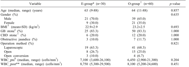
Eosinophilic infiltration of the gallbladder based on his- topathologic examination is a rare entity. Cellular in- filtrates of the gallbladder comprising 90% eosinophils are classified as eosinophilic cholecystitis (EC) (Fig. 1).1-3 The condition is termed lympho-eosinophilic cholecystitis when the infiltrate is comprised of 50-75% eosinophils along with other inflammatory cells in the gallbladder wall.4-6 EC is a rare form of cholecystitis.7 Since EC was first described in 1949 by Albot et al,8 several case reports have been published in the English literature (<50 cases).
The condition likely represents a subgroup of patients with a unique or hypersensitive type of inflammatory re- sponse to altered bile.9
The etiology of EC is not completely understood, but might be associated with a hypersensitivity to antibiotics,
other drugs, herbal medicines, hepatic echinococcosis, or as a variant manifestation of eosinophilic gastroenter- itis.1,10 The diagnosis of EC is usually made based on his- tologic studies following cholecystectomy. The purpose of this study was to evaluate the role of peripheral eosino- philia as a predictable factor associated with EC compared with other types of cholecystitis in patients who under- went cholecystectomies.
METHODS
Patients
Between January 2001 and May 2011, 3539 patients underwent cholecystectomies for cholecystitis in the Department of Surgery, Chonnam National University Hospital. Thirty specimens (0.84%) were confirmed pathologically to be EC. EC is defined as eosinophilic in-
Fig. 1. Histopathology findings of eosinophilic cholecystitis. (A) Dense infiltration of eosinophils ([B] black arrow) involving the gallbladder diffusely in a transmural manner (>90%), suggesting eosinophilic cholecystitis (A: H&E stain, ×40, B: H&E stain, ×200).
filtration of the gallbladder wall consisting of >90%
eosinophils.2
Data from 30 consecutive patients with EC (E-group) were compared with a retrospective control group of 60 patients (other cholecystitis group [O-group]) during the study period. The patients in the two groups were matched for age, gender, body mass index, the presence of chol- elithiasis, jaundice, operative methods, and white blood cell count pre-operatively and 1 day post-operatively. The patients in the E- and O-groups had similar pre-operative assessments and post-operative management.
The clinical presentation (gender, age, and the presence of cholelithiasis), surgical methods, operative findings, histopathologic features, and pre- and post-operative abso- lute eosinophil count were analyzed retrospectively. Eosi- nophilia was defined as a condition in which the absolute eosinophil count in the peripheral blood exceeded ≥600 cells/mm3.11
Statistical analysis
Summary statistics are reported using mean or median values, and standard deviation or range. The statistical evaluation was carried out using SPSS for Windows (version 17.0; SPSS, Inc., Chicago, IL, USA). A Student’s t-test and Mann-Whitney U-test were used for the mean comparison of continuous variables and for ordinal data, respectively, whereas a chi-squared test and Fisher’s exact test were used to compare frequencies of categorical vari-
ables between the groups. To evaluate predictable factors for EC, a multivariate analysis was carried out by binary logistic multiple regression tests using dummy variable.
Significance was defined as a p≤0.05.
RESULTS
The clinicopathologic features of patients are summar- ized in Table 1. Additional parameters, such as pre- and post-operative white blood cell count and operative meth- od, were similar between the E- and O-groups.
Clinical findings of patients with EC
Table 1 lists the clinical features of the 30 patients with EC. The patients with EC were comprised of 21 males (70.0%) and 9 females (30.0%) with a median age of 63 years (range: 9-88 years). The mean body mass index (BMI) was 22.9±2.9 kg/m2.
The clinical manifestations were not specific for EC.
The main presenting complaint was abdominal pain (82.8%) and epigastric discomfort (13.8%). Two patients (6.7%) were asymptomatic.
Cholecystolithiasis (gallbladder stones) and common bile duct stones were noted in 25 (83.3%) and 6 (20.0%) patients, respectively. Three patients (10.0%) had ob- structive jaundice based on the pre-operative laboratory examinations (Table 1), and 6 patients (20.0%) had pre-operative peripheral eosinophilia (Table 2).
Table 3. Features of cases of eosinophilic cholecystitis with allergy or parasite infestation
Case Age (yr) Sex Allergy Parasite Peripheral
eosinophilia Gallstone 1
2
61 34
Male Female
No
Yes (penicillin)
Yes (Clonorchis sinensis) No
Yes No
No Yes Table 2. Data comparison of the eosinophilic cholecystitis group and other cholecystitis group (n=90)
Variable E-group* (n=30) O-group† (n=60) p-value
Eosinophilia_pre‡ (%)
Eos_pre∥ (median, range) (cells/mm3) Eosinophilia_post§ (%)
Eos_post¶ (median, range) (cells/mm3) Eos_diff** (median, range) (cells/mm3)
6 (20.0) 209 (0-1,559)
2 (6.7) 144 (9-801) 72 (1-1,134)
2 (3.3) 147 (0-621)
0 93 (0-490) 65 (1-402)
0.015 0.174 0.109 0.036 0.349
*eosinophilic cholecystitis group, †other cholecystitis group, ‡pre-operative eosinophilia, §post-operative eosinophilia, ∥absolute eosinophil count at pre-operative laboratory findings, ¶absolute eosinophil count at post-operative day 1, **difference in absolute eosinophil count between pre-operative and post-operative day 1
Table 1. Clinical characteristics of patients with cholecystitis (n=90)
Variable E-group* (n=30) O-group† (n=60) p-value
Age (median, range) (years) Gender (%)
Male Female
BMI‡ (mean±SD) (kg/m2) GB stone§ (%)
CBD stone∥ (%) Obstructive jaundice (%) Operation method (%) Laparoscopic Open
Open conversion
WBC_pre¶ (median, range) (cells/mm3) WBC_post** (median, range) (cells/mm3)
63 (9-88) 21 (70.0) 9 (30.0) 22.9±2.9 25 (83.3) 6 (20.0) 3 (10.0) 19 (63.3) 8 (26.7) 3 (10.0) 7,100 (3,600-26,100) 8,750 (5,300-29,500)
64 (11-88) 39 (65.0) 21 (35.0) 23.2±2.5 50 (83.3) 12 (20.0) 7 (11.7) 41 (68.3) 15 (25.0) 4 (6.7)
6,450 (2,900-21,300) 8,100 (5,200-24,600)
0.857 0.635
0.693 1.000 1.000 1.000 0.821
0.204 0.451
*eosinophilic cholecystitis group, †other cholecystitis group, ‡body mass index, §gallbladder stone, ∥common bile duct stone,
¶white blood cell count on pre-operative laboratory findings, **white blood cell count on post-operative day 1
Only one of the 30 patients had a positive history for allergies (penicillin). This one case was due to a parasitic infestation (Clonorchis sinensis). None of the 30 cases were associated with drug therapy, or other pre-existing medical conditions (Table 3).
Comparision of data between the EC and control groups (Table 2)
The median pre-operative absolute eosinophil count was 209 cells/mm3 (range: 0-1,553 cells/mm3) in the E-group and 147 cells/mm3 (range: 0-621 cells/mm3) in the O-group. The median absolute eosinophil count on
post-operative day 1 was 144 cells/mm3 (range: 9-801 cells/mm3) in E-group and 93 cells/mm3 (range: 0-490) in O-group.
In the O-group, only 2 patients (3.3%) had peripheral eosinophilia based on the pre-operative laboratory exami- nations. On the first post-operative day, no patient (0%) demonstrated peripheral eosinophilia as compared to 2 pa- tients (6.7%) in the E-group.
The median absolute eosinophil count between pre-op- erative and post-operative day 1 was 72 cells/mm3 in the E-group and 65 cells/mm3 in the O-group.
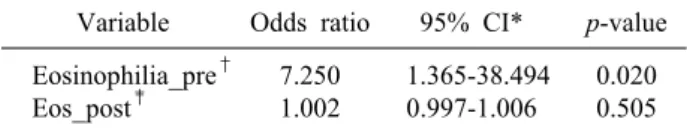
Table 4. Multivariate analysis for predictable factors asso- ciated with eosinophilic cholecystitis
Variable Odds ratio 95% CI* p-value Eosinophilia_pre†
Eos_post‡
7.250 1.002
1.365-38.494 0.997-1.006
0.020 0.505
*95% confidence interval, †pre-operative eosinophilia, ‡ab- solute eosinophil count at post-operative day 1
Univariate and multivariate analyses for pred- ictable factors associated with EC (Table 2) Univariate analysis revealed that pre-operative periph- eral eosinophilia (p=0.015) and the absolute eosinophil count on post-operative day 1 (p=0.036) were significant predictable factors associated with EC (Table 2).
Multivariate analysis revealed that pre-operative periph- eral eosinophilia was an independent predictable factor for EC (odds ratio [OR]=7.250, 1.365 <95% confidence in- terval [CI] <38.494; p=0.020) (Table 4).
DISCUSSION
EC is an uncommon form of cholecystitis with an in- cidence ranging from 0.5-6.5% in cholecystectomy speci- mens (2,7,12). The incidence of EC was 0.5% in a large review of 625 operative cholecystectomy specimens by Fox (2), and 6.4% in a review of 217 specimens by Dabbs.12 In the present study, EC was diagnosed in 30 of 3539 specimens (0.84%), which is comparable to the results of other reported series.
The etiology of EC is obscure, but the suggested ori- gins include allergies, local diathesis involving gallstones, parasites, acalculous cholecystitis, hypereosinophilic syn- drome (HES; in which eosinophils invade tissues, such as the brain, heart, lung, liver, and gallbladder), and eosino- philic gastroenteritis.1,3,12-17 It has been reported that EC might be associated with hypersensitivity to antibiotics, other drugs, and herbal medicines.1,10
A literature review showed that most patients with EC had an idiopathic etiology.1,18 In the present study, only one of the 30 patients had a positive history for allergies (penicillin). This case was due to a parasitic infestation, Clonorchis sinensis. Most patients (26 of 30 patients) had idiopathic EC. None of the 30 patients had an association with drug therapy or other pre-existing medical condi- tions. Therefore, the etiology of EC was not a predictable
factor associated with the population for this study.
In addition, EC has a clinical presentation similar to typical cholecystitis with right upper quadrant pain and an elicited Murphy’s sign. In clinical practice, EC is clin- ically indistinguishable from the most common form of acute cholecystitis (2,13). Therefore, there is no known specific predictable factor for EC and the diagnosis of EC is based on the histopathology of cholecystectomy speci- mens. The characteristic histologic features of EC is trans- mural inflammatory infiltration of the gallbladder wall that is comprised of >90% eosinophils.1,3 Eosinophils are one of the immune system white blood cells components responsible for combating multicellular parasites and in- fections in vertebrates. Along with mast cells, eosinophils also control the mechanisms associated with allergy and asthma. An increase in eosinophils typically occurs in people with parasite infestation of the intestines, collagen vascular disease (rheumatoid arthritis), malignant disease (Hodgkin’s disease), extensive skin disease (exfoliative dermatitis), Addison’s disease, and the use of certain drugs (penicillin).11
It has been reported that a laboratory examination sometimes reveals peripheral blood eosinophilia in pa- tients with EC.3,10,15,16,19-21 Kim1 reported that peripheral eosinophilia occurred in 4 of 15 cases. In contrast, it has been reported that EC has peripheral eosinophilia or spe- cific laboratory features.13,22
In the present study, pre-operative peripheral eosino- philia (≥600 cells/mm3) was noted in 6 of the 30 cases (20%) in the E-group. There was a statistically significant difference between the E-group and the O-group (OR=
7.250, 1.365 <95% CI <38.494; p=0.020). Therefore, if patients with cholecystitis have pre-operative peripheral eosinophilia, there is great potential that the subtype of cholecystitis is EC. The pathogenesis and etiology of EC are not well-understood, but the presence of peripheral eo- sinophilia and abundant eosinophils in the gallbladder wall provide some support that the disease is mediated by a hypersensitivity-type reaction. Although the mechanism for gallbladder recruitment of eosinophils in EC is un- known, Desreumaux et al.23 reported that eosinophil re- cruitment and activation is induced by cytokines, such as interleukin (IL)-3, granulocyte-macrophage colony-stim- ulating factor (GM-CSF), and IL-5, in eosinophilic gastroenteris. Therefore, we suggest that further evalua-
tion is necessary for the molecular mechanism predispos- ing to peripheral eosinophilia and eosinophilic infiltration in patients with EC.
Nevertheless, the results of our study were limited by the non-randomized design, a small number of cases, and the selection bias related to the choice of approach based merely on demographic characteristics. And whether the result of our study is clinically relevant or not remains debatable. Thus, a larger group of patients is necessary to analyze the predictable factors associated with EC.
In summary, EC is a rare entity that is generally found only in cholecystectomy specimens. The etiology is ob- scure, but involves local and systemic eosinophilic in- flammatory reactions. The diagnosis of the EC is usually made based on histologic studies following cholecy- stectomy. In the present study, pre-operative peripheral eosinophilia (≥600 cells/mm3) was found to be a sig- nificant predictable factor for EC. Further researches seem to be necessary to confirm this finding.
REFERENCES
1. Kim YH. Eosinophilic cholecystitis in association with clonorchis sinensis infestation in the common bile duct. Clin Radiol 1999;54:552-554.
2. Fox H, Mainwaring AR. Eosinophilic infiltration of the gallb- ladder. Gastroenterology 1972;63:1049-1052.
3. Felman RH, Sutherland DB, Conklin JL, et al. Eosinophilic chol- ecystitis, appendiceal inflammation, pericarditis, and cephalospor- in-associated eosinophilia. Dig Dis Sci 1994;39:418-422.
4. Sánchez-Pobre P, López-Ríos Moreno F, Colina F, et al. Eosino- philic cholecystitis: an infrequent cause of cholecystectomy.
Gastroenterol Hepatol 1997;20:21-23.
5. Punia RP, Arya S, Jain P, et al. Eosinophilic and lympho-eosino- philic cholecystitis. Indian J Gastroenterol 2003;22:153-154.
6. Hellstrom HR. Eosinophilic and lymphoeosinophilic cholecystitis.
Am J Surg Pathol 1994;18:215-216.
7. Kaji K, Yoshiji H, Yoshikawa M, et al. Eosinophilic cholecystitis
along with pericarditis caused by Ascaris lumbricoides: a case report. World J Gastroenterol 2007;13:3760-3762.
8. Albot G, Poilleux, Olivier C, et al. Les cholecystites a eosino- phils. Presse Med 1949;39:558-559.
9. Malik KA. Eosinophilic cholecystitis: an infrequent cause of cholecystectomy. Pakistan J Med Sci 2010;26:724-725.
10. Adusumilli PS, Lee B, Parekh K, et al. Acalculous eosinophilic cholecystitis from herbal medicine: a review of adverse effects of herbal medicine in surgical patients. Surgery 2002;131:352- 356.
11. Tefferi A. Blood eosinophilia: a new paradigm in disease classi- fication, diagnosis, and treatment. Mayo Clin Proc 2005;80:
75-83.
12. Dabbs DJ. Eosinophilic and lymphoeosinophilic cholecystitis. Am J Surg Pathol 1993;17:497-501.
13. Rosengart TK, Rotterdam H, Ranson JH. Eosinophilic chol- angitis: a self-limited cause of extrahepatic biliary obstruction.
Am J Gastroenterol 1990;85:582-585.
14. Russell CO, Dowling JP, Marshall RD. Acute eosinophilic chol- ecystitis in association with hepatic echinococcosis. Gastroenter- ology 1979;77:758-760.
15. Tenner S, Roston A, Lichtenstein D, et al. Eosinophilic cholan- giopathy. Gastrointest Endosc 1997;45:307-309.
16. al-Abdulla NA, Schulick RD, Regan F. Hypereosinophilic scle- rosing cholangitis: findings using half-Fourier magnetic reso- nance imaging. Hepatogastroenterology 2000;47:359-361.
17. Hepburn A, Coady A, Livingstone J, et al. Eosinophilic chol- ecystitis as a possible late manifestation of the eosinophilia-myal- gia syndrome. Clin Rheumatol 2000;19:470-472.
18. Shakov R, Simoni G, Villacin A, et al. Eosinophilic cholecystitis, with a review of the literature. Ann Clin Lab Sci 2007;37:182- 185.
19. Tajima K, Katagiri T. Deposits of eosinophil granule proteins in eosinophilic cholecystitis and eosinophilic colitis associated with hypereosinophilic syndrome. Dig Dis Sci 1996;41:282-288.
20. Butler TW, Feintuch TA, Caine WP Jr. Eosinophilic cholangitis, lymphadenopathy, and peripheral eosinophilia: a case report. Am J Gastroenterol 1985;80:572-574.
21. Muhlberger F. Morphology of eosinophilic cholecystitis and the problem of its allergic genesis. Int Arch Allergy Appl Immunol 1954;5:434-448.
22. Parry SW, Pelias ME, Browder W. Acalculous hypersensitivity cholecystitis: hypothesis of a new clinicopathologic entity.
Surgery 1988;104:911-916.
23. Desreumaux P, Bloget F, Seguy D, et al. Interleukin 3, gran- ulocyte-macrophage colony-stimulating factor, and interleukin 5 in eosinophilic gastroenteritis. Gastroenterology 1996;110:768- 774.
![Fig. 1. Histopathology findings of eosinophilic cholecystitis. (A) Dense infiltration of eosinophils ([B] black arrow) involving the gallbladder diffusely in a transmural manner (>90%), suggesting eosinophilic cholecystitis (A: H&E stain, ×40, B: H&](https://thumb-ap.123doks.com/thumbv2/123dokinfo/5187031.113050/2.892.88.807.155.427/histopathology-eosinophilic-cholecystitis-infiltration-eosinophils-gallbladder-eosinophilic-cholecystitis.webp)