ABSTRACT
Background: Calprotectin is the major cytosolic protein in neutrophil granulocytes.
Although asthma is known to cause eosinophilic inflammation, some patients with asthma have non-eosinophilic inflammation, which is characterized by local neutrophilic inflammation. The aim of this study was to assess calprotectin expression levels in a mouse model of asthma, and to observe the relationship of serum calprotectin level and clinical variables in patients with asthma.
Methods: Mice were sensitized and challenged with 10 μg and 20 μg of Aspergillus fumigatus, respectively; mice treated with saline were used as a control. The levels of calprotectin were determined using enzyme-linked immunosorbent assay, immunoblotting, and immunohistochemical analysis. The serum levels of calprotectin were also assessed in patients with asthma. The relationship between calprotectin and clinicopathological characteristics was determined.
Results: Calprotectin, S100A8, and S100A9 expression was elevated in the mouse lungs, Calprotectin levels were higher in the serum of patients with asthma (n = 33) compared with those of healthy individuals (n = 28). Calprotectin levels correlated with forced expiratory volume in one second/forced vital capacity (r = −0.215, P = 0.043), smoke amount (r = 0.413, P = 0.017), body mass index (r = −0.445, P = 0.000), and blood neutrophil percentage (r = 0.300, P = 0.004) in patients with asthma.
Conclusion: Our data suggest that calprotectin could potentially be used as a biomarker for asthma.
Keywords: Calprotectin; Asthma; Airway Inflammation
INTRODUCTION
Asthma is a multifactorial chronic lung disease characterized by airflow obstruction and airway inflammation. Asthma is an extremely heterogeneous condition and can be classified into different subgroups, phenotypes, and endotypes.1,2 Asthma can be broadly divided into eosinophilic or non- eosinophilic, based on airway and peripheral blood immune cellular profiles.1,2
Calprotectin is a 36.5 kDa calcium- and zinc-binding protein and is the major cytosolic protein in neutrophil granulocytes.3 Fecal calprotectin levels have been proposed as a
Original Article
Received: Mar 26, 2020 Accepted: Aug 24, 2020 Address for Correspondence:
An-Soo Jang, MD
Division of Allergy and Respiratory Medicine, Department of Internal Medicine, Soonchunhyang University Bucheon Hospital, 170 Jomaru-ro, Wonmi-gu, Bucheon 14584, Republic of Korea.
E-mail: [email protected]
© 2020 The Korean Academy of Medical Sciences.
This is an Open Access article distributed under the terms of the Creative Commons Attribution Non-Commercial License (https://
creativecommons.org/licenses/by-nc/4.0/) which permits unrestricted non-commercial use, distribution, and reproduction in any medium, provided the original work is properly cited.
ORCID iDs Yun-Gi Lee
https://orcid.org/0000-0003-2743-5496 Jisu Hong
https://orcid.org/0000-0002-2452-4824 Pureun-Haneul Lee
https://orcid.org/0000-0002-1431-2393 Junehyuk Lee
https://orcid.org/0000-0003-3958-0172 Sung-Woo Park
https://orcid.org/0000-0003-1498-3389 DoJin Kim
https://orcid.org/0000-0002-1150-9588 An-Soo Jang
https://orcid.org/0000-0001-5343-023X Funding
This research was supported by the Basic Science Research Program through the
Yun-Gi Lee , Jisu Hong , Pureun-Haneul Lee , Junehyuk Lee , Sung-Woo Park , DoJin Kim , and An-Soo Jang
Division of Allergy and Respiratory Medicine, Department of Internal Medicine, Soonchunhyang University Bucheon Hospital, Bucheon, Korea
Serum Calprotectin Is a Potential Marker in Patients with Asthma
Immunology, Allergic
Disorders & Rheumatology
National Research Foundation of Korea (NRF) funded by the Ministry of Education (2017R1D1A1B03027822) and Soonchunhyang University (2019).
Disclosure
The authors have no potential conflicts of interest to disclose.
Author Contributions
Conceptualization: Jang AS; Data curation:
Lee YG, Hong J, Lee PH; Formal analysis: Lee YG, Hong J, Lee PH; Writing - original draft:
Lee YG, Jang AS; Writing - review & editing:
Lee J, Park SW, Kim D, Jang AS.
marker for the diagnosis and follow-up examination of various bowel diseases.3-5 Calprotectin is a heterodimeric complex consisting of S100A8 and S100A9, and has been associated with several inflammatory diseases, including inflammatory bowel disease, arthritis, psoriasis, and pulmonary infection.1,6 S100 proteins comprise a group of damage-associated molecular pattern molecules that bind and activate Toll-like receptor 4 (TLR4) and receptor for advanced glycation end-products (RAGE)1; S100-mediated TLR4 and RAGE activation has been
implicated in a mice model of asthma.7,8 S100A8 and S100A9 are secreted from neutrophils and macrophages, yet their role in inflammatory immune responses remains elusive.
Pre-clinical and clinical studies have indicated an association between calprotectin and asthma. Importantly, S100A8 and S100A9 are upregulated in individuals with asthma compared with healthy individuals, and high levels of S100A8/S100A9 have been associated with more severe, uncontrolled disease phenotypes.9-12 S100A9 levels have been shown to correlate with the percentage of neutrophils in the sputum,12 suggesting that S100A9 may initiate and amplify neutrophilic inflammation in patients with uncontrolled, severe asthma.
Exogenous treatment of S100A8 and S100A9 reduced Th2-mediated responses after allergic airway inflammation1,13,14; however, a recent study using neutralizing antibodies against S100A8 and S100A9 did not prevent asthma in mice.15 The aim of this study was to assess calprotectin expression levels in a mouse model of asthma and to examine the association between calprotectin levels and clinicopathological characteristics in patients with asthma.
METHODS
Animals
All six-week-old female BALB/c mice received a total 10 μg of Aspergillus fumigatus crude antigen (Greer Laboratories, Lenoir, NC, USA) dissolved in 0.2 mL of incomplete Freund's adjuvant (Sigma-Aldrich, St. Louis, MO, USA). Half of this preparation was then deposited in the peritoneal cavity, and the remainder was delivered subcutaneously. Two weeks later, mice received 20 μg of A. fumigatus antigen dissolved in normal saline via the intranasal route.
To induce allergic fungal asthma, A. fumigatus-sensitized mice received the intranasal route 20 μg of A. fumigatus antigen dissolved in normal saline after the third intranasal challenge (Fig. 1A).16 Airway responsiveness was measured, bronchoalveolar lavage fluid (BALF) was collected, and lung tissue was processed for protein and hematoxylin and eosin (H&E) stain, periodic acid-Schiff (PAS) and immunohistochemical (IHC) staining.17
Western blotting
Protein extracts of mouse lung tissue were collected as previously described.17 Proteins were separated by 10% sodium dodecyl-sulfate polyacrylamide gel electrophoresis and transferred onto polyvinylidene fluoride membranes (Millipore, Burlington, MA, USA). The membranes were blocked in Tris-buffered saline containing 5% skimmed milk and 0.1%
Tween-20 for 1 hour at room temperature before incubating with the rabbit anti-calprotectin antibody (1:500; MyBioSource, San Diego, CA, USA) and S100A8 antibody (1:1,000; Abnova, Taipei, Taiwan), S100A9 antibody (1:1,000; Abcam, Cambridge, UK) overnight at 4°C. The membranes were then incubated with an horseradish peroxidase-conjugated secondary antibody for 1 hour at room temperature. Detection was performed using the enhanced chemiluminescence plus Western Blot Detection System (ATTO Corporation, Tokyo, Japan) on X-ray film. The relative protein abundance normalized to β-actin was determined by quantitative densitometry (Sigma-Aldrich).
IHC analysis
Mouse lung formalin-fixed paraffin-embedded sections were subjected to H&E and IHC staining. Calprotectin levels in the mouse lung following A. fumigatus sensitization and challenge were assessed by IHC using an anti-calprotectin antibody.
Patients
All patients had a clinical diagnosis of asthma from the Bucheon Hospital of Soonchunhyang University, which was a clinical diagnosis of asthma according to Global Initiative for Asthma guidelines.18 The biospecimens and clinical data were provided by the biobank of Soonchunhyang University Bucheon Hospital, which is a member of the Korea Biobank Network. Blood sampling for calprotectin was collected at diagnosis before the administration of asthma medications and exacerbation state.17
The clinical characteristics of patients and healthy individuals are presented in Table 1.
Enzyme-linked immunosorbent assays (ELISAs)
Calprotectin levels in serum samples from patients with asthma were determined by ELISA (Hycult Biotech, Wayne, PA, USA). The serum was incubated at room temperature in the ELISA plate followed by the addition of the tracer antibody and a streptavidin-peroxidase- conjugated secondary antibody. TMB solution and stop solution were added, and optical absorbance at 450 nm was read on a microplate reader. The minimum detection limit was set to 1.6 ng/mL for calprotectin according to the manufacturer's recommendations.
0 0 2 4 6
Methacholine, mg/mL
A B
Penh
100 20 5 I.P. 10 µg A. f
in 0.2 mL of Freund's adjuvant
I.N. 20 µg A. f in normal saline (50 uL)
I.N. 20 µg A. f in normal saline (50 uL)
AHR and BAL differential cell count,
lung histology
0 14 15 16 23 25
*
*
* * Sham Fungus
C
10 20 30 40
Sham Total cells
Fungus
*
Cell × 10⁴/mL BALF
0
5 10 15 25
Sham
Macrophages
Fungus
*
Cell × 10⁴/mL BALF
20
0
5 10 15 20
Sham Eosinophils
Fungus
*
Cell × 10⁴/mL BALF
0
0.05 0.10 0.15 0.20
Sham Neutrophils
Fungus
*
Cell × 10⁴/mL BALF
0
Fig. 1. AHR and inflammation in Aspergillus fumigatus sensitization/challenge-mice. (A) A. fumigatus sensitization and challenge protocol. (B) AHR was more increased in A. fumigatus-mice compared to control mice. (C) Total and differential cell count in BALF. BALF was collected on 25 day. Total and differential cell count was increased in A. fumigatus sensitization and challenge mice compared to control mice.
AHR = airway hyperresponsiveness, BALF = bronchoalveolar lavage fluid, I.P. = intraperitoneal, I.N. = intranasal, BAL = bronchoalveolar lavage.
*P < 0.05, A. fumigatus vs. Sham.
Ethics statement
The procedures used and the care of animals were approved by the Institutional Animal Care and Use Committee in Soonchunhyang University (approval No. 2015-06). This study was approved by the Institutional Review Board of Soonchunhyang University Hospital (approval No. SCHBC 2017-12-013-003).
RESULTS
A. fumigatus sensitization/challenge induced airway hyperresponsiveness
(AHR) in mice
A. fumigatus-sensitized/challenged mice had higher AHR and numbers of inflammatory cells in BALF compared with control mice (Fig. 1).
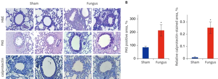
Increased airway inflammation and goblet cell hyperplasia in the lungs of A.
fumigatus-sensitized/challenged mice
Histological examination revealed that the lungs of A. fumigatus-sensitized/challenged mice had numerous focal areas with inflammatory cell infiltrates, as well as peribronchial and intraluminal areas of exudation (Fig. 2A). Moreover, PAS staining showed that A. fumigatus sensitization/challenge resulted in mucosal gland hyperplasia (Fig. 2).
Elevated calprotectin expression in the lungs of A. fumigatus-sensitized/
challenged mice
IHC staining demonstrated that the levels of calprotectin were increased in the lungs of A.
fumigatus-sensitized/challenged mice compared with control mice (Fig. 2). Additionally, western blotting using protein lysates from the lungs of mice showed that A. fumigatus sensitization/
challenge resulted in increased calprotectin, S100A8, and S100A9 protein levels (Fig. 3).
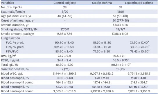
Table 1. Clinical characteristics in control subjects and patients with asthma
Variables Control subjects Stable asthma Exacerbated asthma
No. of subjects 28 33
Sex, male/female 8/20 12/21
Age (of initial visit), yr 46 (44–58) 53 (50–60)
Onset of asthma: age, yr - 50 (37.7–58)
Asthma duration, yr - 4.03 ± 4.56
Smoking status, NS/ES/SM 27/0/1 19/7/7
Smoke amount, pack/yr 2.86 ± 7.56 17.86 ± 8.02
Lung function
FVC, % pred. 90.60 ± 13.40 81.20 ± 18.80 73.90 ± 17.40*
FEV1, % pred. 102.20 ± 13.50 83.94 ± 19.20 73.91 ± 20.70*
FEV1/FVC 85.60 ± 3.40 77.30 ± 9.20 73.42 ± 10.60*
BMI, kg/m2 23.2 ± 2.9 19.5 ± 2.1
PC20, mg/mL 24.4 ± 2.4 16.5 ± 9.75*
Total IgE, kU 42.80 ± 17.70 191.51 ± 311.10*
Skin test positive, % 2 (7.1) 11 (33)
Blood WBC, /µL 5,444.4 ± 1,399.5 9,027.3 ± 2,632.2 9,791.5 ± 3,685.5
Blood eosinophil, % 3.00 ± 2.60 1.78 ± 2.10 2.70 ± 4.10
Blood eosinophil count 164.8 ± 152.1 137.4 ± 144.8 214.1 ± 334.7
Blood neutrophil, % 55.70 ± 9.20 62.88 ± 12.10 68.40 ± 15.50
Blood neutrophil count 3,055.8 ± 1,015.3 5,797.0 ± 2,288.9 7,021.3 ± 3,755.9 Data expressed as mean ± standard deviation, median (interquartile range) or number (%).
BMI = body mass index, ES = ex-smoker, FEV1 = forced expiratory volume in one second, FVC = forced vital capacity, NS = non-smoker, SM = smoker, % pred. = % predicted, PC20 = the concentration of methacholine required to decrease the forced expiratory volume in one second by 20%, IgE = immunoglobulin E, WBC = white blood cell.
*P < 0.01 compared with control subjects.
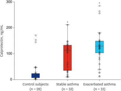
Increased calprotectin levels in the serum of patients with asthma
Thirty-three patients with asthma (mean age, 53 years) and 28 healthy individuals (mean age, 46 years) were recruited, the clinical characteristics of whom are shown in Table 1. The initial percent-predicted forced expiratory volume in one second (FEV1) and forced vital capacity (FVC), as well as the FEV1/FVC ratio, were lower in patients with asthma compared with healthy individuals. Total immunoglobulin E (IgE) and eosinophil blood count were also higher in patients with asthma, while body mass index (BMI) did not differ significantly between the two groups. The duration of asthma was 4.03 ± 4.56 years. Percent-predicted FEV1, percent-predicted FVC, and FEV1/FVC were lower in patients with asthma exacerbations compared with patients with stable asthma, while neutrophil blood count was higher. TheA B
100 200 300
Sham Fungus
*
PAS positive area, %
0
0.1 0.2 0.3
Sham Fungus
*
Relative calprotectin-stained area, %
0
H&E
Sham Fungus
PASIHC calprotectin
Fig. 2. H&E staining, PAS and IHC staining of mouse lung paraffin sections. (A) Top panel: H&E-stained lung tissue contained more inflammatory cell infiltrations in Aspergillus fumigatus-sensitization/challenge mice than control mice. Middle panel: PAS-stained airways contained increased goblet cells in the A. fumigatus- sensitization/challenge mice. Lower panel: IHC-stained lung tissue contained increased calprotectin in A. fumigatus sensitization/challenge mice. (B) The quantitation of goblet cells counts in mouse lung. (C) The quantitation of calprotectin in mouse lung.
H&E = hematoxylin and eosin, PAS = periodic acid-Schiff, IHC = immunohistochemical.
*P < 0.05, Fungus vs. Sham.
0.2 0.4 0.6 1.0
Sham Fungus
*
Calprotectin
0.8
0
0.4 1.2
Sham Fungus
*
S100A8 0.8
0
0.5 1.5
Sham Fungus
S100A9 1.0
0
S100A8 35 kDa
Sham Fungus
S100A9 15 kDa
Calprotectin 55 kDa
β-actin 42 kDa
Fig. 3. Lung protein level of calprotectin as determined by Western blot. Densitometry was determined with 3 immunoblots and normalized to β-actin. Bar graphs are densitometric data (means ± standard deviation).
*P < 0.05 compared to controls.
serum levels of calprotectin were higher in patients with asthma compared with healthy individuals (Fig. 4).
Calprotectin levels were associated with the clinicopathological characteristics of patients with asthma
Calprotectin levels significantly associated with FEV1/FVC (r = −0.215, P = 0.043), smoking (r
= 0.413, P = 0.017), BMI (r = −0.445, P = 0.000), and blood neutrophil proportion (r = 0.300, P = 0.004), and Log PC20 (r = −0.385, P = 0.011) in patients with asthma. Moreover, S100A8, S100A9, and calprotectin expression was increased in human bronchial epithelial cells and mice lungs (Fig. 5).
DISCUSSION
In this study, calprotectin levels were increased in serum from patients with asthma, and correlated with lung function, smoking amount, and blood neutrophil proportion in patients with asthma, suggesting that calprotectin could potentially be used as a biomarker for asthma.
Asthma is known to cause eosinophilic inflammation as well as IgE-mediated mast cell activation. Therefore the inhibition of airway inflammation by inhaled corticosteroids is the cornerstone of asthma therapy.19 However some patients with severe asthma demonstrated airway neutrophilia despite inhaled corticosteroid treatment.20,21 Those patients respond poorly to corticosteroid treatment.22
The increase in neutrophil counts in the sputum of adults with asthma20 and in that of children with acute asthma exacerbations was reported.23 However there is controversy whether solely the presence of eosinophils or neutrophils can be used as a binary disease classifier as there may be overlap between these two phenotypes.
Calprotectin, ng/mL
Exacerbated asthma (n = 33) Stable asthma
(n = 33) Control subjects
(n = 28) 100
200 300
0
*
*
Fig. 4. Calprotectin level in control subjects, stable, and exacerbated asthmatic patients.
*P < 0.05, stable and exacerbated asthma vs. control subjects.
Calprotectin, the heterodimeric protein complex composed of S100A8 and S100A9, is a major calcium-and zinc-binding protein in the cytosol of neutrophils, monocytes, and keratinocytes.24 S100A8 and S100A9 have been associated with cancer metastasis, but also with various severe inflammatory diseases, including psoriasis, atopic dermatitis, atherosclerosis, neurodegenerative diseases, and diabetes.25 Extracellular S100A8 and S100A9 can induce the production and secretion of several inflammatory cytokines and chemokines.26-32 S100A8 and S100A9 are used as a biomarker and predictive indicator of therapeutic responsiveness in various inflammatory diseases.6 Calprotectin is primarily found in the cytosol of neutrophils, and is released upon neutrophil activation, resulting in elevated calprotectin serum levels in patients with inflammatory bowel disease, cystic fibrosis, rheumatoid arthritis, and other conditions associated with local neutrophilic inflammation.33-37
4 10 18
Total WBC, ×103
16 14 12
8 6
0 50 100 150 200 250 300
r = 0.281 P = 0.008
r = 0.413 P = 0.017
r = 0.300 P = 0.004
Calprotectin, ng/mL
0 40
Smoke amount
30 20 10
0 50 100 150 200 250
Calprotectin, ng/mL
0 20
Neutrophils, ×103 15 10
5
0 50 100 150 200 250 300
Calprotectin, ng/mL
r = −0.385 P = 0.011
0.4 1.4
Log(PC 20)
1.2 1.0 0.8 0.6
0 50 100 150 200 250 300
Calprotectin, ng/mL
r = −0.510 P = 0.000
r = −0.215 P = 0.043
10 30
BMI
25 20 15
0 50 100 150 200 250 300
Calprotectin, ng/mL
20 100
Initial FEV1, % 140
80 120
60 40
0 50 100 150 200 250 300
Calprotectin, ng/mL
50 100
Initial FEV1 FVC
90 80 70 60
0 50 100 150 200 250 300
Calprotectin, ng/mL r = −0.445
P = 0.000
Fig. 5. Relationship of calprotectin with WBC, smoke amount, neutrophil proportion, BMI, FEV1%pred. FEV1/FVC, and Log PC20.
WBC = white blood cell, BMI = body mass index, FEV1 = forced expiratory volume in one second, FVC = forced vital capacity, %pred. = % predicted, PC20 = the concentration of methacholine required to decrease the forced expiratory volume in one second by 20%.
There are a few studies that assess S100A8 and S100A9 from serum of asthmatic patients.
S100A8 and S100A9 are involved in innate immune responses under the regulation of TLR4 polymorphisms in baker's asthma pathogenesis, suggesting that serum S100A8 could be a potential biomarker for predicting occupational exposure to wheat flour in bakery workers.38 High levels of fecal calprotectin have been reported in respiratory exacerbation, which may reflect a systemic exacerbation rather than solely lung. Antibiotic treatment has been shown to decrease fecal calprotectin levels, possibly due to its effects on the intestinal microbiome.16 Interestingly, a recent study has shown that interstitial lung disease is independently associated with increased fecal calprotectin levels in systemic sclerosis.39 In this study, serum calprotectin levels were higher in the serum of patients with asthma compared with healthy individuals and correlated with lung function and AHR, suggesting that calprotectin could be a useful biomarker for asthma.
Cigarette smoking and asthma interact to induce important adverse effects on clinical, prognostic and therapeutic outcomes. Prevalence rates for smoking in asthma are relatively close to those found in the general population.40 Smokers with asthma experience worse asthma control than nonsmokers with asthma.40 Mechanisms for the adverse effects of smoking in asthma include altered airway inflammation and corticosteroid insensitivity. Smoking worsens asthma symptoms and morbidity and promotes neutrophilic asthma.40 Cigarette smoke in asthmatic individuals also increased their sputum interleukin-8 levels in parallel to increased neutrophil percentages.41 In our study calprotectin levels correlated with smoke amount and blood neutrophil percentage in patients with asthma, suggesting that smoking increases neutrophil proportion in blood leading to calprotectin secretion. Further studies are needed to provide a more specific approach in patients with asthma with neutrophilic airway inflammation and to help in modulating the use of available conventional therapies.
Obesity42 also alters lung mechanics in asthmatics, but the impact is different for children and adults, likely due to the differential effect of obesity on the central and peripheral airway.
In this study, BMI negatively correlated with calprotectin levels in contrast to lung function and AHR, suggesting that the impact of calprotectin is different for BMI and lung function.
There are limitations to this study. The most significant limitation would be the small sample size of patients with asthma. We did not select the pattern of airway inflammatory cells from sputum, and we observed calprotectin levels between stable and exacerbated state, not therapeutic response. Further studies should be considered for this point.
In conclusion, calprotectin levels were increased in the serum of patients with asthma, and correlated with lung function and AHR, smoke amount, and blood neutrophil percentage in patients with asthma. Calprotectin, S100A8, and S100A9 expression was elevated in human bronchial epithelial cells and mice lungs. Our data suggest that calprotectin could potentially be used as a biomarker for asthma.
REFERENCES
1. Manni ML, Alcorn JF. Calprotectin-g the lung during type 2 allergic airway inflammation. Am J Respir Cell Mol Biol 2019;61(4):405-7.
PUBMED | CROSSREF
2. Carr TF, Zeki AA, Kraft M. Eosinophilic and noneosinophilic asthma. Am J Respir Crit Care Med 2018;197(1):22-37.
PUBMED | CROSSREF
3. Ricciuto A, Griffiths AM. Clinical value of fecal calprotectin. Crit Rev Clin Lab Sci 2019;56(5):307-20.
PUBMED | CROSSREF
4. Manceau H, Chicha-Cattoir V, Puy H, Peoc'h K. Fecal calprotectin in inflammatory bowel diseases: update and perspectives. Clin Chem Lab Med 2017;55(4):474-83.
PUBMED | CROSSREF
5. Lin WC, Wong JM, Tung CC, Lin CP, Chou JW, Wang HY, et al. Fecal calprotectin correlated with endoscopic remission for Asian inflammatory bowel disease patients. World J Gastroenterol 2015;21(48):13566-73.
PUBMED | CROSSREF
6. Wang S, Song R, Wang Z, Jing Z, Wang S, Ma J. S100A8/A9 in inflammation. Front Immunol 2018;9:1298.
PUBMED | CROSSREF
7. Oczypok EA, Milutinovic PS, Alcorn JF, Khare A, Crum LT, Manni ML, et al. Pulmonary receptor for advanced glycation end-products promotes asthma pathogenesis through IL-33 and accumulation of group 2 innate lymphoid cells. J Allergy Clin Immunol 2015;136(3):747-756.e4.
PUBMED | CROSSREF
8. Hammad H, Chieppa M, Perros F, Willart MA, Germain RN, Lambrecht BN. House dust mite allergen induces asthma via Toll-like receptor 4 triggering of airway structural cells. Nat Med 2009;15(4):410-6.
PUBMED | CROSSREF
9. Aoki T, Matsumoto Y, Hirata K, Ochiai K, Okada M, Ichikawa K, et al. Expression profiling of genes related to asthma exacerbations. Clin Exp Allergy 2009;39(2):213-21.
PUBMED | CROSSREF
10. Gray RD, MacGregor G, Noble D, Imrie M, Dewar M, Boyd AC, et al. Sputum proteomics in inflammatory and suppurative respiratory diseases. Am J Respir Crit Care Med 2008;178(5):444-52.
PUBMED | CROSSREF
11. Lee TH, Jang AS, Park JS, Kim TH, Choi YS, Shin HR, et al. Elevation of S100 calcium binding protein A9 in sputum of neutrophilic inflammation in severe uncontrolled asthma. Ann Allergy Asthma Immunol 2013;111(4):268-275.e1.
PUBMED | CROSSREF
12. Lee TH, Chang HS, Bae DJ, Song HJ, Kim MS, Park JS, et al. Role of S100A9 in the development of neutrophilic inflammation in asthmatics and in a murine model. Clin Immunol 2017;183:158-66.
PUBMED | CROSSREF
13. Zhao J, Endoh I, Hsu K, Tedla N, Endoh Y, Geczy CL. S100A8 modulates mast cell function and suppresses eosinophil migration in acute asthma. Antioxid Redox Signal 2011;14(9):1589-600.
PUBMED | CROSSREF
14. Yin LM, Li HY, Zhang QH, Xu YD, Wang Y, Jiang YL, et al. Effects of S100A9 in a rat model of asthma and in isolated tracheal spirals. Biochem Biophys Res Commun 2010;398(3):547-52.
PUBMED | CROSSREF
15. Greenlee KJ, Corry DB, Engler DA, Matsunami RK, Tessier P, Cook RG, et al. Proteomic identification of in vivo substrates for matrix metalloproteinases 2 and 9 reveals a mechanism for resolution of inflammation. J Immunol 2006;177(10):7312-21.
PUBMED | CROSSREF
16. Schuh JM, Blease K, Kunkel SL, Hogaboam CM. Eotaxin/CCL11 is involved in acute, but not chronic, allergic airway responses to Aspergillus fumigatus. Am J Physiol Lung Cell Mol Physiol 2002;283(1):L198-204.
PUBMED | CROSSREF
17. Lee SH, Lee PH, Kim BG, Seo HJ, Baek AR, Park JS, et al. Annexin A1 in plasma from patients with bronchial asthma: its association with lung function. BMC Pulm Med 2018;18(1):1.
PUBMED | CROSSREF
18. Global Initiative for Asthma (GINA). Global strategy for asthma management and prevention 2014. http://
www.ginasthma.org/. Updated 2014. Accessed May 14, 2014.
19. Sur S, Crotty TB, Kephart GM, Hyma BA, Colby TV, Reed CE, et al. Sudden-onset fatal asthma. A distinct entity with few eosinophils and relatively more neutrophils in the airway submucosa? Am Rev Respir Dis 1993;148(3):713-9.
PUBMED | CROSSREF
20. Jatakanon A, Uasuf C, Maziak W, Lim S, Chung KF, Barnes PJ. Neutrophilic inflammation in severe persistent asthma. Am J Respir Crit Care Med 1999;160(5 Pt 1):1532-9.
PUBMED | CROSSREF
21. Louis R, Lau LC, Bron AO, Roldaan AC, Radermecker M, Djukanović R. The relationship between airways inflammation and asthma severity. Am J Respir Crit Care Med 2000;161(1):9-16.
PUBMED | CROSSREF
22. Green RH, Brightling CE, Woltmann G, Parker D, Wardlaw AJ, Pavord ID. Analysis of induced sputum in adults with asthma: identification of subgroup with isolated sputum neutrophilia and poor response to inhaled corticosteroids. Thorax 2002;57(10):875-9.
PUBMED | CROSSREF
23. Norzila MZ, Fakes K, Henry RL, Simpson J, Gibson PG. Interleukin-8 secretion and neutrophil
recruitment accompanies induced sputum eosinophil activation in children with acute asthma. Am J Respir Crit Care Med 2000;161(3 Pt 1):769-74.
PUBMED | CROSSREF
24. Semprini S, Capon F, Tacconelli A, Giardina E, Orecchia A, Mingarelli R, et al. Evidence for differential S100 gene over-expression in psoriatic patients from genetically heterogeneous pedigrees. Hum Genet 2002;111(4-5):310-3.
PUBMED | CROSSREF
25. Kinoshita R, Sato H, Yamauchi A, Takahashi Y, Inoue Y, Sumardika IW, et al. Newly developed anti-S100A8/A9 monoclonal antibody efficiently prevents lung tropic cancer metastasis. Int J Cancer 2019;145(2):569-75.
PUBMED | CROSSREF
26. Chernov AV, Dolkas J, Hoang K, Angert M, Srikrishna G, Vogl T, et al. The calcium-binding proteins S100A8 and S100A9 initiate the early inflammatory program in injured peripheral nerves. J Biol Chem 2015;290(18):11771-84.
PUBMED | CROSSREF
27. Eisenblaetter M, Flores-Borja F, Lee JJ, Wefers C, Smith H, Hueting R, et al. Visualization of tumor- immune interaction - Target-specific imaging of S100A8/A9 reveals pre-metastatic niche establishment.
Theranostics 2017;7(9):2392-401.
PUBMED | CROSSREF
28. Gopal R, Monin L, Torres D, Slight S, Mehra S, McKenna KC, et al. S100A8/A9 proteins mediate neutrophilic inflammation and lung pathology during tuberculosis. Am J Respir Crit Care Med 2013;188(9):1137-46.
PUBMED | CROSSREF
29. Cremers NA, van den Bosch MH, van Dalen S, Di Ceglie I, Ascone G, van de Loo F, et al. S100A8/A9 increases the mobilization of pro-inflammatory Ly6Chigh monocytes to the synovium during experimental osteoarthritis. Arthritis Res Ther 2017;19(1):217.
PUBMED | CROSSREF
30. Schiopu A, Cotoi OS. S100A8 and S100A9: DAMPs at the crossroads between innate immunity, traditional risk factors, and cardiovascular disease. Mediators Inflamm 2013;2013:828354.
PUBMED | CROSSREF
31. Wu Y, Li Y, Zhang C, A X, Wang Y, Cui W, et al. S100A8/A9 released by CD11b+Gr1+ neutrophils activates cardiac fibroblasts to initiate angiotensin II-Induced cardiac inflammation and injury. Hypertension 2014;63(6):1241-50.
PUBMED | CROSSREF
32. Huang H, Huang Q, Tang T, Gu L, Du J, Li Z, et al. Clinical significance of calcium-binding protein S100A8 and S100A9 expression in non-small cell lung cancer. Thorac Cancer 2018;9(7):800-4.
PUBMED | CROSSREF
33. Wilkerson EM, Johansson MW, Hebert AS, Westphall MS, Mathur SK, Jarjour NN, et al. The peripheral blood eosinophil proteome. J Proteome Res 2016;15(5):1524-33.
PUBMED | CROSSREF
34. Kalla R, Kennedy NA, Ventham NT, Boyapati RK, Adams AT, Nimmo ER, et al. Serum calprotectin:
a novel diagnostic and prognostic marker in inflammatory bowel diseases. Am J Gastroenterol 2016;111(12):1796-805.
PUBMED | CROSSREF
35. Gray RD, Imrie M, Boyd AC, Porteous D, Innes JA, Greening AP. Sputum and serum calprotectin are useful biomarkers during CF exacerbation. J Cyst Fibros 2010;9(3):193-8.
PUBMED | CROSSREF
36. Kopeć-Mędrek M, Widuchowska M, Kucharz EJ. Calprotectin in rheumatic diseases: a review. Reumatologia 2016;54(6):306-9.
PUBMED | CROSSREF
37. Decaesteker T, Seys S, Hox V, Dilissen E, Marijsse G, Manhaeghe L, et al. Serum and sputum calprotectin, a reflection of neutrophilic airway inflammation in asthmatics after high-altitude exposure. Clin Exp Allergy 2017;47(12):1675-7.
PUBMED | CROSSREF
38. Pham DL, Yoon MG, Ban GY, Kim SH, Kim MA, Ye YM, et al. Serum S100A8 and S100A9 enhance innate immune responses in the pathogenesis of Baker's asthma. Int Arch Allergy Immunol 2015;168(2):138-46.
PUBMED | CROSSREF
39. Caimmi C, Bertoldo E, Venturini A, Caramaschi P, Frulloni L, Ciccocioppo R, et al. Relationship between increased fecal calprotectin levels and interstitial lung disease in systemic sclerosis. J Rheumatol 2019;46(3):274-8.
PUBMED | CROSSREF
40. Polosa R, Thomson NC. Smoking and asthma: dangerous liaisons. Eur Respir J 2013;41(3):716-26.
PUBMED | CROSSREF
41. Chalmers GW, MacLeod KJ, Thomson L, Little SA, McSharry C, Thomson NC. Smoking and airway inflammation in patients with mild asthma. Chest 2001;120(6):1917-22.
PUBMED | CROSSREF
42. Khalid F, Holguin F. A review of obesity and asthma across the life span. J Asthma 2018;55(12):1286-300.
PUBMED | CROSSREF