서론
상실된 치아를 회복하는 방법으로 Bra�nemark 등1은 처음으로 골유착성 임플란트를 시도하였으며 이후 임플란트는 결손치 아를 가진 환자의 구강기능 회복을 위해 널리 사용되고 있다.
이제 임플란트는 치아결손부를 회복하는 주요한 치과술식의 하나이며 장기간 평가에서도 좋은 결과를 나타내고 있다.2,3
임플란트 성공의 중요한 요소중의 하나는 임플란트 안정성 이다. 임플란트의 초기 안정성은 임플란트의 성공과 실패의 주요한 판단기준이며 즉시 식립의 가이드 역할을 해주고, 임 플란트 보철물 하중시기를 결정하는데 도움을 주어 어느 시기 에 보철물에 교합력을 가해야 하는지 객관적인 기준이 된다.
또한 임플란트 장기간 예후평가에도 매우 중요하다.4,5
임플란트 안정성은 임플란트가 주변골과 접촉됨으로써 얻 을 수 있는 일차안정성과 골치유와 골형성결과에 의한 이차안 정성으로 구분할 수 있는데 일차안정성은 임플란트 식립 부위 의 골질과 골량, 외과적 술식, 임플란트의 길이, 직경, 형태, 식 립방법, 디자인 및 표면 거칠기의 결과 등에 영향을 받는다.4,6,7 최근에는 임플란트의 안정성을 위해 임플란트 디자인과 임플 란트 표면처리의 개선이 이루어져 왔으나 이러한 개선에도 불
구하고, 환자에게 식립된 임플란트의 안정성을 판단하여 하중 시기를 결정하는 것은 매우 어렵다. 현재 임플란트의 안정성 은 타진반응, Periotest�, Dental fine tester�, 촉진을 통한 동요도 검진, 방사선 판독을 통한 골질의 평가, 식립회전력(Insertion torque)등을 이용하여 평가한다.4,8,9그 외의 비침습적인 방법으로 Meredith 등은 생체내 임플란트의 안정성을 정량적으로 측정 하기 위한 공진주파수분석법(resonance frequency analysis, RFA)을 개발하였으며, 임상에서 식립 직후 초기안정성을 평가하기에 좋은 방법이다.4,10,11이중에서 식립회전력(Insertion torque) 과 공진 주파수분석법으로 얻는 임플란트 안정성지수(Implant Stability Quotient, ISQ)는 수치화할 수 있는 장점이 있다.12-14또한 공진주파 수분석법을 이용한 임플란트 안정성 평가는 재현성이 높고 안 정적으로 임플란트 초기고정에 영향을 미치는 요소를 파악할 수 있다.15,16
그 동안 임플란트의 길이, 직경, 디자인 등 특성에 따른 임플 란트의 초기 안정성 연구에서 Saadoun과 LeGall17은 깊게 식립된 임플란트가 골유착을 위한 표면적이 넓기 때문에 높은 성공률 을 보인다고 하였으며, Jaffin과 Berman18은 Lekholm과 Zarb의 골질 분류법 중 골질이 Type I, II, III의 경우 실패율이 3%였지만 가장 연한 골질인 Type IV실패율이 35%로 나타났다고 보고한 것과
임플란트의 직경, 길이 및 디자인변화가 임플란트 안정성지수(ISQ)에 미치는 영향
이정열∙이원창∙김민수∙김종은∙신상완*
고려대학교 임상치의학대학원, 임상치의학연구소
연구 목적: 이 연구의 목적은 임플란트의 직경, 길이 및 디자인의 변화가 임플란트 안정성 지수에 미치는 영향을 비교하는 것이다.
연구 재료 및 방법: 골질차이에 의한 변수를 제거하기 위해 상악골 평균밀도와 유사한 균일한(0.48g /cm3) 밀도를 가진 Polyuretane foam blocks (Sawbones�, Pacific Research Laboratories Inc, Vashon, Washington)을 이용, 임플란트(Implantium�, Dentium, Seoul, Korea)를 다양한 직경(∅ 3.8, ∅ 4.3 및 ∅ 4.8)과 길이(8, 10 및 12 mm)로 식립하여 그 변화가 임 플란트 안정성 지수(Implant Stability Quotient, ISQ)에 미치는 영향을 비교하였다. 또 같은 직경과 길이(∅4.3 × 10 mm)에서 submerged와 non-submerged (SimplelineII�, Dentium, Seoul, Korea) 디자인이 ISQ 에 미치는 영향을 비교하였다. 식립 회전력의 영향을 배제하기 위해 동일한 35 N의 Torque로 각 실험군당 10개씩 총 60개의 임플란트를 식립하였다.
Osstell™ mentor(Integration Diagnostic AB, Sweden)를 이용하여 공진주파수를 측정한 후 ISQ 값으로 기록하였고, 그 결과를one-way ANOVA와 Tukey HSD test로 분석하였다(α=.05).
결과:1. 임플란트 직경의 변화는 ISQ에 영향을 미치지 않았으나(P>.05), 임플란트 길이가 증가함에 따라 ISQ도 증가하였다(P<.001). 2. 임플란트 디자인의 변화는 ISQ와 유의 한 상관관계를 보여Submerged 디자인의 ISQ가non-submerged 디자인보다 높게 나타났다(P<.05).
결론:임플란트 안정성을 높이기 위해서는 가능한 길이가 긴 임플란트를 식립하는 것이 유리하며, 같은 길이의 임플란트에서 볼 때 Non-submerged 디자인보다 submerged 디 자인이 더 높은 ISQ를 얻을 수 있을 것으로 생각된다. (대한치과보철학회지 2012;50:292-8)
주요단어:식립 회전력; 임플란트 안정성 지수(ISQ); 임플란트 길이 및 직경; 공명 주파수 분석법
*교신저자: 신신상상완완
152-703 서울특별시 구로구 구로동길 97 고대부속 구로병원 치과 보철과 02-2626-1922: e-mail,[email protected] 원고접수일: 2012년 9월 18일 / 원고최종수정일: 2012년 10월 9일 / 원고채택일: 2012년 10월 10일
2012 대한치과보철학회
이 글은 크리에이티브 커먼즈 코리아 저작자표시-비영 리 3.0 대한민국 라이선스에 따라 이용하실 수 있습니다.
c cc
같이 골질이 임플란트를 매식하기에 좋지 않은 경우이거나 과 도한 골흡수가 있는 경우에는 초기 안정을 얻기가 어려워 실 패할 가능성이 높은 것으로 알려져 왔다. Langer 등19은 골질과 골량이 부족한 경우 직경이 큰 임플란트를 사용할 것을 제안 하였으며, Ivanoff 등20의 후향적 연구에 의하면 직경 3.75 mm 임 플란트의 실패율이 5%였으나 직경 5.0 mm 임플란트에서는 실 패율이 18%라고 보고하였다.
사체 골조직에서나 동물실험인 경우 주로 식립부위의 골질 이 다양하고 식립환경에 따라 임플란트 안정성지수에 영향을 줄 수 있었고,21-23또 골질이 일정한 모형골을 이용한 연구에서 는 서로 다른 식립회전력이 ISQ 값에 대해 임플란트의 길이나 직경 등의 영향을 파악하는 데 혼란을 주는 요인으로 작용할 수 있었다.24-27
따라서 이 연구에서는 골질의 차이로 인한 변수를 제거하기 위해 밀도가 균일한 모형골을 이용하여 조건을 균일하게 하 고, 임플란트 식립시 식립회전력을 35 N으로 일정하게 유지함 으로써 식립회전력의 변화로 인한 임플란트 안정성지수에의 영향을 배제하여, 식립된 임플란트에서 임플란트 길이, 직경 의 변화에 따른 임플란트 안정성지수의 차이와, submerged와 non-submerged 디자인의 변화에 따라 임플란트 안정성지수에 미치는 영향을 평가하였다.
연구 재료 및 방법 1. 연구 재료
1) 모형골 선택
임플란트 식립은 밀도가 균일한 폴리우레탄재질의 모형골 (Sawbones�, Pacific Research Laboratories Inc, Vashon, Washington)을 사용하였고 그 밀도는 상악골의 평균밀도와 유사한 0.48 g/cm3 를 선택하였다(상악구치부 평균 bone density는 0.31 g/cm3이고, 상 악전치부는 0.55 g/cm3이다) (Fig. 1, Table 1).28
2) Implant fixture
임플란트는Implantium�(Dentium, Seoul, Korea)을 사용하였고, nonsubmerged 디자인은Simpleline II�(Dentium, Seoul, Korea; gingi- val height 2.2 mm)를 사용하였다. 각 군마다 10개씩 사용하여 총 60개의 임플란트를 사용하였다(Fig. 2).
3) 임플란트 안정성지수(ISQ)측정
OsstellTMmentor (Integration Diagnostics AB, Sweden)를 사용하여 공진주파수 분석법으로 임플란트 안정성지수를 측정하였다.
Table 1. Average material properties (Sawbones�, Pacific Research Laboratories Inc, Vashon, Washington)
Compressive Tensile Shear
Density
Strength Modulus Strength Modulus Strength Modulus
pcf g/cc
MPa MPa MPa MPa MPa MPa
5* 0.08 0.6 16 1.0 32 0.59 7.1
10* 0.16 2.2 58 2.1 86 1.6 19
15* 0.24 4.9 123 3.7 173 2.8 33
20* 0.32 8.4 210 5.6 284 4.3 49
25* 0.40 12.9 317 8.8 399 5.9 68
30* 0.48 18 445 12 592 7.6 87
40* 0.64 31 759 19 1000 11 130
50* 0.80 48 1148 27 1469 16 178
*pcf (pound per cubit feet), **Mpa(mega pascal)
Fig. 1. Artificial bone block (Sawbone�) used in this experiment. Fig. 2. Implant fixtures used in this experiment(A: Implantium�, B: Simpleline II�, Dentium, Seoul, Korea).
A B
2. 연구 방법
1) 임플란트 식립
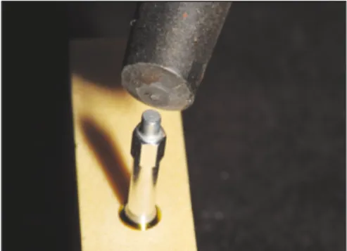
식립시의 오차를 줄이기 위해 35 N의 동일한 식립회전력 (Insertion torque)으로 식립하였으며, 식립술식(Drilling procedure) 의 조절을 통하여 임플란트 Platform이 인공골 상부에 식립됨과 동시에 35 N의 식립회전력(Insertion torque)이 되도록 표준화하였 다. 임플란트의 식립은 임플란트 진료용 엔진인 INTRA surg300 (KaVo, Biberach, Germany)을 이용하였다(Fig. 3).
2) 공진주파수 측정
식립후 ISQ 측정시 떨림에 의해 오차가 발생하는 것을 예방 하기 위해 인공골을 단단히 고정한 후 Smartpeg을 연결하고 OsstellTMmentor를 이용하여 공진주파수를 MD 방향으로 3회, BL방향으로 3회씩 측정하여 평균값을 얻었다. 측정된 값은 임 플란트 안정성지수(ISQ)로 기록하였다.
실험에 사용된 임플란트의 제원은 각각∅4.3 mm × 10 mm,
∅4.3 mm × 8 mm, ∅4.3 mm × 12 mm, ∅3.8 mm × 10 mm, ∅4.8 mm × 10 mm 로 임플란트 길이 및 직경의 변화가 ISQ 에 미치 는 영향과, ∅4.3 mm × 10 mm submerged 디자인과∅4.3 mm × 10 mm non-submerged 디자인의 변화가 ISQ 에 미치는 영향을 평가 하였다(Table 2).
3) 통계 분석
실험에서 얻어진 모든 계측항목에 대해 평균값과 표준편차 를 구하고 SPSS statistics ver 12.0 (SPSS Inc, Chicago, IL, USA)을 이 용하여 분석하였다. 실험군 간의 차이는one-way ANOVA 를 사 용하였고, 사후검정으로 Tukey HSD test로 분석하였다(α=.05).
결과
1. 임플란트 직경의 변화에 따른 ISQ
임플란트 직경 4.3 mm, 길이 10 mm 임플란트를 기준으로 하 여 직경의 변화를 주어 직경 3.8 mm, 길이 10 mm와 직경 4.8 mm, 길이 10 mm의 ISQ를 비교하였다. 직경 3.8 mm 임플란트에 서 ISQ 가 가장 높게 나타났고 오히려 직경 4.3 mm에서 ISQ 지수 가 낮게 나타났으나 통계학적으로 유의성이 없었다(P>.05) (Fig. 4, Table 3).
2. 임플란트 길이의 변화에 따른 ISQ
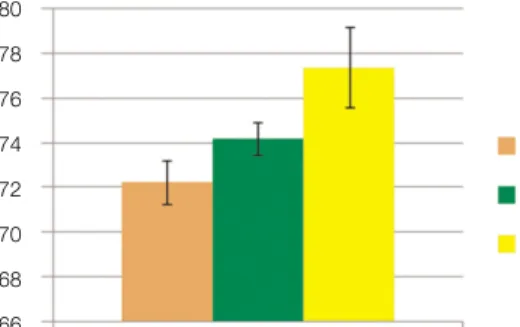
ISQ는 동일한 직경 4.3 mm 임플란트에서 길이에 따라서 길이 가 길어질수록 증가하였다. 직경 4.3 mm, 길이8 mm 임플란트에 서 가장 낮은 ISQ를 나타냈으며 길이 12 mm 임플란트에서 가장 높은 수치를 보였다(P<.001) (Fig. 5, Table 4).
Fig. 3. Measurement of ISQ value using resornance frequency analysis.
Table 2. Implant used in this experiment (n = 60 implant)
Type Size (mm) N
∅4.3 × 8 10
∅4.3 × 10 10
Submerged ∅4.3 × 12 10
∅3.8 × 10 10
∅4.8 × 10 10
Non-submerged ∅4.3 × 10 10
Fig. 4. ISQ values (Mean ± SD) depending upon implant diameter.
76
75
74
73
72
ISQ
Implnat diameter
∅3.8
∅4.3
∅4.8
Table 3. Mean ISQ values of implant diameter
Diameter (mm) ISQ (Mean ± SD)
∅3.8 75.1 ± 0.51
∅4.3 74.2 ± 0.75
∅4.8 74.7 ± 1.08
3. 임플란트 디자인의 변화에 따른 ISQ
동일한 직경(4.3 mm)과 길이(10 mm)의 임플란트에서 sub- merged 디자인의 ISQ가 non-submerged 디자인보다 높게 나타났다 (P<.05) (Fig. 6, Table 5).
고찰
골유착성 임플란트가 도입된 이래 골유착을 위해서 반드시 3 - 6개월간의 치유기간이 을 가졌으나, 최근에는 임플란트의 높은 임상적 성공률과 기술의 발달로 치료기간을 줄여 환자의 심미적, 기능적 손상을 회복시켜 심리적,정신적 안정을 제공 하려는 시도가 많이 이루어지고 있다.
임플란트 초기 안정성은 임플란트 식립 직후에 얻어져 골과 임플란트 사이의 접촉양상에 의해 영향을 받으며 이후에 얻어 지는 골유착과정에 많은 영향을 미치므로 임플란트 성공의 중 요한 요소라고 할 수 있다. 특히 골질이나 골량이 부족한 부위 에서 임플란트 안정성은 성공의 중요한 척도라고 할 수 있
다.26,27따라서, 임플란트의 성공여부는 시술자가 임플란트의
안정성을 이해하고 얼마나 조절할 수 있는가에 따라 달라질 수 있다.
Meredith4가 임플란트의 안정성을 평가하기 위해 개발한 공진 주파수 분석법은 객관적이고 비침습적인 방법으로 인정받고 있다.9공진주파수 분석은 임플란트의 초기고정을 공진주파수 를 이용하여 측정하는 방식으로 ISQ라는 임플란트 안정성 지
수로 나타내며 ISQ 값이 보통 65이상이면 즉시 부하(Immediate loading)가 가능한 것으로 보고되고 있다.29공진주파수 값이 감 소하는 경우 고정 정도의 감소와 연관이 있어 임플란트 실패 의 가능성이 높다고 판단할 수 있다. 그래서 공진주파수 측정 은 임플란트 성공과 실패를 예측하기 위해 많이 사용되고 있 다. 초기에는 RFA 값을 Hz로 표시하였으나 OsstellTMmentor로 상품화된 뒤 ISQ란 수치로 1 - 100까지 표시하게 되어 있다. 공진 주파수측정으로 임플란트 식립시 초기안정성과 치유기간 동 안의 안정성을 평가할 수 있어 실패가능성이 높은 임플란트를 조기에 발견하고 임플란트 보철후의 안정성을 평가하는 것이 어느 정도 가능하게 되었다.
ISQ 값이 임플란트의 초기 안정성을 충분히 나타낸다는 점 에서 어느 정도 동의가 이루어 지고 있다고 볼 수 있다. 이런 초 기 안정성의 확보를 위해 여러 연구들에서 직경이 큰 임플란 트는 지지골과의 접촉면적을 증가시키고 주변골에 가해지는 응력을 감소시켜 초기 안정성을 얻는데 유리한 것으로 보고되 고 있다.20,30그러나 초기 안정성은 골질에 따라 달라질 수 있기 때문에 다양한 골질에서 초기안정성에 미치는 영향을 파악하 는데 한계가 있었다.
즉 골질의 차이가 주는 영향은 술자가 조절할 수 없는 부분 이지만, 임플란트의 크기나 길이 또는 다양한 변화요인 들이 초기 안정성에 영향을 줄 수 있으므로 그에 대한 다양한 연구 가 이루어져 왔다. Krennmair와 Waldenberger31는 성공률을 높이기 위해 직경뿐만 아니라 충분한 길이의 임플란트를 선택해야 한 다고 하였다. 그러나 Langer 등19과 Ivanoff 등20은 골질이 불리한 Fig. 5. ISQ values (Mean ± SD) depending upon implant length.
80 78 76 74 72 70 68 66
ISQ
Implnat length
L8 L10 L12
Fig. 6. ISQ values (Mean ± SD) depending upon implant design.
76
74
72
70
68
66
ISQ
Implnat design
Submerged Non-submerged
Table 4. Mean ISQ values of implant length
Implant length (mm) ISQ (Mean ± SD)
L 8.0 72.2 ± 0.95*
L 10.0 74.2 ± 0.75*
L 12.0 77.4 ± 1.79*
*statistically significant (P<.001).
Table 5. Mean ISQ values of implant design
Implant design ISQ (Mean ± SD)
submerged 74.2 ± 0.75 **
non submerged 70.85 ± 1.68**
**statistically significant (P<.05).
부위에 식립되거나 실패한 임플란트를 대체하기 위해 사용된 직경이 크고 길이가 짧은 임플란트의 성공률이 낮다고 보고하 였다. Grunder 등32은 짧고 가는 임플란트에서 골흡수량과 실패 율이 높았다고 하였으며, Bilhan 등33은 원통형보다는 원추형의 굵은 직경의 임플란트가 일차안정성이 높다고 하였고, Kido 등34은 골밀도가 증가할수록, 직경이 굵을수록 임플란트의 안 정도가 증가한다고 보고하였다. 그러나 Ohta 등12은 임플란트 의 직경은 ISQ값에 유의한 영향을 주지 않는다고 하였고, 단지 식립회전력이 서로 상관관계를 보인다고 하였다. 또 Al-Nawas 등26과 Turkyilmaz 등27도 식립회전력이 클수록 ISQ값도 높아진다 고 하였다.
이렇게 여러 연구의 결과에서 볼 때 ISQ에 영향을 주는 요인 에 대한 기존의 실험에서는 설정된 독립변수 이외의 다른 요 인들이 결과에 작용하여 영향요소의 순수한 효과를 알려주지 는 못하고 있다. 특히 기존의 ISQ에 영향을 미치는 요소 중 식 립 회전력의 영향이 매우 큰데, 골질이나 식립환경에 따라 다 양한 식립회전력이 가해진 채로 ISQ가 측정되어 다른 요소들 의 영향을 변별하기 어려웠다.
따라서 이 연구에서는 시범 연구(Pilot study)를 통해서 최종적 인 35 N의 식립회전력을 얻기 위해 인공골에 적합하게 조절된 외과적 술식(surgical protocol)을 만들어 동일한 힘으로 식립함으 로써 식립 회전력의 영향을 배제하소 ISQ를 측정하였다.
실험 결과 임플란트의 직경의 변화에 따른 ISQ는 유의한 차 이를 보이지 않았다. 임플란트의 길이변화에 따른 ISQ는 임플 란트 길이가 길어질수록 즉, 길이 8 mm 임플란트보다 12 mm 임 플란트에서 더 높은 안정성 지수 수치를 나타났으며 통계학적 으로 유의성이 있었다(P<.001). 이는 Sim과 Lang15의 연구에서와 같은 결론이었다. 임플란트의 디자인변화에 따른 ISQ는 동일 한 직경과 길이의 임플란트에서 submerged 디자인의 ISQ가 non- submerge 디자인보다 높게 나타났는데, 여기서 동일한 골접촉 면적과 접촉 길이에서도 non-submerged 디자인이 갖는 경부의 차이가 ISQ 값을 낮게 보여준다는 점이 흥미롭다.
이상의 실험결과로 볼 때, 높은 ISQ를 얻기 위해서는 가능한 길이가 긴 임플란트를 식립하는 것이 유리하며, non-submerged 디자인보다 Submerged 디자인이 더 높은 ISQ를 얻을 수 있을 것 으로 판단된다.
물론 공진주파수 분석법이 측정시 식립회전력 뿐만 아니라 다른 여러 가지 요인이 결과에 영향을 줄 수 있겠으나, 이 실험 은 그런 영향력을 최대한 배제하고 실험에서 제기한 변수에 의한 순수한 영향에 의한 결과를 도출하였다는 점이 의의가 있다고 생각된다. 이런 결과를 바탕으로 in vivo 실험에서도 변 수를 표준화하여 초기 안정도 평가가 필요할 것으로 생각된 다. 이번 연구에 기초하여 향후 임상적 적용에서 임플란트 길 이와 임플란트 디자인의 적절한 선택에 중요한 기준을 제공해 줄 것으로 판단되었으며, 향후 임플란트 안정성의 표준화된 정량분석을 위한 계속적인 연구가 진행되어야 할 것으로 생각 된다.
결론
임플란트 안정성을 높이기 위해서는 가능한 길이가 긴 임플 란트를 식립하는 것이 유리하며, 같은 길이의 임플란트에서 볼 때 Non-submerged디자인보다 submerged 디자인이 더 높은 ISQ를 얻을 수 있을 것으로 생각된다.
참고문헌
1. Bra�nemark PI, Adell R, Breine U, Hansson BO, Lindstro¨m J, Ohlsson A. Intra-osseous anchorage of dental prostheses. I.
Experimental studies. Scand J Plast Reconstr Surg 1969;3:81-100.
2. Adell R, Lekholm U, Rockler B, Bra�nemark PI. A 15-year study of osseointegrated implants in the treatment of the eden- tulous jaw. Int J Oral Surg 1981;10:387-416.
3. Adell R, Eriksson B, Lekholm U, Bra�nemark PI, Jemt T. Long- term follow-up study of osseointegrated implants in the treatment of totally edentulous jaws. Int J Oral Maxillofac Implants 1990;5:347-59.
4. Meredith N. Assessment of implant stability as a prognostic de- terminant. Int J Prosthodont 1998;11:491-501.
5. Atsumi M, Park SH, Wang HL. Methods used to assess implant stability: current status. Int J Oral Maxillofac Implants 2007;22:
743-54.
6. Javed F, Almas K, Crespi R, Romanos GE. Implant surface mor- phology and primary stability: is there a connection? Implant Dent 2011;20:40-6.
7. Trisi P, De Benedittis S, Perfetti G, Berardi D. Primary stabili- ty, insertion torque and bone density of cylindric implant ad mod- um Branemark: is there a relationship? An in vitro study. Clin Oral Implants Res 2011;22:567-70.
8. Friberg B, Sennerby L, Meredith N, Lekholm U. A comparison between cutting torque and resonance frequency measurements of maxillary implants. A 20-month clinical study. Int J Oral Maxillofac Surg 1999;28:297-303.
9. Sennerby L, Meredith N. Resonance frequency analysis: mea- suring implant stability and osseointegration. Compend Contin Educ Dent 1998;19:493-8, 500, 502.
10. Meredith N, Alleyne D, Cawley P. Quantitative determination of the stability of the implant-tissue interface using resonance fre- quency analysis. Clin Oral Implants Res 1996;7:261-7.
11. Friberg B, Sennerby L, Linden B, Gro¨ndahl K, Lekholm U. Stability measurements of one-stage Bra�nemark implants during healing in mandibles. A clinical resonance frequency analysis study. Int J Oral Maxillofac Surg 1999;28:266-72.
12. Ohta K, Takechi M, Minami M, Shigeishi H, Hiraoka M, Nishimura M, Kamata N. Influence of factors related to implant stability detected by wireless resonance frequency analysis de- vice. J Oral Rehabil 2010;37:131-7.
13. Lachmann S, Ja¨ger B, Axmann D, Gomez-Roman G, Groten M, Weber H. Resonance frequency analysis and damping capacity assessment. Part I: an in vitro study on measurement reliability and a method of comparison in the determination of primary den- tal implant stability. Clin Oral Implants Res 2006;17:75-9.
14. Lachmann S, Laval JY, Axmann D, Weber H. Influence of implant geometry on primary insertion stability and simulated peri- implant bone loss: an in vitro study using resonance frequency analysis and damping capacity assessment. Int J Oral Maxillofac Implants 2011;26:347-55.
15. Sim CP, Lang NP. Factors influencing resonance frequency analysis assessed by Osstell mentor during implant tissue inte- gration: I. Instrument positioning, bone structure, implant length.
Clin Oral Implants Res 2010;21:598-604.
16. Han J, Lulic M, Lang NP. Factors influencing resonance frequency analysis assessed by Osstell mentor during implant tissue inte- gration: II. Implant surface modifications and implant diameter.
Clin Oral Implants Res 2010;21:605-11.
17. Saadoun AP, LeGall ML. Clinical results and guidelines on Steri-Oss endosseous implants. Int J Periodontics Restorative Dent 1992;12:486-95.
18. Jaffin RA, Berman CL. The excessive loss of Branemark fixtures in type IV bone: a 5-year analysis. J Periodontol 1991;62:2-4.
19. Langer B, Langer L, Herrmann I, Jorneus L. The wide fix- ture: a solution for special bone situations and a rescue for the com- promised implant. Part 1. Int J Oral Maxillofac Implants 1993;8:400-8.
20. Ivanoff CJ, Sennerby L, Johansson C, Rangert B, Lekholm U.
Influence of implant diameters on the integration of screw im- plants. An experimental study in rabbits. Int J Oral Maxillofac Surg 1997;26:141-8.
21. Chan HL, El-Kholy K, Fu JH, Galindo-Moreno P, Wang HL.
Implant primary stability determined by resonance frequency analy- sis in surgically created defects: a pilot cadaver study. Implant Dent 2010;19:509-19.
22. Fenner M, Vairaktaris E, Stockmann P, Schlegel KA, Neukam FW, Nkenke E. Influence of residual alveolar bone height on im- plant stability in the maxilla: an experimental animal study.
Clin Oral Implants Res 2009;20:751-5.
23. Abrahamsson I, Linder E, Lang NP. Implant stability in relation to osseointegration: an experimental study in the Labrador dog.
Clin Oral Implants Res 2009;20:313-8.
24. Tabassum A, Meijer GJ, Wolke JG, Jansen JA. Influence of sur- gical technique and surface roughness on the primary stability of an implant in artificial bone with different cortical thickness: a
laboratory study. Clin Oral Implants Res 2010;21:213-20.
25. Tabassum A, Meijer GJ, Wolke JG, Jansen JA. Influence of the surgical technique and surface roughness on the primary stability of an implant in artificial bone with a density equivalent to maxillary bone: a laboratory study. Clin Oral Implants Res 2009;20:327-32.
26. Al-Nawas B, Wagner W, Gro¨tz KA. Insertion torque and reso- nance frequency analysis of dental implant systems in an animal model with loaded implants. Int J Oral Maxillofac Implants 2006;21:726-32.
27. Turkyilmaz I, To¨zu¨m TF, Tumer C, Ozbek EN. Assessment of correlation between computerized tomography values of the bone, and maximum torque and resonance frequency values at dental implant placement. J Oral Rehabil 2006;33:881-8.
28. Devlin H, Horner K, Ledgerton D. A comparison of maxillary and mandibular bone mineral densities. J Prosthet Dent 1998;79:323-7.
29. Isoda K, Ayukawa Y, Tsukiyama Y, Sogo M, Matsushita Y, Koyano K. Relationship between the bone density estimated by cone-beam computed tomography and the primary stability of dental implants.
Clin Oral Implants Res 2012;23:832-6.
30. Bardyn T, Ge′det P, Hallermann W, Bu¨chler P. Quantifying the influence of bone density and thickness on resonance frequen- cy analysis: an in vitro study of biomechanical test materials. Int J Oral Maxillofac Implants 2009;24:1006-14.
31. Krennmair G, Waldenberger O. Clinical analysis of wide-diameter frialit-2 implants. Int J Oral Maxillofac Implants 2004;19:710- 5.
32. Grunder U, Polizzi G, Goene′R, Hatano N, Henry P, Jackson WJ, Kawamura K, Ko¨hler S, Renouard F, Rosenberg R, Triplett G, Werbitt M, Lithner B. A 3-year prospective multicenter follow- up report on the immediate and delayed-immediate placement of implants. Int J Oral Maxillofac Implants 1999;14:210-6.
33. Bilhan H, Geckili O, Mumcu E, Bozdag E, Su¨nbu¨loğlu E, Kutay O. Influence of surgical technique, implant shape and di- ameter on the primary stability in cancellous bone. J Oral Rehabil 2010;37:900-7.
34. Kido H, Schulz EE, Kumar A, Lozada J, Saha S. Implant diameter and bone density: effect on initial stability and pull-out resistance.
J Oral Implantol 1997;23:163-9.
The influence of implant diameter, length and design changes on implant stability quotient (ISQ) value in artificial bone
Jeong-Yol Lee, DDS, PhD, Won-Chang Lee, DDS, Min-Soo Kim, DDS, Jong-Eun Kim, DDS,MSD, Sang-Wan Shin*, DDS, PhD Postgraduate School of Clinical Dentistry, Institute for Clinical Dental Research, Korea University, Seoul, Korea
Purpose: The purpose of this study is to ascertain the stability of the implant by comparing the effects of the change of implant diameter, length and design on implant stability quotient. Materials and methods: To remove the variable due to the difference of bone quality, the uniform density (0.48 g/cm3) Polyuretane foam blocks (Sawbones�, Pacific Research Laboratories Inc, Vashon, Washington) were used. Implants (Implantium�, Dentium, Seoul, Korea) were placed with varying diameters (∅3.8, ∅4.3 and ∅4.8) and length (8 mm, 10 mm and 12 mm), to assess the effect on implant stability index (ISQ). Also the influence of the design of the submerged and the non-submerged (SimplelineII�, Dentium, Seoul, Korea) on ISQ was evaluated. To exclude the influence of insertion torque, a total of 60 implants (n = 10) were placed with same torque to 35 N.
Using OsstellTMmentor (Integration Diagnostic AB, Sweden) ISQ values were recorded after measuring the resonant frequency, one-way ANOVA and Tukey HSD test results were analyzed. (α=0.05). Results: 1. The change of the diameter of the implant did not affect the ISQ (P>.05), but the increase of implant length increased the ISQ(P<.001).
2. The change in implant design were correlated with the ISQ, and the ISQ of submerged design was significantly higher than that of the non-submerged design(P<.05).
Conclusion: In order to increase implant stability, the longer implant is better to be selected, and on the same length of implant, submerged design is thought to be able to get a higher ISQ than the non-submerged. (J Korean Acad Prosthodont 2012;50:292-8)
Key words: Insertion torque; Implant Stability Quotient (ISQ) value; length and type of implant; RFA (resonance frequency analysis)
*Corresponding Author: Sang-Wan Shin
Institute for Clinical Dental Research, Korea University Hospital, 97 Gurodong-gil, Guro-gu, Seoul, 152-703, Korea +82 2 2626 1922: e-mail, [email protected]
Article history
Received September 18, 2012 / Last Revision October 9, 2012 / Accepted October 10, 2012
c cc
2012 The Korean Academy of Prosthodontics
This is an Open Access article distributed under the terms of the Creative Commons Attribution Non-Commercial License (http://creativecommons.org/licenses/by-nc/3.0) which permits unrestricted non-commercial use, distribution, and reproduction in any medium, provided the original work is properly cited.