INTRODUCTION
Intestinal lymphangiectasia is a relatively rare disorder char- acterized by dilated intestinal submucosal lymphatics with a loss of lymph into the bowel lumen (1). It has a clinical impor- tance in that hypoproteinemia, edema, lymphopenia, and hypogammagloblinemia can be frequently associated. It is classified into two groups according to its causes; primary type (idiopathic), which has no particular causes, and secondary type, which has underlying causes (2). In Korea, only two cases of intestinal lymphangiectasia have been reported in Korean literatures (3, 4). So far, to our knowledge, there are no reports of intestinal lymphangiectasia associated with cirrhotic ascites.
Until now, there is no specific treatment of proteining-los- ing enteropathy from intestinal lymphangiectasia. However, Bac et al. (5) reported that octreotide was effective in one patient with the condition. Here we report a case of protein- losing enteropathy induced by intestinal lymphangiectasia in liver cirrhosis patient who was successfully treated with octreotide with a review of the literature.
CASE REPORT
A 47-yr-old male patient was admitted to our hospital with complaints of abdominal pain and diarrhea. Nine years before the admission he had been diagnosed as chronic hepatitis B virus infection, and eight months before admission, he had
been diagnosed as liver cirrhosis, but he received no specific treatment. On admission, he complained of diffuse abdominal cramping pain and watery diarrhea. His blood pressure was 100/50 mmHg; heart rate 72 beats/min; respiratory rate 24 breaths/min; and body temperature 38.4℃. He showed an acutely-ill appearance. Physical examination showed mild abdominal distention, diffuse abdominal tenderness, but no rebound tenderness. Laboratory findings were as follows: WBC 2,700/ L (lymphocyte 810/ L), Hb 9.1 g/dL, platelet 53,000/
L, ESR 24 mm/hr, total protein 3.9 g/dL, albumin 2.3 g/dL, total cholesterol 94 mg/dL, ALT/AST 16/57 IU/dL, total bilirubin 1.2 mg/dL, creatinine 1.1 mg/dL, prothrombin time 65%, HBsAg positive, HBsAb negative, HBeAg negative, HBeAb positive, and normal urine analysis. Serum immuno- globulin levels included a normal IgA level of 246 mg/dL (82-453 mg/dL), a normal IgM level of 107 mg/dL (46-304 mg/dL), and a low IgG level of 537 mg/dL (751-1,560 mg/
dL). Ascites showed yellowish color and analysis showed WBC 1,300/ L (neutrophil 75%), albumin 0.2 g/dL, triglyceride 8 mg/dL and LDH 36 U/L. Chest radiography finding showed right pleural effusion. Simple abdomen showed mild para- lytic ileus. Abdominal ultrasonography showed moderate splenomegaly, ascites, and liver cirrhosis. Cardiovascular assess- ment showed no signs of pericarditis or valvular abnormali- ties. Therefore, we diagnosed the patient as having a sponta- neous bacterial peritonitis associated with liver cirrhosis and started antibiotics therapy, diuretics, and albumin supply.
Notwithstanding daily continuous albumin infusion, hypoal- Hang Lak Lee, Dong Soo Han,
Jin Bae Kim, Yong Chul Jeon, Joo Hyun Sohn, Joon Soo Hahm
Department of Internal Medicine, Hanyang University Kuri Hospital, Guri, Korea
Address for correspondence Dong Soo Han, M.D.
Department of Gastroenterology, Hanyang University Kuri Hospital, 249-1 Gyomun-dong, Guri 471-701, Korea
Tel : +82.31-560-2226, Fax : +82.31-555-2998 E-mail: [email protected]
466 J Korean Med Sci 2004; 19: 466-9
ISSN 1011-8934
Copyright � The Korean Academy of Medical Sciences
Successful Treatment of Protein-Losing Enteropathy Induced by
Intestinal Lymphangiectasia in a Liver Cirrhosis Patient with Octreotide : A Case Report
A 47-yr-old man with hepatitis B virus associated liver cirrhosis was admitted to our hospital with diarrhea and generalized edema and diagnosed as protein-losing en- teropathy due to intestinal lymphangiectasia by intestinal biopsy and 99mTc albumin scan. During hospitalization, he received subcutaneous octreotide therapy. After 2 weeks of octreotide therapy, follow-up albumin scan showed no albumin leakage, and the serum albumin level was sustained. We speculate that liver cirrhosis can be a cause of intestinal lymphangiectasia and administration of octreotide should be considered for patients with intestinal lymphangiectasia whose clinical and bio- chemical abnormalities do not respond to a low-fat diet.
Key Words : Lymphagiectasis, Intestinal; Protein-Losing Enteropathies; Octreotide
Received : 4 June 2003 Accepted : 4 August 2003
Protein-Losing Enteropathy treated with Octreotide in a Liver Cirrhosis Patient 467
buminemia was sustained. So we suspected that another caus- es of hypoalbuminemia might be present and started relevant investigation. With a suspicion of protein-losing enteropathy, albumin scan was performed. It showed protein-losing at je- junum after 2 hr of injection and subsequently moved distal jejunum, ileum, and colon (Fig. 1). In addition, 1 anti-trypsin clearance was increased at 50.03 mL/day (nomal <30 mL/day).
Small bowel series and colonocopy showed a nonspecific find-
ing. Then we performed duodenoscopy for the inspection of the suspected lesion on albumin scan. Duodenoscopy showed serosanguinous exudates in the duodenal mucosa, mucosal erosion, snow flake appearances, and erythema in the duode- nal third portion (Fig. 2). Histological examination of a biopsy specimen from the duodenum showed markedly dilated lym- phatics in the lamina propria (Fig. 3). Based on the microscopic findings, we could make a diagnosis of protein-losing enteropa-
Fig. 1.Albumin scan shows protein-losing at the jejunum level after 2 hr of injection and subsequently moved distal jejunum, ileum, and colon.
2 HRS 3 HRS 4 HRS
5 HRS 6 HRS 7 HRS
Fig. 2.Duodenoscopy shows serosanguinous exudates in the duo- denal mucosa, mucosal erosion, and erythema in the duodenal 3rd portion.
Fig. 3.Histological examination of a biopsy from the duodenum. (A) None the markedly dilated lymphatics in the lamina propria (H&E stain,
×100). (B) The dilated lymphatics are more clearly shown (H&E stain, ×200).
A B
468 H.L. Lee, D.S. Han, J.B. Kim, et al.
thy induced by intestinal lymphangiectasia.
Since clinical and chemical abnormalities did not respond to low-fat diet, on the 24th day of hospitalization, we started subcutaneous octreotide therapy on the patient (0.1 mg three times daily). After two weeks of octreotide therapy, follow-up albumin scan showed no albumin leakage for three hours after injection. After octreotide therapy, the serum albumin level increased from 2.2 g/dL to 2.7 g/dL after 50 g albumin infu- sion. It was consistent with the albumin scan finding. Dur- ing octreotide therapy, the serum albumin level was maintained at 2.8 g/dL without albumin infusion. During follow-up, we stopped octreotide therapy for economic problem of the pati- ent. After one month, the serum albumin level decreased to 1.9 g/dL with continuous albumin infusion and the patient’s symptoms recurred with exacerbation of laboratory findings.
So the octreotide therapy was resumed. After initiation of octreotide therapy, the serum albumin level increased with time. Until now his serum albumin level has maintained, and there have been no apparent adverse effects from octreotide treatment.
DISCUSSION
Intestinal lymphangiectasia is characterized by obstruction of the intestinal lymphatics, and the increased lymphatic pres- sure can cause protein-losing enteropathy and malabsorption.
Intestinal lymphangiectasia is a primary disorder in cases of malformation of lymphatic vessels at the intestinal level or in other areas of the body. It can also occur secondary to diseases that may cause intestinal lymphatic obstruction, for example, abdominal or retroperitoneal fibrosis (6). The most common causes of secondary intestinal lymphangiectasia are cardiac diseases and chemotherapeutic, infectious, or toxic substances that are associated with inflammatory processes that may cause retroperitoneal lymph node enlargement. Therefore, all of above condition should be included as a differential diagno- sis in intestinal lymphangiectasia. None of these conditions were present in our patient. However, CT or lymphangiogram may be needed in our case to exclude other possible condi- tions. Intestinal lymphangiectasia primarily affects children and young adults. It usually presents with features of a pro- tein-losing enteropathy; diarrhea, edema, and hypoalbumin-
emia. Chyluria, chylothorax, chylous ascites, lymphocytopenia and hypocalcemia have also been recognized (7, 11). Endo- scopic findings of intestinal lymphangiectasia include a scat- tered snowflake-like lesion in the mucosa (8). Our case showed a similar snowflake appearance on endoscopy. Consumption of a high-fat meal during the evening before endoscopic eval- uation may make these findings more apparent. The diagno- sis of intestinal lymphangiectasia is generally established by a biopsy of the intestinal mucosa, which demonstrates dilat- ed mucosal and submucosal lymphatic channels. Because the lesions are frequently patchy, multiple biopsies may be nec- essary to demonstrate the lymphatic lesion. A low-fat diet with medium-chain triglycerides has been recommended as the initial management of intestinal lymphagiectasia (7). How- ever, the patient’s compliance to low-fat diets is not always good, and refractory cases are occasionally observed with low- fat diet. So several intervention strategies have been reported to date, such as corticosteroid, antiplasmin therapy, cytotoxic drug, and even surgery, but the results have not been satisfac- tory (5, 9). And there are some reports that protein-losing enteropathy due to portal hypertension may be improved by placement of a transjugular intrahepatic portosystemic shunt (11, 13).
Octreotide is a long-acting somatostatin analog that has been shown to suppress gastrointestinal motility and hormone secretion in the pituitary gland, pancreas, and intestine. It also reduces the splanchnic blood flow, decreases endogenous fluid secretion in the jejunum, reduces lymph fluid excretion, and inhibits the absorption of triglycerides (10). There have been only three patients who were treated with octreotide for pro- tein-losing enteropathy from intestinal lymphangiectasia (2, 5, 7). The mechanism of action of octreotide in intestinal lym- phangiectasia involves reduction of intestinal blood flow, inhi- bition of triglyceride absorption, and reduction of lymph flow.
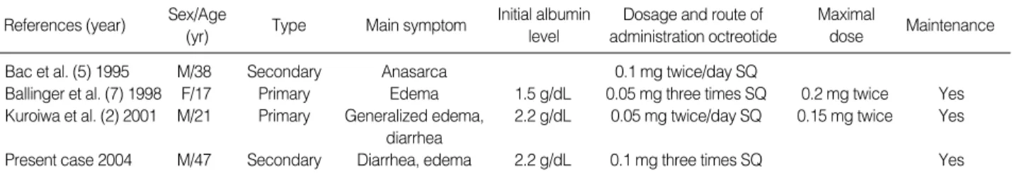
The true impact of somatostatin and octreotide on portal hemodynamics remains controversial, and variable changes in portal pressure and intravariceal pressure in response to these drugs have been reported in different studies (14, 15). So, in our case such as intestinal lymphagiectasia combined with portal hypertension, octreotide may effect on portal pressure and therefore improve protein losing. The clinical character- istics in these cases are summarized in Table 1. All of three cases including our case show the efficacy of octreotide in in-
References (year) Sex/Age
(yr) Type Main symptom Initial albumin level
Dosage and route of administration octreotide
Maximal
dose Maintenance
Bac et al. (5) 1995 M/38 Secondary Anasarca 0.1 mg twice/day SQ
Ballinger et al. (7) 1998 F/17 Primary Edema 1.5 g/dL 0.05 mg three times SQ 0.2 mg twice Yes Kuroiwa et al. (2) 2001 M/21 Primary Generalized edema, 2.2 g/dL 0.05 mg twice/day SQ 0.15 mg twice Yes
diarrhea
Present case 2004 M/47 Secondary Diarrhea, edema 2.2 g/dL 0.1 mg three times SQ Yes
Table 1.Cases of intestinal lymphangiectasia treated with octreotide in the literature
*SQ: subcutaneous.
Protein-Losing Enteropathy treated with Octreotide in a Liver Cirrhosis Patient 469
testinal lymphangiectasia, however, long-term administration of octreotide seems to be required. No adverse effects have been documented in octreotide treatment.
To our knowledge there have been no previous reports of intestinal lymphangiectasia associated with cirrhotic ascites.
We think that the lesion in our patient had been acquired from impaired lymph flow, considering the late onset of dis- ease and the fact that the lymphangiectasia appeared to be associated with liver cirrhosis, a disease known to alter lymph flow. Thus we think that intestinal lymphangiectasia should be suspected as a differential diagnosis in refractory hypoal- buminemia in liver cirrhosis patients.
There are several important aspects in our case. First, it is the first case of intestinal lymphangiectasia developing in liver cirrhosis patient. We speculate that liver cirrhosis can be a cause of intestinal lymphangiectasia though the previous men- tioned mechanism. Second, through it is a relatively rare dis- ease, we should consider intestinal lymphangiectasia as a dif- ferential diagnosis in a patient with protein-losing enteropa- thy. Third, when we experience continuous hypoalbuminemia despite albumin infusion in a liver cirrhosis patient, we should suspect that presence of another cause of hypo-albuminemia.
Although there can be an economic problem and side effects of long-term therapy of octreotide, the administration of oc- treotide should be considered for patients with intestinal lym- phangiectasia whose clinical and biochemical abnormalities do not respond to a low-fat diet. The mechanism of action is not entirely clear but appears to involve reduction in gut protein loss. Further investigation on the mechanism of action of octreotide is needed. And more fundamental strategies should be introduced in the management of intestinal lym- phangiectasia.
REFERENCES
1. Sleisenger MH, Fordtran JS. Gastrointestinal disease: pathophysi- ology, diagnosis, management. 6th ed. Philadelphia: WB Saunders, 1998.
2. Kuroiwa G, Takayama T, Sato Y, Takahashi Y, Fujita T, Nobuoka A, Kukitsu T, Kato J, Sakamaki S, Niitsu Y. Primary intestinal lym-
phangiectasia successfully treated with octreotide. J Gastroenterol 2001; 36: 129-32.
3. Lee SH, Chang YW, Bak SK, Han JY, Kim JH, Jung YH, Lee BW, Han YS, Dong SH, Kim HJ, Kim BH, Lee JI, Chang R. A case of duodenal lymphangiectasia. Korean J Gastroenterol 2003; 41: 59-63.
4. Park GS, Kwak JY, Kim JS, Kwon TC, Jo YJ. A case of primary Intestinal lymphangiectasia. Korean J Gastrointest Endosc 1999; 19:
634-42.
5. Bac DJ, Van Hagen PM, Postema PT, ten Bokum AM, Zondervan PE, van Blankenstein M. Octreotide for protein-losing enteropathy with intestinal lymphangiectasia. Lancet 1995; 345: 1639.
6. Salvia G, Cascioli CF, Ciccimarra F, Terrin G, Cucchiara S. A case of protein-losing enteropathy caused by intestinal lymphangiectasia in a preterm infant. Pediatrics 2001; 107: 416-7.
7. Ballinger AB, Farthing MJ. Octreotide in the treatment of intestinal lymphangiectasia. Eur J Gastroenterol Hepatol 1998; 10: 699-702.
8. Patel AS, DeRidder PH. Endoscopic appearance and significance of functional lymphangiectasia of the duodenal mucosa. Gastrointest Endosc 1990; 36: 376-8.
9. Mine K, Matsubayashi S, Nakai Y, Nakagawa T. Intestinal lymphangi- ectasia markedly improved with antiplasmin therapy. Gastroentero- logy 1989; 96: 1596-9.
10. Lamberts SW, van der Lely AJ, de Herder WW, Hofland LJ. Octreo- tide. N Engl J Med 1996; 334: 246-54.
11. Heresbach D, Raoul JL, Genetet N, Noret P, Siproudhis L, Ramee MP, Bretagne JF, Gosselin M. Immunological study in primary intesti- nal lymphangiectasia. Digestion 1994; 55: 59-64.
12. Dousset B, Legmann P, Soubrane O, Chaussade S, Couturier D, Houssin D, Calmus Y. Protein-losing enteropathy secondary to hep- atic venous outflow obstruction after liver transplantation. J Hepa- tol 1997; 27: 206-10.
13. Stanley AJ, Gilmour HM, Ghosh S, Ferguson A, McGilchrist AJ.
Transjugular intrahepatic portosystemic shunt as a treatment for pro- tein-losing enteropathy caused by portal hypertension. Gastroenterol- ogy 1996; 111: 1679-82.
14. Garcia-Pagan JC, Escorsell A, Moitinho E, Bosch J. Influence of phar- macological agents on portal hemodynamics: basis for its use in the treatment of portal hypertension. Seminar Liver Dis 1999; 19: 427-38.
15. D’Amico G, Pagliaro L, Bosch J. Pharmacological treatment of portal hypertension: an evidence-based approach. Seminar Liver Dis 1999;
19: 475-505.