Bioconversion of Ginsenoside Rb 1 to Compound K using Leuconostoc lactis DC201
Jin-Ying Piao, Yeon-Ju Kim, Lin-Hu Quan, Dong-Uk Yang, Jin-Woo Min, Seon-Heui Son
1, Sang-Mok Kim
1and Deok-Chun Yang*
Korean Ginseng Center for Most Valuable Product and Ginseng Genetic Resource Bank, Kyung Hee University, Yongin 446-701, Korea
1
Seoul Main Customs, Seoul 135-702, Korea
Abstract - Ginseng (Panax ginseng) is frequently used in Asian countries as a traditional medicine. The major components
of ginseng are ginsenosides. Among these, ginsenoside compound K has been reported to prevent the formation of malignancy and metastasis of cancer by blocking the formation of tumor and suppressing the invasion of cancer cells. In this study, ginsenoside Rb
1was converted into compound K, via secreted β -glucosidase enzyme from the Leuconostoc lactis DC201 isolated, which was extracted from Kimchi. The strain DC201 was suspended and cultured in MRS broth at 37℃.
Subsequently, the residue from the cultured broth supernatant was precipitated with EtOH and then dissolved in 20 mM sodium phosphate buffer (pH 6.0) to obtain an enzyme liquid. Meanwhile, the crude enzyme solution was mixed with ginsenoside Rb
1at a ratio of 1:4 (v/v).The reaction was carried out at 30℃ and 190 rpm for 72 hours, and then analyzed by TLC and HPLC. The result showed that ginsenoside Rb
1was transformed into compound K after 72 hours post reaction.
Key words - Bioconversion, Ginsenoside Rb
1, Compound K, Leuconostoc lactis DC201
*Corresponding author. E-mail : [email protected]
Introduction
Panax ginseng is semi-shady and perennial herb, and belong to the family Araliaceae. The Genus Panax is originated from Greek words, Pan means all and ax means curing. It has been cultivated and its highly valued root used for medicinal purposes.
The amphipathic molecule is composed of a glycone and an aglycone. The triterpenoids in ginseng saponins include oleanane with 5 rings, and dammarane with 4 rings. Especially a dammarane triterpenoid is well known as major pharmaceutical activity.
If the ginseng saponins are taken orally, the nonpolar alkaloid is absorbed, but most of saponins are not absorbed effectively. These remained and non-digested saponins are degraded by intestinal bacteria such as Lactobacillus and Leuconostoc. The sugar moiety is used by bacteria and the remained aglycone is dissolved in blood. The dissolved minor saponins are, show highly pharmaceutical effects.
Based on the intestinal microbial community, the absorption pattern of major saponins is quite different and specific to individuals (Karikura et al., 1991; Takino et al., 1994;
Hasegawa et al., 1995). Therefore the conversion of the major saponins to minor saponins which is more absorbable and has higher biological activity, is meaningful.
Odashima (1985) reported that ginsenoside Rh
2inhibits the growth of lung cancer, carcinogenic B-16, and HELA cells in vitro. Lee (1999) showed in vitro that IH-901 (compound K) suppresses the metastatic character of HL-60 leukaemia, PC-14 lung cancer, MKN-45 gastric cancer, and Hep-G2 liver cancer cells and that compound K induces cell death in malignant bone marrow cells. Compound K, ginsenoside Rh
2, and Rh
1are also reported to exert anti-allergic effects (Shin et al., 2003; Zhou et al., 2006). The method of conversion for major saponins to minor saponins includes acid treatment, base treatment, heat treatment, and enzyme treatment. Especially the conversion by microbes is processed in moderated condition such as neutral, room temperature, pressure of the atmosphere, and the reaction is specific to the substrate (Chi et al., 2005a;
Chi et al., 2005b; Park et al., 2001).
Kimchi is a kind of fermented food and good source to lactic acid bacteria (LAB). LAB contributes a singular quality to the taste of Kimchi, as they do in dairy products, such as cheese and fermented milk. A number of type strains have been isolated from Kimchi. In this study, the strain Leuconostoc lactis DC201 was isolated from cabbage Kimchi and characterized by a polyphasic approach, which included a physiological analysis and a phylogenetic tree based on 16S rRNA gene sequence.
We used a crude extract of this β -glucosidase-producing lactic acid strain to convert ginsenoside Rb
1to compound K and to determine optimal conditions for this bioconversion.
Our finding may be applied in the large scale production of compound K, a relatively potent ginseng derivative.
Materials and Methods
Sample preparation
Kimchi samples were collected from Kyung-gi province in Korea, and saponin standards (Rb
1, Rd, F
2, compound K and Rh
2) were donated by the Ginseng Genetic Resource Bank, in Korea. Ginsenoside Rb
1was purified and analyzed with NMR (400 MHz Varian Inova AS 400, Varian USA) in this laboratory.
Saponins were detected using thin-layer chromatography on silica gel (60 F
254TLC plate, Merck) and analyzed by HPLC (HPLC system NS 3000i, Futecs Co., Ltd.).
Taxonomy of the strain Leuconostoc lactis DC201 Lactic acid bacteria were isolated from various types of Kimchi. The Kimchi suspension was spread on MRS agar plates (Difco) after serial dilution with saline solution. Single colonies were selected and transferred to new plates for isolation. After incubation at 37℃ for 2-3 days, the isolated strains were identified by partial 16S rRNA gene sequence, and either cultured further on MRS agar (BD) for immediate study or stored as a suspension in 25% (w/v) glycerol at -70℃.
The isolates were tested for β -glucosidase activity on MRS agar containing esculin. The 16S rRNA gene was amplified from the chromosomal DNA using the universal bacterial primer set fD1 and rP1 (Weisburg et al., 1991), and the purified PCR product was sequenced by Genotech, in Daejeon, Korea (Kim et al., 2005). The partial sequence of the 16S rRNA
gene was compiled with SeqMan software (DNASTAR Inc.).
The 16S rRNA gene sequences of the related taxa were obtained from GenBank and edited using the BioEdit program (Hall, 1999). Multiple alignments were performed with CLUSTAL X (Thompson et al., 1997). Evolutionary distances were calculated using the Kimura two-parameter model (Kimura, 1983). The phylogenetic tree was constructed by the neighbor- joining method (Saitou et al., 1987). Bootstrap analysis with 1,000 replicates was also conducted to obtain confidence levels for the branches.
Growth profiles on different culture media
Various media, including de Man, Rogosa and Sharpe (MRS), Luria-Bertani (LB), nutrient broth (NB), and tryptic soy broth (TSB), were used to culture cells and test enzyme activity. For a seed culture, a single colony was inoculated into MRS broth and incubated at 37℃ in shaking incubator (190 rpm) until the optical density at 600 nm reached 1.0.
From this suspension, a 500 µl aliquot was inoculated into 50 ml liquid medium, such as MRS, LB, NB, or TSB. The strain was then incubated at 37℃ for 48 hrs in shaking incubator (190 rpm). A sample was withdrawn every 2 hrs to read the OD
600.
Effect of temperature on growth
Leuconostoc lactis DC201 was incubated in MRS until the OD
600reached 1.0, then diluted 100-fold into fresh MRS broth and subdivided for culture at increasing growth temperatures (25℃, 30℃, 37℃, and 45℃). The cultures were incubated for 24 hrs with mixing at 190 rpm. Growth was monitored at OD
600.
Effect of pH on growth
Culture vessels containing MRS broth were individually adjusted with 0.5 N HCl and 0.5 N NaOH to pH 5.0, 5.5, 6.0, 6.5, 7.0, 7.5, and 8.0, respectively. The strain DC201 was preincubated in MRS broth to OD
600= 1.0, then diluted 100 fold into MRS broth at a pre-adjusted pH. The cultures were incubated at 37℃ for 24 hrs with 190 rpm. Growth was monitored at OD
600.
Preparation of crude enzyme
Growth and appearance on esculin-MRS agar confirmed
Fig. 1. Phylogenetic tree based on 16S rRNA gene sequences, showing phylogenetic relationships among Leuconostoc lactis DC201 and other related type strains.
β -glucosidase activity in a single colony isolated from the Kimchi. This isolate was thereafter referred to Leuconostoc lactis DC201. For crude enzyme preparation, DC201 was grown in MRS broth at 37℃ with mixing (190 rpm) until the OD
600reached 1.0. The culture broth was centrifuged at 15,000 g and 4℃ for 10 min. Cell-free supernatant was mixed with 4 volumes cold ethanol and placed on ice for 40 min to precipitate protein. After centrifugation under 4℃ within 40 minutes, the precipitate was dissolved in 20 mM sodium phosphate buffer (pH 6.0), and used as crude enzyme solution.
Conversion of ginsenoside Rb
1Crude enzyme prepared from strain DC201 was mixed with 0.2 mM ginsenoside Rb
1(1:4, v/v) and reacted for 72 hrs at 30℃ with stirring (190 rpm). A 1.5 ml sample was removed from the reaction tube every 24 hrs and extracted with water- saturated butanol. The extract was dissolved and applied to a TLC plate (Silica gel, Merck), and developed with the solvent system CHCl
3/CH
3OH/H
2O (65:35:10, v/v, lower phase).
HPLC analysis
A 1.5 ml sample was removed from the enzymatic reaction tube and extracted with water-saturated butanol. The extract was dissolved in methanol, filtered through a 0.45 µm membrane, and then analyzed by HPLC using an RP-18 column (250 × 4.6 mm, ID 5 μm) under UV detector at 203 nm. The injection volume was 25 µl. The mobile phase consisted of acetonitrile (solvent A) and water (solvent B) applied by gradient at a flow rate of 1.6 ml/min. Gradient conditions were as follows:
solvent A/solvent B (15/85, 21/79, 58/42, 90/10, 90/10, 15/85,
15/85) with run times (0-5, 5-25, 25-70, 70-72, 72-82, 82-84, and 84-100 min), respectively.
Results and Discussion
Identification of the strain Leuconostoc lactis DC201 The 16S rRNA gene sequences of the strain and related taxa were obtained from GenBank, edited by BioEdit, and multi-aligned by CLUSTAL X software. The phylogenetic tree was constructed for strain DC201 with the neighbor- joining method, and bootstrap analysis was conducted with 1,000 replicates to obtain confidence levels for the branches.
This tree shows that the strain DC201 is closely related to Leuconostoc lactis (99%) (Fig. 1).
Growth profiles of DC201 in different media
A 24-hr culture grown from one colony in MRS broth was diluted 100-fold into LB, NB, TSB, and MRS, and incubated at 37℃ with stirring. Based on a plot of OD
600versus time, MRS broth supported optimal growth, followed by TSB>
LB>NB. Cultures in MRS broth reached log phase at 4-6 hr after inoculation. After late log phase, the accumulative cell mass in MRS was greater than those in TSB (Data not shown).
Effect of temperature on growth
A stationary phase MRS culture broth (OD
600= 1.0) of
Leuconostoc lactis DC201 was diluted 100-fold into MRS
broth and subdivided for incubation at various temperatures
(25℃, 30℃, 37℃, 45℃). Growth was monitored at 600 nm
for 24 hrs with stirring. DC201 showed optimal growth at
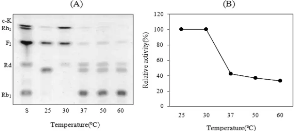
Fig. 2. TLC analysis according to temperature (25-60℃) on the enzymatic conversion of ginsenoside Rb
1to compound K by Leuconostoc lactis DC201 (A), and corresponding effects on β-glucosidase activity (B).
Fig. 3. TLC analysis according to pH (4.0-10.0) on the conversion of ginsenoside Rb
1to compound K by Leuconostoc lactis DC201 (A), and corresponding effects on β-glucosidase activity (B).
37℃. The strain grew in MRS broth at 25-37℃, but not above temperatures 45℃ (data not shown).
Effect of pH on growth
A stationary phase culture (OD
600= 1.0) of the strain DC201 in MRS broth was diluted 100-fold into MRS media that were pre-adjusted to pH 5.0, 5.5, 6.0, 6.5, 7.0, 7.5, and 8.0. Growth was monitored at 600 nm for 24 hrs at 37℃, with stirring. Leuconostoc lactis DC201 grew at pH 6.5-8.0 (data not shown). For further work, MRS broth was not adjusted because its pH is about pH 6.5.
Effect of temperature on the enzyme reaction
The ginsenoside Rb
1was mixed with crude enzyme solution and placed at different temperatures (25℃, 30℃, 37℃, 50℃, and 60℃) for 60 hrs (Fig. 2). Based on the TLC analysis, the
reaction temperature dramatically affected on the production of compound K (Fig. 2 A). The rate was highest at 30℃, and lower at 25℃ and at 37-60℃. HPLC analysis showed a similar pattern to TLC result (Fig. 2 B). Enzymes prepared from Candida peltata, Rhizopus japonicas, and Paecilomyces thermophila show optimal reaction temperatures of 50℃
(Saha and Bothast, 1996), 45℃ (Kim, 1989), and 75℃ (Yang et al., 2008), respectively. Enzymes from strain Lactobacillus brevis LH8 show an optimal temperature of 30℃ (Quan et al., 2008), similar to that of DC201.
Effect of pH on the enzyme reaction
The optimal pH for the reaction of Leuconostoc lactis
DC201 crude enzyme with ginsenoside Rb
1was pH 6.0-8.0,
the enzyme has no activity below pH 4.0. The final product
was analyzed by HPLC (Fig. 3 B). Corresponding enzyme
Fig. 4. Time course of ginsenoside Rb
1bioconversion by Leuconostoc lactis DC201 crude enzyme at 30℃ and pH6.0.
The TLC analysis was developed in CHCI
3/MeOH/H
2O (65:35:10, v/v, lower phase). S: saponin standards.
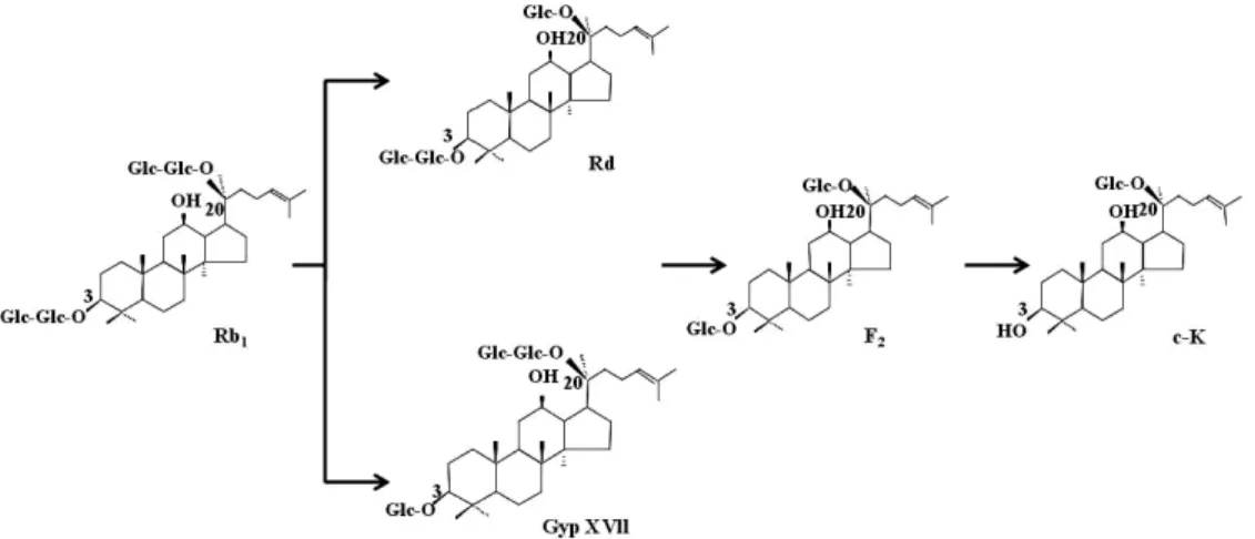
Fig. 6. Proposed pathway of ginsenoside Rb
1transformation to compound K by the Leuconostoc lactis DC201 crude enzyme.
Fig. 5. HPLC profile of ginsenoside Rb
1metabolites produced by Leuconostoc lactis DC201 crude enzyme.
The crude enzyme was reacted with 0.2 mM ginsenoside Rb
1for 24 hrs (A), 48 hrs (B), and 72 hrs (C), extracted with n-butanol, evaporated in vacuo, dissolved in methanol, and analyzed by HPLC.
activities in other species showed pH optimum for conversion at pH 6.2 (Paecilomyces thermophila) (Yang et al., 2008), pH4.8-5.0 (Rhizopus japonicus) (Kim, 1989), pH 6.0-11.0 (Lactobacillus brevis LH8) (Quan et al., 2008), and pH 5.0 (Candida peltata) (Saha et al., 1996).
The time course of ginsenoside bioconversion
During the reaction of strain DC201 crude enzyme with ginsenoside Rb
1, each sample was retrieved for TLC analysis every 24 hrs for 3 days. The level of ginsenoside Rb
1decreased, and levels of gypenoside XVII, ginsenoside Rd, and F
2increased. By 72 hrs, all ginsenoside Rb
1was converted to compound K (Fig. 4).
Analysis of Biotransformation pathway by HPLC
The optimum temperature and pH for the enzyme reaction
of Leuconostoc lactis DC201 with ginsenoside Rb
1was
25°-30℃, pH 6.0-8.0, respectively. With time, the level of
ginsenoside Rb
1decreased, and levels of gypenoside XVII,
ginsenoside Rd, and F
2increased. By 72 hrs, all ginsenoside
Rb
1was converted to compound K (Fig. 4). The reaction of
ginsenoside Rb
1with crude enzyme was sampled at 24 hrs
intervals for analysis by HPLC (Fig. 5). The ginsenoside Rb
1peak (retention time, 44 min) decreased, and the ginsenoside Rd (retention time, 51 min) and gypenoside XVII (retention time, 54 min) peaks increased. The Rb
1conversion proceeded from Rb
1→ Rd, GypXVII → F
2→ compound K (Fig. 6). The HPLC analysis showed that, in 72 min of reaction time, all of the F
2was converted to compound K. The proposed pathway is as follows: GypXVII → F
2→ compound , which indicate that enzymes hydrolyze one glucose molecules at C-3 and two glucose molecules at C-10 of Gypenoside XVll. The major component include protopanaxadiol ginsenoside, such as Rb
1and Rb
2, and have been shown to be metabolized by human intestinal bacterial to their final derivative, such as compound K. Recently, ginsenoside compound K has been reported to induce tumor cell apoptosis, inhibit tumor metastasis, and restrain tumor invasion.
In this study, we isolated a lactic acid bacteria producing β -glucosidase traditional food Kimchi, and the isolated strain was identified by 16S rRNA sequences. We confirmed that a crude enzyme preparation from Leuconostoc lactis DC201 can convert ginsenoside Rb
1to compound K.
Acknowledgements
This work was supported by a grant from the Next- Generation BioGreen 21 Program (SSAC, grant#: PJ008204), Rural Development Administration, Republic of Korea.
Literature Cited
Chi, H. and G.E. Ji. 2005a. Transformation of ginsenosides Rb
1and Re from Panax ginseng by food microorganisms.
Biotechnol Lett. 27(11):765-771.
Chi, H., D.H. Kim and G.E. Ji. 2005b. Transformation of ginsenosides Rb
2and Rc from Panax ginseng by food microorganisms. Biol. Pharm. Bull. 28(11):2102-2105.
Hall, T.A. 1999. BioEdit: a user-friendly biological sequence alignment editor and analysis program for Windows 95/98/
NT. Nucleic Acids Symp. Ser. 41:95-98.
Hasegawa, H., J.H. Sung, S. Matsumiya and M. Uchiyama.
1995. Main ginseng saponin metabolites formed by intestinal
bacteria. Planta Med. 62:453-457.
Karikura, M., T. Miyase, H. Tanizawa, T. Taniyama and Y.
Takino. 1991. Studies on absorption, distribution, excretion and metabolism of ginseng saponins. VII. Comparison of the decomposition modes of ginsenoside-Rb
1and Rb
2in the digestive tract of rats. Chem. Pharm. Bull. Tokyo, Japan.
39(9):2357-2361.
Kim, M.K., W.T. Im, H. Ohta, M. Lee and S.T. Lee. 2005.
Sphingopyxis granuli sp. nov., a ß-glucosidase-producing bacterium in the family Sphingomonadaceae in α-4 subclass of the Proteobacteria. J. Microbiol. 43:152-157.
Kim, S.D. 1989. Production of the convertible enzyme of ginsenoside Rb
1by Rhizopus japonicus. Korean J. Mycology 17(1):31-34.
Kimura, M. 1983. The Neutral Theory of Molecular Evolution.
Cambridge University Press, Cambridge, UK.
Lee, S.J., J.H. Sung, S.J. Lee, C.K. Moon and B.H. Lee. 1999.
Antitumor activity of a novel ginseng saponin metabolite in human pulmonary adenocarcinoma cells resistant to cisplatin.
Cancer Letters 144(1):39-43.
Odashima S., T. Ohta, H. Kohno, T. Matsuda, I. Kitagawa, H.
Abe and S. Arichi. 1985. Control of phenotypic expression of cultured B16 melanoma cells by plant glycosides. Cancer Res. 45:2781-2784.
Park, S.Y., E.A. Bae, J.H. Sung, S.K. Lee and D.H. Kim.
2001. Purification and characterization of ginsenoside Rb
1- metabolizing beta-glucosidase from Fusobacterium K-60, a human intestinal anaerobic bacterium. Biosci Biotechnol Biochem. 65(5):1163-1169.
Saha, B.C. and R.J. Bothast. 1996. Production, purification, and characterization of a highly glucose-tolerant novel β - glucosidase from Candida peltata. Appl. Environ. Microbiol.
62:3165-3170.
Saitou, N. and M. Nei. 1987. The neighbor-joining method: a new method for reconstructing phylogenetic trees. Mol.
Biol. Evol. 4:406-425.
Shin, J.E., E.K. Park, E.J. Kim, Y.H. Hong, K.T. Lee and D.H. Kim. 2003. Cytotoxicity of compound K (IH-901) and ginsenoside Rh
2, main biotransformants of ginseng saponins by bifidobacteria, against some tumor cells. J. Ginseng. Res.
27:129-134.
Takino, Y. 1994. Studies on the pharmacodynamics of ginsenoside- Rg
1, -Rb
1and -Rb
2in rats. YakugakuZasshi 114(8):550-564.
Thompson, J.D., T.J. Gibson, F. Plewniak, F. Jeanmougin and
D.G. Higgins. 1997. The CLUSTAL_X windows interface:
flexible trategies for multiple sequence alignment aided by quality analysis tools. Nucleic Acids Res. 25:4876-4882.
Quan, L.H., Z.Q. Liang, H.B. Kim, S.H. Kim, S.Y. Kim, Y.D.
Noh and D.C. Yang. 2008. Conversion of ginsenoside Rd to compound K by crude enzymes extracted from Lactobacillus brevis LH8. J. Ginseng Res. 32(3):226-231.
Weisburg, W.G., S.M. Barns, D.A. Pelletier and D.J. Lane.
1991. 16S ribosomal DNA amplification for phylogenetic study. J. Bacteriol. 173:697-703.
Yang, S., Z. Jiang, Q. Yan and H. Zhu. 2008. Characterization of a thermo stable extacellular β -glucosidase with activities of exoglucanase and transglycosylation from Paecilomyces thermophila. J. Agric. Food Chem. 56:602-608.
Zhou, W., M.Q. Feng, J.Y. Li and P. Zhou. 2006. Studies on the preparation, crystal structure and bioactivity of ginsenoside compound K. J. Asian Natural Products Res. 8(6):519-527.
(Received 7 November 2011 ; Revised 21 December 2011 ; Accepted 23 December 2011)