Received: 5 July, 2017 Revised: 25 August, 2017 Accepted: 25 August, 2017
Ⓒ The Korean Society of Mycology
This is an Open Access article distributed under the terms of the Creative Commons Attrib- ution Non-Commercial License (http://creative- commons.org/licenses/by-nc/4.0/) which permits unrestricted non-commercial use, distribution, and reproduction in any medium, provided the original work is properly cited.
Kor. J. Mycol. 2017 September, 45(3): 167-174 https://doi.org/10.4489/KJM.20170021
pISSN : 0253-651X eISSN : 2383-5249
OPEN ACCESS
RESEARCH ARTICLE
Two New Records of Ascomycetes from Crop Field Soils in Korea
Mahesh Adhikari
1, Hyun Seung Kim
1, Sun Kumar Gurung
1, Setu Bazie
1, Hyun Gu Lee
1, Hyang Burm Lee
2, Youn Su Lee
1*1
Division of Biological Resource Sciences, Kangwon National University, Chuncheon 24341, Korea
2
Division of Food Technology, Biotechnology & Agrochemistry, College of Agriculture and Life Sciences, Chonnam National University, Gwangju 61186, Korea
*Corresponding author: [email protected]
Abstract
In the ongoing survey of fungal diversity in Korea, two Ascomycetes species, namely Rhinocladiella similis and Toxicocladosporium irritans, were isolated in 2016. These species were identified based on the internal transcribed spacer region and morphological characteristics.
This is the first report of these species in Korea, and the morphological characteristics and images of the fungi are presented.
Keywords: Ascomycetes, Internal transcribed spacer, Phylogeny
Introduction
Sac fungi or Ascomycetes are the largest phylum of fungi, with over 64,000 species [1].
‘Ascus’ is the distinct characteristic of this group of fungi. However, some species in this fungal group are asexual, and thus there is no formation of asci and ascospores.
Rhinocladiella is a widely distributed fungus that can be found in soil, herbaceous substrates, and decaying wood. Rhinocladiella similis belongs to the Eurotiomycetes class within the order Chaetothyriales of the Herpotrichiellaceae family. The typical morphology of Rhinocladiella consists of a profusely branched conidial apparatus of the same texture and pale-brown pigmentation as its mycelium [2]. Rhinocladiella is a genus of melanized fungi that can cause chromoblastomycosis. This order contains several clinically relevant species of the genera Exophialia, Cladophialophora, Fonsecaea, and Phialophora, which are possible etiologic agents of chromoblastomycosis and/or phaeohyphomycosis [3, 4].
The genus Toxicocladosporium (Cladosporiaceae, Capnodiales) was described by Crous et al. [5] as harboring cladosporium-like fungi consisting of distinct “dark, thick-walled conidial and conidiophore septa, and lacking the typical coronate Cladosporium scar type.”
Toxicocladosporium is widely distributed and has the capacity to colonize distinct
substrates and plant families like Cladosporium. The type species of T. irritans was isolated
from moldy paint in Suriname and named “irritans” because of the production of several volatile metabolites in culture that irritated skin exposed to the fungus [5]. The main objective of this study was to (i) describe the newly recorded isolates, R. similis and T.
irritans, morphologically and molecularly, and (ii) compare the morphological features of these newly recorded isolates with those of previously reported isolates.
Materials and Methods
Sampling and isolation
Soil samples were collected from various locations in Gyeongnam, Gyeongsangnam-do, Korea. The GPS location was 35.071097 N, 127.580510 E for isolate KNU16-146 and 35.235877 N, 128.414059 E for isolate KNU16-332. Fungal isolation was performed using the conventional dilution technique [6]. After dilution, isolates were cultured on potato dextrose agar (PDA; Difco, Detroit, MI, USA) supplemented with 100 µg chloramphenicol (bacteriostat/L PDA) for 5~7 days at 25°C until growth of a fungal colony was observed.
For further use, the isolate was preserved at 20°C on PDA slants.
Morphological characterization
The fungal macro-morphological characteristics were studied on PDA. The fungal specimens were single point inoculated and were incubated at 25°C for 7 days in darkness.
Colony characteristics were recorded, and fungal materials were examined using an Olympus BX50F-3 light microscope (Olympus, Tokyo, Japan). For the micro-morphological examination, microscopic mounts of all isolates were made from colonies grown on PDA in lactic acid with a drop of alcohol added to remove air bubbles and excess conidia.
Photomicrographs of the isolates were obtained with an HK 3.1 CMOS digital camera (KOPTIC, Seoul, Korea) attached to an Olympus BX50F-3 microscope. The microscopic structures of the isolates were also examined using a scanning electron microscope (LEO Model 1450VP Variable Pressure Scanning Electron Microscope; Carl Zeiss, Oberkochen, Germany).
Genomic DNA extraction, PCR amplification, sequencing, and phylogenetic analysis
For molecular genetic identification, isolates were grown on PDA for a week, and total
genomic DNA was extracted using DNeasy Plant Mini Kit (Qiagen, Germantown, MD,
USA) following the manufacturer’s instructions. The internal transcribed spacer (ITS)
region was amplified using primers ITS1 (5'-TCCGTAGGTGAACCTGCG-3') and ITS4
(5'-TCCTCCGCTTATTGATATGC-3') [7], and the amplified PCR products were sequenced
using an ABI Prism 3730 DNA analyzer (Applied Biosystems, Foster City, CA, USA). All
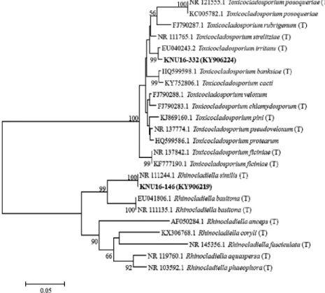
sequence information was analyzed using the BLAST program National Center for Biotechnology Information (NCBI, http://www.ncbi.nlm.nih.gov/blast/). A phylogenetic tree was constructed using the neighbor-joining method and MEGA6.0 software [8].
Statistical confidence in the tree topology was evaluated based on a bootstrap analysis with 1,000 replicates. The newly recorded fungal isolates were deposited in the National Institute of Biological Resources (NIBR), Korea, under deposition numbers NIBRFG 0000499480 and NIBRFG0000499478 for isolates KNU16-146 and KNU16-332 respectively.
The sequences of isolate KNU16-146 and KNU16-332 were also deposited in GenBank under accession numbers KY906219 and KY9066224, respectively (Table 1).
Table 1. Sequences of Rhinocladiella similis and Toxicocladosporium irritans along with their National Institute of Biological Resources and GenBank accession numbers
Isolates NIBR no. GenBank
accession no.
Closest GenBank library strain Similarity (%) KNU16-146 NIBRFG0000499480 KY906219 Rhinocladiella similis 100 KNU16-332 NIBRFG0000499478 KY9066224 Toxicocladosporium irritans 99
Results
Morphology and micromorphology of isolate KNU16-146
Rhinocladiella similis de Hoog & Caligiorne, Journal of Clinical Microbiology 41 (10):
4777 (2003)
The colony on PDA was slightly brown on the front side and black on the back side of the
Fig. 1. Morphology of the isolate Rhinocladiella similis (KNU16-146) grown for 7 days on
potato dextrose agar. A, obverse colony; B, reverse colony; C, D, simple microscopic images of
conidiophores and conidia; E, F, scanning electron micrographs of conidiophores and conidia.
plate. It grew moderately, attaining a diameter of 60~65 mm in 7 days at 25°C (Fig. 1A, 1B). The colony was floccose. Sporulation was moderate to dense, the surface was rough, and the form was irregular. Diffusible pigments were not produced. Hyphae were pale olivaceous to brown and 1.3 mm wide. Conidiogenous cells were profusely branched, and the conidial color was brown. Conidiogeneous cells were cylindrical, 11~19 by 2 µm apically (Fig. 1C~1F). Conidia were subhyaline, noncatenate, cylindrical, and narrowed toward the base, and 3~6 by 1.3 µm of diameter. Budding cells were broadly ellipsoidal with a size of 1~5 µm. Morphology comparison of our study isolate (KNU16-146) with the previously reported R. similis isolate has been mentioned in Table 2.
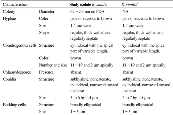
Table 2. Comparison of the morphologies of isolate KNU16-146 and a previously described isolate of Rhinocladiella similis
Characteristics Study isolate R. similis R. similis
aColony Diameter 65 ∼70 mm on PDA NA
Hyphae Color pale olivaceous to brown pale olivaceous to brown
Size 1.4 µm wide 1.5 µm wide
Shape regular, thick walled and regularly septate
regular, thick walled and regularly septate Conidiogenous cells Structure cylindrical with the apical
part of variable length.
cylindrical with the apical part of variable length.
Color brown brown
Number and size 11∼19 and 2 µm apically 11∼19 and 2 µm apically
Chlamydospores Presence absent absent
Conidia Structure subhyaline, noncatenate, cylindrical, narrowed toward the base
subhyaline, noncatenate, cylindrical, narrowed toward the base
Size 3 to 6 by 1.4 µm 4 to 7 by 1.5 µm
Budding cells Structure broadly ellipsoidal broadly ellipsoidal
Size 1∼5 µm 1∼5 µm
PDA, potato dextrose agar; NA, Not available.
a
Source of description [2].
Morphology and micromorphology of isolate KNU16-332
Toxicocladosporium irritans Crous & U. Braun, Studies in Mycology 58: 39 (2007)
The colony color on PDA was black on both the front and back sides of the plate. It grew
moderately, attaining a diameter of 65~70 mm in 7 days at 25°C. (Fig. 2A, 2B). Sporulation
was moderate to dense, the form was irregular, the texture was floccose, and the surface
was rough. It had branched mycelium that were finely verruculose. Conidiophores were
macronematous and solitary. Conidiogenous cells were terminal or lateral with a slight
taper towards the apex. Conidia were catenulate, ellipsoid to ovoid, and 0.4~1 µm wide
(Fig. 2C, 2F). Morphology comparison of our study isolate (KNU16-332) with the
previously reported T. irritans isolate has been mentioned in Table 3.
Fig. 2. Morphology of the isolate Toxicocladosporium irritans (KNU16-332) grown for 7 days on potato dextrose agar. A, obverse colony; B, reverse colony; C, D, simple microscopic images of conidiophores and conidia; E, F, scanning electron micrographs of conidiophores and conidia.
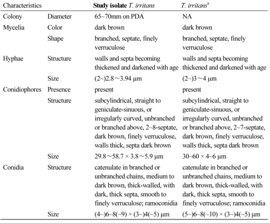
Table 3. Comparison of the morphologies of isolate KNU16-332 and a previously described isolate of Toxicocladosporium irritans
Characteristics Study isolate T. irritans T. irritans
aColony Diameter 65~70mm on PDA NA
Mycelia Color dark brown dark brown
Shape branched, septate, finely verruculose
branched, septate, finely verruculose
Hyphae Structure walls and septa becoming thickened and darkened with age
walls and septa becoming thickened and darkened with age
Size (2~)2.8∼3.94 µm (2~)3∼4 µm
Conidiophores Presence present present
Structure subcylindrical, straight to geniculate-sinuous, or irregularly curved, unbranched or branched above, 2~8-septate, dark brown, finely verruculose, walls thick, septa dark brown
subcylindrical, straight to geniculate-sinuous, or irregularly curved, unbranched or branched above, 2~7-septate, dark brown, finely verruculose, walls thick, septa dark brown
Size 29.8∼58.7 × 3.8∼5.9 µm 30~60 × 4~6 µm
Conidia Structure catenulate in branched or unbranched chains, medium to dark brown, thick-walled, with dark, thick septa, smooth to finely verruculose; ramoconidia
catenulate in branched or unbranched chains, medium to dark brown, thick-walled, with dark, thick septa, smooth to finely verruculose; ramoconidia Size (4~)6~8(~9) × (3~)4(~5) µm (5~)6~8(~10) × (3~)4(~5) µm PDA, potato dextrose agar; NA, Not available
a