of rotifers such as Brachionus calyciflorus was proposed by Luo et al.(2006). However, it was also stressed out that the identification and classification based only on the morphological characteristics are not appropriate for the members of chlorellacean genera(Pröschold et al. 2010).
Hence, Luo et al.(2010) proposed a new system of generic separation for these groups based on molecular phylogeny of the combined SSU rRNA and ITS sequences and com- pensatory base changes(CBC) in helix III of ITS2. These
https://doi.org/10.11626/KJEB.2020.38.1.061
INTRODUCTION
Micractinium(Chlorophyta, Trebouxiophyceae, Chlo- rellales) was first described in 1858 by Fresenius(Luo et al.
2006) and this genus is known to have spherical to ovoid cells and possess a parietal and cup-shaped chloroplast with a distinct pyrenoid. Since it shares highly similar mor- phological features with the genus Chlorella, morphologi- cal discrimination by the bristle formation in the presence
Original article
First record of a marine microalgal species, Micractinium singularis (Trebouxiophyceae) isolated from Janghang Harbor, Korea
Seung-Woo Jo1,2, Nam Seon Kang3, Hyunsik Chae2, Jung A Lee3, Kyeong Mi Kim3, Moongeun Yoon3, Ji Won Hong3,* and Ho-Sung Yoon1,2
1Department of Energy Science, Kyungpook National University, Daegu 41566, Republic of Korea
2School of Life Sciences, Kyungpook National University, Daegu 41566, Republic of Korea
3Department of Taxonomy and Systematics, National Marine Biodiversity Institute of Korea, Seocheon 33662, Republic of Korea
Korean J. Environ. Biol.
38(1) : 61-70(2020) ISSN 1226-9999(print) ISSN 2287-7851(online)
Korean Journal of Environmental Biology
* Corresponding author Ji Won Hong
Tel. 041-950-0743
E-mail. [email protected]
Received: 7 January 2020 Revised: 16 February 2020
Revision accepted: 17 February 2020
Abstract: A eukaryotic microalga was isolated from seawater in Janghang Harbor, Korea and its morphological, molecular, and physiological characteristics were investigated.
Due to its simple morphology, no distinctive characters were found by morphological observation, such as light microscope or scanning/transmission electron microscopy(S/
TEM). However, molecular phylogenetic evidence inferred from the concatenated small subunit(SSU) 18S rRNA and internal transcribed spacer(ITS) sequence data indicated that the isolate belonged to the newly described Micractinium singularis. Furthermore, it was clustered with Antarctic Micractinium strains and it also showed a psychrotolerant property, surviving at temperatures as low as 5°C. However, its optimal growth temperatures range from 15°C to 25°C, indicating that this microalga is a mesophile.
Additionally, gas chromatography-mass spectrometry(GC/MS) analysis showed that the isolate was rich in nutritionally important omega-3 polyunsaturated fatty acid, and high-performance liquid chromatography analysis(HPLC) revealed that the high-value antioxidant lutein was biosynthesized as an accessory pigment by this microalga, with glucose as the major monosaccharide. Therefore, in this study, a Korean marine M.
singularis species was discovered, characterized, and described. It was subsequently added to the national culture collections.
Keywords: first record, Janghang Harbor, Korean marine mic roalga, Micractinium sin
gularis
approaches have been reliable standard methods for genus differentiation between Chlorella and Micractinium.
Yet, species-level delimitation within the genus Micrac
tinium has been hampered by the lack of available diag- nostic morphological keys as well as their plasticity in res- ponse to both abiotic and biotic stresses. As a result, there are only 16 taxonomically accepted species even though 25 Micractinium species have been described so far(Guiry and Guiry 2019). Currently, no comprehensive taxonomic keys exist which allow for reliable species identification in this group. Consequently, many attempts have been made to find diagnostic morphological characters that can be used for determination of Micractinium species in accordance to molecular phylogeny.
In this study, a unicellular microalga belonging to the genus Micractinium was isolated from Janghang Harbor in Korea and a pure culture was established. Its phylogenetic position based on the concatenated SSU rRNA and ITS sequence analyses revealed that the isolate was clustered with the recently described Antarctic M. singularis strain KSF0094(Chae et al. 2019) and possessed a cold-tolerant property. In conclusion of the results, we report informa- tion on the first record of this species in Korea and its mor- phological, molecular, and chemotaxonomic features.
MATERIALS AND METHODS
1. Sample collection and isolation of microalga Seawater samples were collected from Janghang Harbor in Sinchang-ri, Janghang-eup, Seocheon-gun, Chungc- heongnam-do, Korea in March of 2016. The location and physico-chemical data of the sampling site were given in Table 1. Water samples were filtered on 25μm mesh net to remove grazing organisms and aliquots(100μL) of the samples were spread onto 1%(w/v) NaCl(Duksan, Ansan, Korea) BG-11 agar plates(UTEX, Austin, TX, USA) sup- plemented with 100μg mL-1 imipenem(Sigma-Aldrich, St.
Louis, MO, USA) to suppress bacterial growth and gener- ate pure cultures(Hong et al. 2015). The plates were then
incubated at 10°C in a growth chamber(FLI-2010A, Eyela, Tokyo, Japan) with cool fluorescent light(approximately 30μmole m-1 s-1) in a light:dark cycle(16:8 h) until mi- croalgal colonies were formed. Single colonies were asepti- cally streaked onto fresh BG-11 agar plates supplemented with 20μg mL-1 imipenem and this step was repeated until an axenic was produced.
2. Morphological identification
A single colony was transferred in 100mL BG-11 medi- um and the flasks were autotrophically incubated at 10°C.
Well-grown live cells were observed by an upright micro- scope(Microscope Axio Imager.A2, Carl Zeiss, Göttingen, Germany). The Brachionus biotest was performed in or- der to induce bristle formation in the Micractinium isolate according to the method developed by Luo et al.(2005).
Brachionus calyciflorus cysts(TB21) were obtained from MicroBioTests(Gent, Belgium).
For SEM, 10mL aliquots of cultures at approximately 1,000cells mL-1 were fixed for 10 min in osmium tetroxide (OsO4, Electron Microscopy Sciences, Hatfield, PA, USA) at a final concentration of 2%(v/v). The fixed cells were collected on a 3μm pore size, polycarbonate membrane filter(Whatman, Kent, UK) and washed three times with distilled water to remove residual media components. The membranes were dehydrated in an ethanol series(Merck, Darmstadt, Germany) and immediately dried using a criti- cal point dryer(CPD 003, Bal-Tec, Balzers, Liechtenstein).
The dried filters were mounted on a stub and coated with platinum in a low vacuum sputter coater(FM ACE200, Leica, Wetzlar, Germany). Surface morphology was ob- served with a field emission scanning electron microscopy (FE-SEM, SUPRA 55VP, Carl Zeiss, Jena, Germany).
For TEM, cells were transferred to a 10mL tube and fixed in 2.5%(v/v) glutaraldehyde for 2hrs and the content was concentrated at 1,610g for 10min in a Vision Centri- fuge VS-5500(Bucheon, Korea). The resulting pellet was subsequently transferred to a 1.5 mL tube and rinsed in 0.2 M sodium cacodylate buffer(Electron Microscopy Scienc- es) at pH 7.4. After several rinses in 0.2M sodium caco-
Table 1. Description of the sampling site
Depth(m) Temperature(°C) Salinity(PSUa) Latitude Longitude
0.3 7.7 22.5 36°00ʹ23.96ʺN 126°41ʹ23.52ʺE
aPSU: practical salinity unit.
dylate buffer, cells were post-fixed for 90min in 1%(w/v) OsO4 in deionized water. The pellet was then embedded in agar(Duksan). Dehydration was performed in a grad- ed ethanol series(50, 60, 70, 80, 90, and 100% ethanol, followed by two changes in 100% ethanol). The material was embedded in Spurr’s resin(Electron Microscopy Sci- ences). Sections were prepared on an EM UC7 ultramicro- tome(Leica) and stained with 3%(w/v) aqueous uranyl acetate(Electron Microscopy Sciences) followed by lead citrate(Electron Microscopy Sciences). The sections were visualized on an H-7650 TEM(Hitachi, Tokyo, Japan) us- ing a voltage of 100kV.
3. Molecular identification
For molecular analysis, genomic DNA was extracted us- ing a DNeasy Plant Mini kit(Qiagen, Hilden, Germany).
The primer sets NS1 and NS8 and ITS1 and ITS4(White et al. 1990) were used to amplify the SSU 18S rRNA and ITS region, respectively. Synthesis of the primers employed in this study and the DNA sequencing were conducted at the Macrogen facility(Daejeon, Korea). Phylogenetic anal- ysis was performed with the string sequences of SSU-ITS1- 5,8S-ITS created according to Luo et al.(2010) using the software package Molecular Evolutionary Genetics Anal- ysis(MEGA) version 7.0(Kumar et al. 2016). The com- bined data set of the isolate was aligned with those of the 11 chlorellacean microalgae strains based on the previous publications(Hoshina et al. 2010; Luo et al. 2010; Chae et al. 2019) using ClustalW incorporated in MEGA 7.0. pack- age(Kumar et al. 2016), and closely related sequences were downloaded from the National Center for Biotechnology Information(NCBI) database, manually trimmed, and aligned with MEGA software using the ClustalW tool. The best-fit nucleotide-substitution model(Kimura 2-parame- ter+a discrete Gamma distribution with 5 rate categories, K2+G) was selected using MEGA 7.0 based on Bayesian
information criterion. This model was used to build a max- imum likelihood(ML) phylogenetic tree with 1,000 boot- strap replicates. M. conductrix CCAP211/83 was used as an outgroup. DNA sequences obtained in this study were de- posited in the NCBI under accession numbers KY655270 and KY655271(Table 2). The ITS2 secondary structures were constructed using Mfold(Zuker 2003) and 4SALE (Seibel et al. 2006, 2008) according to Chae et al.(2019).
4. Optimal growth condition determination A single colony of strain MM0003 from a pure culture was aseptically streaked onto BG-11 agar plates in trip- licate and incubated for 14 days. Survival and growth of MM0003 cells maintained at temperatures ranging from 5°C to 35°C(at intervals of 5°C) were examined to deter- mine the optimum culture temperature. Salt tolerance test was conducted at 20°C using BG-11 agar supplemented with 0.0M, 0.5M, 1.0M, 1.5M, and 2.0M NaCl(Daejung, Siheung, Korea) respectively.
5. GC/MS analysis
The isolates were autotrophically grown in BG-11 medi- um for 20 days and cells were harvested by centrifugation at 2,063g(1580R, Labogene, Daejeon, Korea) for lipid analysis. Then samples were freeze-dried and pulverized to enhance extraction efficiency. Lipid extraction was performed using the method developed by Breuer et al.
(2013). The FAME composition was analyzed using a 7890A gas chromatograph equipped with a 5975C mass selective detector(Agilent, Santa Clara, CA, USA) accord- ing to our previous publication(Kang et al. 2019). Com- pound identification was performed by matching the mass spectra with those in the Wiley/NBS registry of mass spec- tral data. The searches with a match value higher than 90%
were considered valid.
Table 2. Results from BLAST searches (searched date: 22 December 2019) using the 18S rRNA and ITS sequences of Micractinium singu- laris MM0003
Marker gene Accession No. Length(bp) Closest match
(GenBank accession No.) Overlap(%) Sequence similarity(%)
18S rRNA KY655270 1,770 Micractinium sp.
KNUA034 (KM243325) 100 100.00
ITS KY655271 724 Micractinium sp.
KNUA036 (KT883910) 100 97.65
6. Biomass characterization
Freeze-dried biomass samples were pulverized with a mortar and pestle and sieved through ASTM No. 230 mesh(opening=63μm). Ultimate analysis was conduct- ed in order to determine the carbon(C), hydrogen(H), nitrogen(N), and sulfur(S) contents using a Flash 2000 elemental analyzer(Thermo Fisher Scientific, Milan, Italy) in duplicate. Oxygen(O) was calculated by sub- tracting the sum of percent of C, H, N, S and ash from 100%. Gross calorific value(GCV) was estimated by the following equation developed by Given et al.(1986):
[GCV=0.3278C+1.419H+0.09257S-0.1379O+0.637 (MJ kg-1)]. Protein content was calculated from the N content in the ultimate analysis by using the conversion factor of ×6.25(Mariotti et al. 2008).
Proximate analysis was carried out on a DTG-60A ther- mal analyzer(Shimadzu, Kyoto, Japan). Platinum pans were used to contain 30mg of α-alumina(α-Al2O3) pow- der(Shimadzu) as a reference material and approximately 10mg of each sample, respectively. Nitrogen(>99.999%, N2) was supplied as the carrier gas at a rate of 25mL min-1 to protect the microalgae powder from oxidation. Samples were heated from 50°C to 900°C at a rate of 10°C min-1. Thermogravimetric analysis(TGA) data were analyzed by ta60 Ver. 2.21 software(Shimadzu).
7. HPLC analysis
Pigment extraction was performed using the method developed by Zapata et al.(2000). Briefly, freeze-dried microalgal biomass was extracted in 90% HPLC-grade ac- etone(Daejung) and filtered through a Whatman polytet- rafluoroethylene(PTFE) syringe filter with a pore size of 0.2μm(Whatman, Florham Park, NJ, USA). Samples were then analyzed on an Agilent 1260 Infinity HPLC system(Agilent, Waldbronn, Germany) equipped with a Discovery C18 column(25cm×4.6 mm, 5μm; Supelco, Bellefonte, PA, USA) at 33°C based on the method used in our previous publication(Jang et al. 2017). HPLC-grade methanol and ammonium acetate were purchased from Daejung and Fluka(Sigma-Aldrich), respectively. Pigment standards such as β-carotene, chlorophyll(Chl)-a, Chl-b, lutein, and violaxanthin were obtained from Sigma-Al- drich.
8. Carbohydrate analysis
For monosaccharide analysis, 50mg of freeze-dried bio-
mass samples were hydrolyzed in 2.5mL 2 N sulfuric acid (Sigma-Aldrich) at 94°C for 3h. When the reaction tubes were cooled to room temperature, a drop of 40% calcium carbonate(Sigma-Aldrich) was added to the hydrolysates.
Samples were filtered through a 0.2μm PTFE filter(What- man) and analyzed on a Prominence Modular HPLC system(Shimadzu) with a Sugar-Pak I column(10μm, 6.5 mm×300mm; Waters, Milford, MA, USA) according to our previous publication(Kang et al. 2019). All monosac- charide standards(arabinose, fructose, fucose, galactose, glucose, lactose, maltose, mannitol, mannose, rhamnose, ribose, sorbitol, sucrose, and xylose) were obtained from Sigma-Aldrich. Monosaccharide contents in mg g-1 dry weight(DW) biomass were quantified by calculating the total peak areas of each monosaccharide derived from a calibration curve.
RESULTS
1. Morphology of the isolate
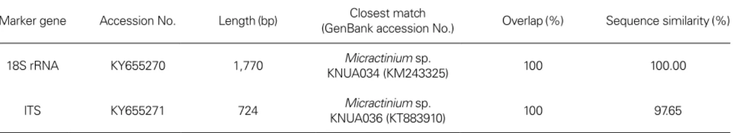
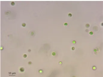
As shown in Fig. 1 and Fig. 2, the algal cells were soli- tary and round to slightly ellipsoid in shape and their siz- es ranged from approximately 3 to 5μm in diameter. No bristles were observed in the isolate in the presence of B.
calyciflorus. Cytological observation showed that the cells had a predominant cup-shaped chloroplast(C) containing one pyrenoid(P) and multiple layers of thylakoids(Fig. 3).
The nucleus(N) was also observed located in the center of the microalga. Starch(S) and mitochondrion(M) were also found in the cells(Fig. 3).
Fig. 1. Light micrograph of M. singularis MM0003.
2. Phylogenetic position determined by genetic markers
Molecular identification results were summarized in Ta- ble 2. The 18S rRNA sequence of the isolate was identical to Micractinium sp. KNUA034(KM243325), but the ITS sequence of strain MM0003 was only 97.65% homolo- gous to that of Micractinium sp. KNUA034(KM243327).
As illustrated in Fig. 4, strain MM0003 was clustered with other 3 Antarctic Micractinium species such as M. singu
laris KSF0094(MN414469), M. variabile KSF0085(MN 414468), and Micractinium sp. KNUA034(KM243325
+KM243327). The ITS2 secondary structure of 3 Mi
cractinium species exhibited typical four helices and pyr- imidine-pyrimidine bulge on helix II, the conserved se- quence TGGT(UGGU) on the 5ʹ side of helix III(Mai and Coleman 1997), and a C-G pairing on the top of helix III(Fig. 5). Overall secondary structures of M. singularis MM0003 and KSF0094 were very similar to each other and CBC or hemi-CBC was not found in these strains.
However, M. singularis MM0003 and M. variabile KSF 0085 showed differences in helix I structure and these results were in agree with those obtained in the previous publication(Chae et al. 2019).
3. Optimal growth temperatures and NaCl tolerance for the isolate
As shown in Table 3, the isolate could survive and grow in a temperature range of 5-25°C whilst their optimal growth temperature was 15-25°C. It could not survive over 30°C and delayed growth was also observed at 5°C.
Also, the isolate was able to withstand up to 0.5M NaCl even though growth was suppressed in the presence of NaCl. It did not survive at a concentration over 1.0M NaCl.
Fig. 2. FE-SEM images of M. singularis MM0003.
Fig. 3. TEM ultrastructure images of M. singularis MM0003. C:
chloroplast; M: mitochondrion, N: nucleus; P: pyrenoid; S: starch.
4. Fatty acid composition
The major fatty acids of strain MM0003 were C16:0(26.1
±0.2%), C16:4 ω3(12.6±0.3%), and C18:3 ω3(30.9±
0.3%). Summarized characteristics for the strain are given in Table 4.
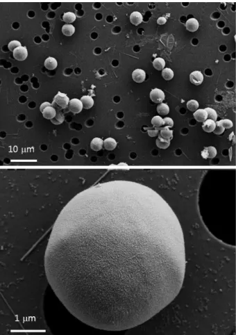
Fig. 4. The phylogenetic relationship between strain MM0003 and its closely related species based on the concatenated SSU 18S rRNA and ITS sequences using the K2+G model with M. conductrix as an outgroup. The tree was generated by the ML method using 1,000 bootstrap replicates. The scale bar represents a 1% difference in the nucleotide sequences.
Fig. 5. ITS2 secondary structure for M. singularis MM0003, M. singularis KSF0094, and M. variabile KSF0085. The key CBCs at the tip of helix III are indicated in a red box.
Table 3. Growth of strain MM0003 at various temperatures
Temperature(°C) 5 10 15 20 25 30 35
Growtha + ++ +++ +++ +++ - -
a +++: good growth; ++: moderate growth; +: poor growth; -: no growth.
5. Biomass properties
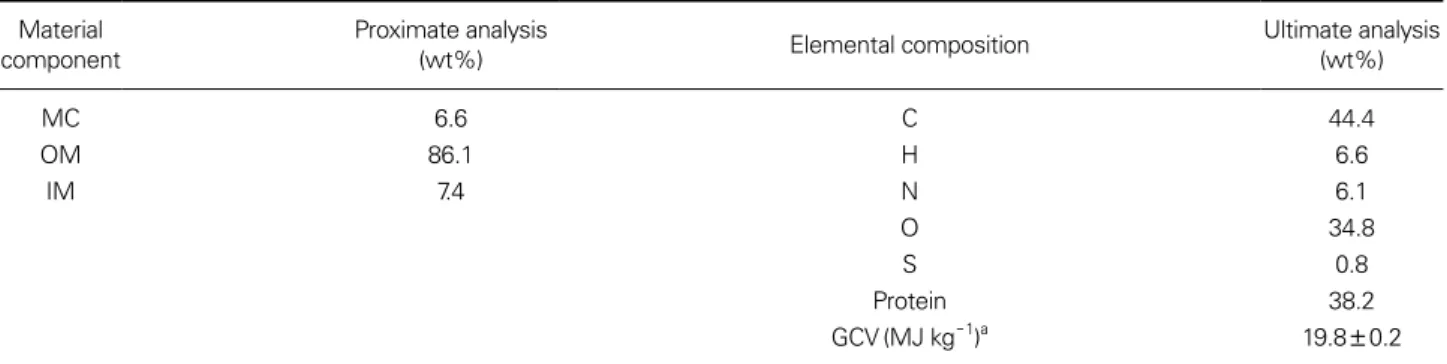
In proximate analysis by TGA, the moisture content (MC) is determined by the mass loss before 110°C under
N2 atmosphere, the organic matter(OM) refers to the mass loss between 110-900°C under N2 as a result of thermal decomposition, and the remaining mass represents the inorganic matter(IM). The TGA profile is shown in Fig.
6 and the material composition of strain MM0003 is pre- sented in Table 6. The GCV and protein content based on the ultimate analysis were 19.8MJ kg-1 and 38.2%, respec- tively(Table 5).
Table 4. Lipid profile of strain MM0003
Component Content(%)a Note
8-Heptadecene(C17H34) 0.4±0.0 - Neophytadiene(C20H38) 0.6±0.0 - Palmitic acid(C16:0) 21.6±0.2 SFA(major) Palmitoleic acid(C16:1 ω7) 2.8±0.1 - Hexadecadienoic acid(C16:2 ω6) 1.2±0.1 - Hexadecatrienoic acid(C16:3 ω3) 2.4±0.0
Hexadecatetraenoic acid(C16:4 ω3) 12.6±0.3 Omega-3 PUFA (major)
Stearic acid(C18:0) 1.0±0.0 -
Oleic acid(C18:1 ω9) 2.6±0.1 -
Linoleic acid(C18:2 ω6) 7.6±0.3 - α-Linolenic acid(C18:3 ω3) 30.9±0.3 Omega-3 PFUA
(major) Eicosatetraenoic acid(C20:4) 0.8±0.0 -
aValues represent the average±standard deviation of three independent experiments.
Table 5. Proximate and ultimate analysis results of M. singularis MM0003 Material
component Proximate analysis
(wt%) Elemental composition Ultimate analysis
(wt%)
MC 6.6 C 44.4
OM 86.1 H 6.6
IM 7.4 N 6.1
O 34.8
S 0.8
Protein 38.2
GCV(MJ kg-1)a 19.8±0.2
aValue represents the average±standard deviation of two independent experiments.
Fig. 6. TGA profile of M. singularis MM0003.
Weight(%)
Temperature(°C)
Table 6. Pigment profile of strain MM0003
Peak number Retention time Compound Content(%)a
1 18.2 Unknown 6.5±0.1
2 19.8 Unknown 4.3±0.0
3 25.3 Lutein 30.8±0.0
5 28.8 Chl-b 13.4±0.1
6 32.0 Chl-a 38.2±0.1
7 40.9 β-carotene 6.8±0.0
aValues represent the average±standard deviation of three independent experiments.
6. Pigment composition
The pigment profile of M. singularis MM0003 is report- ed in Table 6. The major pigments of the isolate were Chl-a (38.2%±0.1%), lutein(30.8%±0.0%), Chl-b(13.4%±
0.1%) and β-carotene(6.8±0.0%). The other minor peaks were not identified.
7. Monosaccharide composition
The carbohydrate composition of the isolate is summa- rized in Table 7. Its major monosaccharides were glucose (88.8%, 14.8mg g-1 DW) and a small portion of minor sugars(mannose 9.9% and mannitol 1.2%) were also de- tected.
8. Deposition of the isolate
Stain MM0003 obtained in this study was deposited in the National Marine Biodiversity Institute of Korea(MA- BIK) and the Korean Collection for Type Cultures(KCTC) under the accession numbers of MABIK-LP-00000134 and KCTC 13290BP, respectively.
DISCUSSION
In this study, a psychrotolerant Korean M. singularis strain was axenically isolated and its identity was analyzed by morphological, molecular, and physiological approach- es. Establishment of a pure culture was achieved by the combination of imipenem treatment and physical separa- tion technique. As the isolate only had very unsophisticat- ed cell structures such as the chloroplast, pyrenoid, nucle- us, and mitochondrion, it was unable to distinguish strain MM0003 from Chlorella spp. by traditional morphological criteria.
However, it was able to determine the phylogenetic po-
sition of the isolate by the sequence analyses of SSU and ITS regions. As shown in Fig. 4, the isolate showed close relationship with the newly described Antarctic Micractin
ium species(Chae et al. 2019). In addition, the key CBCs in the ITS2 secondary structure also confirmed that strain MM0003 belonged to the genus Micractinium(Hoshina et al. 2010). Strain MM0003 exhibited typical four-fingered hands(helices I through IV) and a C-G pairing on the top of helix III(Fig. 5). The predicted ITS2 secondary struc- ture diagrams for the closely related Micractinium species also confirmed that strain MM0003 possessed similar sec- ondary structure to M. singularis KSF0094 isolated from Deception Island, South Shetland in Antarctica(Chae et al.
2019). Furthermore, strain MM0003 exhibited the clos- est affinity with the non-bristle-forming M. singularis KSF0094. Since both Antarctic Micractinium strains did not produce bristle under grazing pressure and possess solitary habit, it can be concluded that they share similar morphological, physiological, and molecular characteris- tics with each other. Hence, strain MM0003 was identified as M. singularis and this is the first report of this species in Korea.
The isolate was capable of surviving and propagating as low as at 5°C showing its cryotolerant properties. Howev- er, the optimum growth temperature was 15-25°C which indicates that this Korean M. singularis strain is mesophilic.
It has been reported that the maintenance of membrane fluidity by the unsaturation of the fatty acids in membrane lipids under cold temperatures is one of the main adap- tation strategies of polar microalgae(Osipova et al. 2009;
Boelen et al. 2013). Analysis of the cellular fatty acid com- position of strain MM0003 also revealed that it was rich in C16:0(21.6%) saturated fatty acid(SFA) and C18:3 ω3 (30.9%) and C16:4 ω3(30.6%) polyunsaturated fatty acids (PUFAs). Beneficial health effects of essential omega-3 PUFAs were well documented by Mehta et al.(2009).
Currently omega-3 PUFAs are derived from mainly marine biological resources, such as fish oil, and various commer- cial products are available worldwide. Given the current ocean pollution and overfishing issues, as well as global climate change, the sustainability of marine fish as a safe resource of omega-3 PUFAs for human consumption is in great doubt(Kang 2011; Jeromson et al. 2015). Therefore, this marine micro alga may serve as potential biological resource that can replace fish-based oil for sustainable and pollution-free omega-3 PUFAs production.
The OM can be defined as the part of solid fuel that is driven-off as a gas by heating and typical biomass gener-
Table 7. Monosaccharide composition of strain MM0003
Compound Content
%a mg g-1 DW
Glucose 88.8±3.6 14.8
Mannose 9.9±0.7 2.9
Mannitol 1.2±0.1 0.6
aValues represent the average±standard deviation of three independent experiments.
ally has a OM content of up to 80%(crop residue: 63- 80%; wood: 72-78%). The OM content of this microalga (86.1%) used in this study was higher than the ranges of plant- and wood-based biomass feedstocks. The GCV was also calculated to understand the potential of algal biomass as a biofuel feedstock(Table 5). The results showed that the GCV was within the range of the terrestrial energy crops(17.0-20.0MJ kg-1)(Ross et al. 2008). Since fine particulate matters have become a national concern in re- cent years, some of coal-burning power stations in Korea have converted to biomass-burning stations and many plants are considering this move in the near future. Hence, microalgae pellet made of mass-cultivated microalgae bio- mass would be an excellent mixed combustion biofuel for these coal power stations.
Pigment analysis results indicated that strain MM0003 was able to autotrophically biosynthesize Chl-a, lutein, and Chl-b as its major pigments. In particular, lutein is an extensively used antioxidant carotenoid in the food, phar- maceutical, and cosmetics industries and it has recently received increased attention due to the beneficial effects on eye health(Buscemi et al. 2018). Currently, commercial sources are mainly extracted from marigold flowers, but the lutein content in Tagetes erecta is low and it was also reported that the lutein yields are very low(Ausich 1997;
Piccaglia et al. 1998; Del Campo et al. 2007; Vechpanich and Shotipruk 2011). Therefore, mass-cultivated biomass of M. singularis MM0003 under controlled conditions could serve as an alternative source of lutein.
It was reported that quantitative and qualitative analyses of simple sugars in microalgae are essential steps for the optimal utilization of microalgal biomass(Ortiz-Tena et al. 2016). Carbohydrate analysis revealed that the major monosaccharide of the isolate was glucose(88.8±3.6%) and mannose(9.9±0.7%) and mannitol(1.2±0.1%) were also identified as minor components. Since glucose is a preferred carbon source for Saccharomyces cerevisiae, resid- ual biomass of strain MM0003 after extraction of valuable components from microalgae could be hydrolyzed into fer- mentable sugars for bioethanol production via fermenta- tion. This finding would also contribute to a better under- standing of the diversity of microalgal monosaccharides.
The physiological characteristics reported in this study offer in-depth insight into chemotaxonomic markers, as well as morphological and molecular characteristics, of the isolated M. singularis strain. Moreover, the results may serve as foundation for the future improvements in the production of high-value products.
In this study, we provided the first record of M. singularis in Korea on the basis of the morphological, molecular, and physiological data. It can be concluded that this marine microalga may serve as a potential biological resource for producing biochemicals of commercial interest as well as a promising candidate for further phylogenetic and evolu- tionary studies in both industrial and academic fields. Also, there are still a large number of domestic Trebouxiophyce- ae remained unknown(Kim et al. 2018), further efforts are required to explore the diversity of Trebouxiophyceae in Korea.
ACKNOWLEDGEMENTS
This work was supported by the Efficient Securement of Marine Bioresources and Taxonomic Research(2020 M00100) funded by the National Marine Biodiversity In- stitute of Korea(MABIK).
REFERENCES
Ausich RL. 1997. Commercial opportunities for carotenoid pro- duction by biotechnology. Pure Appl. Chem. 69:2169-2173.
Boelen P, R van Dijk, JSS Damsté, WIC Rijpstra and AG Buma.
2013. On the potential application of polar and temperate marine microalgae for EPA and DHA production. AMB Ex- press 3:1-9.
Breuer G, WAC Evers, JH de Vree, DMM Kleinegris, DE Mar- tens, RH Wijffels and PP Lamers. 2013. Analysis of fatty acid content and composition in microalgae. J. Vis. Exp. 80:
e50628.
Buscemi S, D Corleo, F Di Pace, M Petroni, A Satriano and G Marchesini. 2018. The effect of lutein on eye and extra-eye health. Nutrients 10:1321.
Chae H, S Lim, HS Kim, HG Choi and JH Kim. 2019. Morphology and phylogenetic relationships of Micractinium(Chlorella- ceae, Trebouxiophyceae) taxa, including three new species from Antarctica. Algae 34:267-275.
Del Campo JA, M Garcia -Gonzalez and MG Guerrero. 2007.
Outdoor cultivation of microalgae for carotenoid production:
Current state and perspectives. Appl. Microbiol. Biotechnol.
74:1163-1174.
Given PH, D Weldon and JH Zoeller. 1986. Calculation of calorific values of coals from ultimate analyses: theoretical basis and geochemical implications. Fuel 65:849-854.
Guiry MD and GM Guiry. 2019. AlgaeBase. World-wide electron-
ic publication, National University of Ireland, Galway. http://
www.algaebase.org; searched on 29 December 2019.
Hong JW, SW Jo, HW Cho, SW Nam, W Shin, KM Park, KI Lee and HS Yoon. 2015. Phylogeny, morphology, and physiology of Micractinium strains isolated from shallow ephemeral freshwater in Antarctica. Phycol. Res. 3:212-218.
Hoshina R, M Iwataki and N Imamura. 2010. Chlorella variabilis and Micractinium reisseri sp. nov.(Chlorellaceae, Trebouxio- phyceae): Redescription of the endosymbiotic green algae of Paramecium bursaria(Peniculia, Oligohymenophorea) in the 120th year. Phycol. Res. 58:188-201.
Jang HS, NS Kang, KM Kim, BH Jeon, JS Park and JW Hong.
2017. Description and application of a marine microalga Aux- enochlorella protothecoides isolated from Ulleung-do. J. Life Sci. 27:1152-1160.
Jeromson S, IJ Gallagher, SD Galloway and DL Hamilton. 2015.
Omega-3 fatty acids and skeletal muscle health. Mar. Drugs 13:6977-7004.
Kang JX. 2011. Omega-3: A link between global climate change and human health. Biotechnol. Adv. 29:388-390.
Kang NS, JA Lee, HS Jang, KM Kim, ES Kim, M Yoon and JW Hong. 2019. First record of a marine microalgal species, Chlorella gloriosa(Trebouxiophyceae) isolated from the Dok- do Islands, Korea. Korean J. Environ. Biol. 37:527-535.
Kim MR, JH Kim, DH Kim and OM Lee. 2018. Eight taxa of newly recorded species of Chlorophytes(Chlorophyceae and Trebouxiophyceae, Chlorophyta) in Korea. Korean J. Environ.
Biol. 36:277-284.
Kumar S, G Stecher and K Tamura. 2016. MEGA7: Molecular evolutionary genetics analysis version 7.0 for bigger datasets.
Mol. Biol. Evol. 33:1870-1874.
Mai JC and AW Coleman. 1997. The internal transcribed spacer 2 exhibits a common secondary structure in green algae and flowering plants. J. Mol. Evol. 44:258-271.
Mariotti F, D Tomé and PP Mirand. 2008. Converting nitrogen into protein-beyond 6.25 and Jones’ factors. Crit. Rev. Food Sci.
Nutr. 48:177-184.
Mehta LR, RH Dworkin and SR Schwid. 2009. Polyunsaturated fatty acids and their potential therapeutic role in multiple sclerosis. Nat. Clin. Pract. Neurol. 5:82-92.
Luo W, L Krienitz, S Pflugmacher and N Walz. 2005. Genus and species concept in Chlorella and Micractinium(Chlorophy- ta, Chlorellaceae): genotype versus phenotypical variability under ecosystem conditions. Verh. Int. Ver. Theor. Angew.
Limnol. 29:170-173.
Luo W, S Pflugmacher, T Pröschold, N Walz and L Krienitz. 2006.
Genotype versus phenotype variability in Chlorella and Mi-
cractinium(Chlorophyta, Trebouxiophyceae). Protist 157:315- 333.
Luo W, T Pröschold, C Bock and L Krienitz. 2010. Generic concept in Chlorella-related coccoid green algae(Chlorophyta, Tre- bouxiophyceae). Plant Biol. 12:545-553.
Ortiz -Tena JG, B Rühmann, D Schieder and V Sieber. 2016.
Revealing the diversity of algal monosaccharides: fast car- bohydrate fingerprinting of microalgae using crude biomass and showcasing sugar distribution in Chlorella vulgaris by biomass fractionation. Algal Res. 17:227-235.
Osipova S, L Dudareva, N Bondarenko, A Nasarova, N Sokolova, L Obolkina, O Glyzina and O Timoshkin. 2009. Temporal vari- ation in fatty acid composition of Ulothrix Zonata(Chlorophyta) from ice and benthic communities of Lake Baikal. Phycologia 48:130-135.
Piccaglia R, M Marotti and S Grandi. 1998. Lutein and lutein es- ter content in different types of Tagetes patula and T. erecta.
Ind. Crops Prod. 8:45-51.
Pröschold T, C Bock, W Luo and L Krienitz. 2010. Polyphyletic dis- tribution of bristle formation in Chlorellaceae: Micractinium, Diacanthos, Didymogenes and Hegewaldia gen. nov.(Tre- bouxiophyceae, Chlorophyta). Phycol. Res. 58:1-8.
Ross AB, JM Jones, ML Kubacki and T Bridgeman. 2008. Clas- sification of macroalgae as fuel and its thermochemical be- haviour. Bioresour. Technol. 99:6494-6504.
Seibel PN, T Müller, T Dandekar, J Schultz and M Wolf. 2006.
4SALE: a tool for synchronous RNA sequence and second- ary structure alignment and editing. BMC Bioinformatics 7:498.
Seibel PN, T Müller, T Dandekar and M Wolf. 2008. Synchronous visual analysis and editing of RNA sequence and secondary structure alignments using 4SALE. BMC Res. Notes 1:91.
White TJ, T Bruns, S Lee and J Taylor. 1990. Amplification and di- rect sequencing of fungal ribosomal RNA genes for phyloge- netics. pp. 315-322. In PCR Protocols: A Guide to Methods and Applications(Innis MA, DH Gelfand, JJ Sninsky and TJ White eds.). Academic Press, San Diego, CA, USA.
Vechpanich J and A Shotipruk. 2011. Recovery of free lutein from Tagetes erecta: Determination of suitable saponification and crystallization conditions. Sep. Sci. Technol. 46:265-271.
Zapata M, F Rodríguez and JL Garrido. 2000. Separation of chlorophylls and carotenoids from marine phytoplankton: A new HPLC method using a reversed phase C8 column and pyridine -containing mobile phases. Mar. Ecol. Prog. Ser.
195:29-45.
Zuker M. 2003. Mfold web server for nucleic acid folding and hybridization prediction. Nucleic Acids Res. 32:3406-3415.