Honokiol Inhibits Nitric Oxide-Induced Apoptosis in Rabbit Articular Chondrocytes via PI-3K/AKT Pathway
Won Kil Lee and Song Ja Kim*
Department of Biological Sciences, College of Natural Sciences, Kongju National University, Gongju 314-701, Korea Received July 29, 2010 /Accepted October 9, 2010
Honokiol is a small molecular weight ligand originally isolated from the Chinese medicinal herb
Magnolia officinalis, a plant used in traditional Chinese and Japanese medicine [9]. In a previous study,the effects of honokiol were shown to have anti-angiogenic, anti-invasive and anti-proliferative activ- ities in a variety of cancers [1,3,4,11,13,17,24,29,30]. We showed previously that direct production of nitric oxide (NO) by treatment of NO donor, sodium nitroprusside (SNP), led to apoptosis in rabbit articular chondrocytes [15,16]. This study confirmed that NO-induced apoptosis was suppressed by honokiol treatment in a dose-dependent manner as determined by cell phenotype, MTT assay, Western blot analysis and FACS analysis in articular chondrocytes. Treatment of honokiol inhibited SNP-induced expression of p53 as well as DNA fragmentation in articular chondrocytes, but increased expressionof pro-caspase-3. Inhibition of SNP-induced apoptosis by honokiol treatment was rescued by LY294002, the specific inhibitors of phosphoinositide 3-kinase (PI-3K) in articular chondrocytes.
Our results indicate that honokiol inhibits NO-induced apoptosis via PI-3K/AKT pathway in rabbit articular chondrocytes.
Key words : Honokiol, apoptosis, PI-3K/AKT pathway
*Corresponding author
*Tel:+82-41-850-8507, Fax:+82-41-850-0927
*E-mail : [email protected]
Introduction
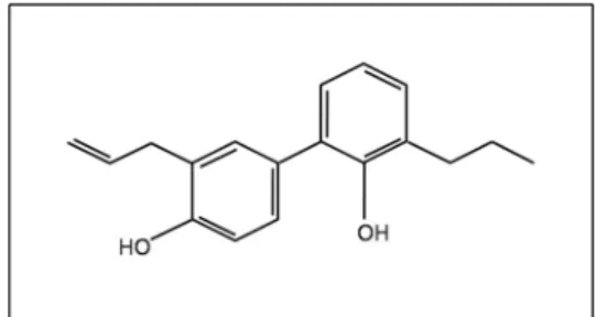
Honokiol [C
18H
18O
2, molecular weight=266.33] (Fig. 1) is TNFα-related a small molecular weight ligand,originally iso- lated from the Chinese medicinal herb ‘Magnolia officinalis’, a plant used in traditional Chinese and Japanese medicine of many diseases [9]. Honokiol, one of the major phenolic constituents of magnolia bark, have several pharmacological effects such as anti-oxidant [20], anti-thrombosis [26], an- ti-bacterial, xanthine oxidase inhibition, anti-tumor, anti-pla- telet aggregation, anti-inflammatory effects [19], anti-ar- rhythmic [18] and anxiolytic effect [28]. Previous reports have demonstrated that honokiol exhibited remarkable in- hibitory effects on mouse skin tumor promotion in an in vivo two-stage carcinogenesis, and inhibited the growth of human leukemic HL-60 cells [8]. Moreover, honokiol has in- duced apoptosis which characterized by DNA fragmentation and apoptotic bodies in human lymphoid leukemia Molt 4B cell [11].
Chondrocytes in cartilage are differentiated from mesen- chymal cells during embryonic development. Differentiated chondrocytes, which are the only cell type found in normal
mature cartilage, synthesize sufficient amounts of carti- lage-specific extracellular matrix (ECM) to maintain matrix integrity. This homeostasis is demolished in degenerative diseases, such as osteoarthritis (OA) and rheumatoid arthri- tis (RA) [7,23]. Arthritis is characterized by structural and biochemical changes in chondrocytes and cartilage, includ- ing degradation of cartilage matrix, insufficient synthesis of ECM caused loss of chondrocyte phenotype.
It is now generally accepted that proinflammatory cyto- kines, such as interleukin-1 beta (IL-1β) and tumor necrosis factor-alpha (TNFα), play a predominant role in structural and biochemical alterations in chondrocyte and cartilage [7].
One of the leading mechanisms by which cytokines elicit their effects on cartilage involves the stimulation of nitric oxide (NO) production via inducible NO synthase [2,21].
Although NO-induced cartilage destruction is caused by
Fig. 1. Molecular structure of honokiol. (C18H18O2, MW=266.33)
various factors, increased apoptotic cell death [5,10,31] and loss of differentiated phenotype of articular chondrocytes [6,25] appear to be important contributors. Our previously results showed that direct production of NO by treatment of NO donor, sodium nitroprusside (SNP), in primary cul- tured articular chondrocytes, led to apoptosis, dediffer- entiation, and cyclooxygenase (COX)-2 expression via a com- plex protein kinase signaling cascade involving mi- togen-activated protein (MAP) kinase and protein kinase C (PKC) [14-16].
However, the regulatory mechanism of NO-induced apoptosis has not been clearly elucidated yet. This study ad- dressed the effects of honokiol on the regulation of NO-in- duced apoptosis by SNP, and suggests that inhibition of No-induced apoptosis by honokiol is required for PI-3 Kinase-dependent pathway.
Materials and Methods Cell culture
Rabbit articular chondrocytes were isolated from cartilage slices of 2-week-old New Zealand white rabbits by enzy- matic digestion as described previously [33]. Cartilage slices were dissociated enzymatically for 6 hr in 0.2% collagenase type II (381 U/mg solid, Sigma, Louis, MO) in Dulbecco's modified Eagle's medium (DMEM) (Gibco-BRL, Gaithersburg, MD). Individual cells were suspended in DMEM supple- mented with 10% (v/v) fetal bovine-calf serum, 50 g/ml streptomycin, and 50 units/ml penicillin, after which and they were then plated on culture dishes at a density of 5×10
4cells/cm
2. The medium was changed every 2 days after seed- ing, and cells reached confluence in approximately 5 days.
The 3.5 day cell cultures were treated with honokiol.
Honokiol was purchased from Wako (Wako, Osaka) and a stock solution (MW: 266.33, 100 mM) in DMSO prepared and stored at 4℃ The following pharmacological agents were added 1 hr prior to honokiol, LY294002 to inhibit PI-3 kinase.
LY294002 war obtained from BioMol (BioMol, PA).
Western blot analysis
Whole cell lysates were prepared by extracting proteins using a buffer containing 50 mM Tris-HCl, pH 7.4, 150 mM NaCl, 1% Nonidet P-40, and 0.1% sodium dodecylsulfate (SDS), supplemented with protease inhibitors [10 g/ml leu- peptin, 10 g/ml pepstatin A, 10 g/ml aprotinin and 1 mM of 4-(2-aminoethyl) benzenesulfonyl fluoride] and phospha- tase inhibitors (1 mM NaF and 1 mM Na
3VO
4). The lysa-
teswere size-fractionated by SDS-polyacrylamide gel electro- phoresis and transferred to a nitrocellulose membrane. The nitrocellulose sheet was then blocked with 3% non-fat dry milk in Tris-buffered saline. Expression of caspase-3 and pAkt were detected using antibody purchased from Cell Signaling Technology (Danvers, MA), and p53 and Actinwere detected using antibodies purchased from Santa Cruz Biotechnology (Santa Cruz, CA). Blots were developed using a peroxidase-conjugated secondary antibody and vi- sualized with an ECL system.
Cell Proliferation assay
We used the MTT assay to quantify the proliferation of cells treated with honokiol and with SNP. Cells were seeded in 96-well plates at a density of 2×10
4cells per well for 24 hr. Cells were treated with honokiol and were treated with SNP. 10 μl MTT reagent 1 was added to the cells per well.
The plate was incubated for 4 hr at 37℃until purple for- mazan crystal developed. And, 100 μl MTT reagent 2 was added to the cells per well. After overnight in incubator, the absorbance at 600 nm was read and four wells were ex- amined with a spectrophotometer for each treatment.
Cell cycle distribution by FACS analysis
Cell cycle distribution was assessed by staining DNA con- tent with propidium iodide as previously described method [12] with some modifications. Briefly, chondrocytes were plated at a density of 2×10
5cells per 35-mm culture dish and incubated for 24 hr. Fresh complete media containing seri- ally diluted honokiol were replaced to culture dishes and further incubated for 24 hr. After incubation, both adherent and floating cells were harvested and fixed with 80% ethanol in PBS overnight. Fixed cells were incubated with RNase A (50 μg/ ml) for 25 min prior to staining nucleic acid with propidium iodide (50 μg/ml) for 5 min. The DNA content of 2×10
4cells in each group was analyzed by flow cytometer
(Partec GmbH, Münster, Germany) and the results weredemonstrated as histograms of DNA content. Quantification of the distinct cell cycle phases was calculated using the FloMax program.
DNA fragmentation
Cellular DNA was extracted after treatment of indicated honokiol with SNP for 24 hr. Briefly, whole cells were wash- ed with PBS and resuspended with lysis buffer containing 50 mM Tris-HCl (pH 7.5), 20 mM EDTA, and 1% NP-40.
After centrifugation at 3,000 rpm for 5 min, 10 μl of 10%
Fig. 2. NO-induced apoptosis were inhibited Honokiol dose-dependent manner in rabbit articular chondrocytes. Articular chon- drocytes were untreated (control) or treated with 1 mM SNP and 10 μM of honokiol plus 1 mM SNP for 24 hr. The apoptotic cells death was determined by phase-contrast microscope (magnification, 200X) (A). Primary chondrocytes apoptosis were determined by MTT assay (B) and expression of p53 and actin was determined by western blot analysis. Actin was used as loading control (C). It was determined that the protein levels of p53 were subsequently quantified by densitometric analysis (D). Statistically significant differences between control and other treatment: *p<0.005, **p<0.0001.
SDS and 10 μl of 50 mg/ml RNase A were added to the supernatants, and then incubated at 56°C for 2 hr.
2Subsequently, 10 μl of proteinase K (2.5 mg/ml) was added and further incubated at 37°C for 2 hr. DNA was allowed to precipitate with 0.5 volume of 10 M ammonium acetate and 2.5 volume of cold ethanol of total volume at −70°C for overnight. Extracted DNA was dissolved in 25 μl of 10 mM Tris-HCl buffer (pH 8.0) containing 1 mM EDTA. Two micrograms of DNA samples were resolved electrophoreti- cally on a 2% agarose gel and visualized under UV transilluminator.
Data analyses and statistics
The results are expressed as the means±S.E. values calcu- lated from the specified number of determinations. A Student’s t-test was used to compare individual treatments with their respective control values. A probability of p<0.05
was taken as denoting a significant difference.
Result
Honokiol suppress NO-induced apoptosis in rabbit articular chondrocytes
NO mediates the regulation and survival of chondrocyte
phenotype by inducing apoptosis. To assess anti-apoptosis
effect of honokiol in NO-induced apoptotic cells, various
does (1 μM - 10 μM) of honokiol and 1 mM SNP were treated
in cells. The cell morphology observed by phase-contrast mi-
croscope showed that NO-induced apoptosis was inhibited
by honokiol treatment (Fig. 2A). Honokiol significantly
blocked NO-induced apoptotic death of chondrocytes in a
dose-dependent manner, consistent with the pattern of cell
morphology (Fig. 2B). As anticipated, p53 expression also
decreased in a dose-dependent manner as determined by
Fig. 3. Honokiol inhibited NO-induced apoptosis in rabbit articular chondrocytes. Chondrocytes were untreated (control) or treated with 1 mM SNP and 10 μM of honokiol plus 1 mM SNP for 24 hr. The apoptotic cells death was determined by FACS analsis (A). Each phase of cell cycle was calculated using FloMax program (B). The cells were fixed with 80% ethanol in PBS, stained with propridium iodide (PI).
Fig. 4. Honokiol suppressed expression of p53 and caspase-3. Also it protected NO-induced DNA damagein rabbit articular chondrocytes. Articular chondrocytes were untreated (control) or treated with 1 mM SNP and 10 μM of honokiol plus 1 mM SNP for 24 hr. Expression of p53 and Caspase-3 inhibited honokiol treatment by western blot analysis. Actin was used as loading control (A). Actin was a protein loading control. It was determined that the protein levels of p53, pro-case- pase-3 and pAKT were subsequently quantified by densitometric analysis (B). The DNA damage protection of honokiol was determined by DNA fragmentation (C).
Fig. 5. Inhibitory effect of NO-induced apoptosis by honokiol treatment is required for PI-3K/AKT-dependent pathway. Primary cultured chondrocytes were untreated (control), treated with 1 mM SNP and 10 μM honokiol and 10 μM LY294002 for 24 hr. The apoptic cells death was determined by phase-contrast microscope (magnification, 200X) (A). Apoptosis of chondrocytes were determined by MTT assay (B) and expression of p53, caspase-3, pAKT and actin was determined by western blot analysis. Actin was used as loading control (C). It was determined that the protein levels of p53, pro-casepase-3 and pAKT were subsequently quantified by densitometric analysis (D). Statistically significant differences between control and other treatment: *p<0.005, **p<0.0001.
Western blot analysis quantified by densitometric analysis respectively (Fig. 2C and 2D).
Apoptotic cell death was blocked in chondrocytes with SNP (1 mM) and honokiol (10 μM) for 24 hr (Fig. 3A). This result was demonstrated as histograms of DNA content. The distribution of cells in each phase of cell cycle was calculated using FloMax program (Fig. 3B, upper and lower panel). As shown in Fig. 3B, about 34% of the cells were increased at sub G1 phase and 46% of the cells decreased at G1 phase after treatment with SNP alone compared with those in controls. When SNP was combined with honokiol, the cells of sub G1 phase were dramatically decreased and the cells of G1 phase were increased compared with those in SNP treatment cells. The results indicate that induction of apopto- sis was occured by transition of G1 cells to sub G1 cells.
To elucidate the mechanism of honokiol-regulated apop- tosis, caspase-3, an executioner of cell death, and p53, a sig- naling molecule upstream of caspase-3, were examined, and
phosphorylation of AKT, known as a survival signaling mol- ecule downstream of PI-3K was also investigated in rabbit articular chondrocytes (Fig. 4A). SNP-induced p53 was blocked by treatment of honokiol, effectively. As anticipated, pro-caspase-3 and pAKT expression was rescued by treat- ment of honokiol. It was determined that the protein levels of p53, pro-casepase-3 and pAKT were subsequently quanti- fied by densitometric analysis (Fig. 4B). Consistent with the result of Fig. 4A, honokiol significantly blocked NO-induced apoptotic cell death, as determined by DNA fragmentation (Fig. 4C). These results indicate that honokiol effectively re- covered NO caused apoptotic cell death in rabbit articular chondrocytes.
Inhibitory effect of honokiol on the NO-induced apoptosis is required for PI-3K/AKT-dependent pathway
To further investigate anti-apoptotic effects regulated by
honokiol, LY294002, a well known signaling molecule up-
stream of PI-3K/AKT was treated in chondrocytes to block activation of PI-3K/AKT. In consequence, anti-apoptotic ef- fects of honokiol were significantly inhibited in chon- drocytes (Fig. 5A), and these inhibition by LY294002 in- creased NO-induced apoptosis (Fig. 5B). LY294002 increases suppression of the p53 which honokiol leads. However in- crease of pro-caspase-3 is inhibited by LY294002 in articular chondrocytes (Fig. 5C). These protein levels were quantified by densitometric analysis (Fig. 5D). These findings collec- tively suggest that PI3K/AKT is required for inhibition of NO-induced apoptosis by honokiol in articular chondrocytes.
Discussion
Recently, alternative mechanisms of action have been pro- posed to account for the anti-tumor effect of honokiol and many studies have been initiated to explore the anti-tumor efficacy of honokiol. Also, the growth inhibition of tumor by honokiol is highly related to cell cycle arrest at the G0/G1 phase and induction of apoptosis [27].
NO mediates the regulation and survivalof chondrocyte phenotype by inducing dedifferentiation and apoptosis. Our previous results showed that apoptosis of chondrocyte caused by direct production of NO with the SNP is regulated by opposite functions of two mitogen-activated protein kin- ase subtypes, extracellular signal-regulated kinase-1/-2 (ERK-1/-2) and p38 kinase, in association with the elevation of p53 protein level, caspase-3 activation, and differentiation status [14-16]. SNP treatment stimulated activation of both ERK-1/-2 and p38 kinase. The activated ERK-1/-2 plays a role as an inhibitory signal for NO-induced apoptosis, whereas p38 kinase functions as a signal for the maintenance of the differentiated status and an inducing signal for apop- tosis of chondrocytes. And, we also reported that NO pro- duction in articular chondrocytes inhibits cell survival sig- naling, including apoptosis [22]. IGF-1 inhibits NO-induced apoptosis and dedifferentiation by modulating actin cytoske- letal architecture. Actin cytoskeleton regulates these func- tions by modulating PI-3K, AKT, MAPK, and PKC-α and -
signaling. Thus, inhibition of NO-induced apoptosis by disruption of the actin cytoskeleton is consistent with the suppression of the apoptotic signaling pathway, such as acti- vation of p38 kinase, inhibition of PKC-α and -
, NF-κ B activation, p53 accumulation, and caspase-3 activation.
These data additionally indicate that disruption of the actin cytoskeleton blocks NO-induced inhibition of the PI-3K/
AKT. Moreover, inhibition of the PI-3K/AKT pathways is required for the blockage of p38 kinase activation, inhibition of PKC-α and -
, NF-kB activation, accumulation of p53, and caspase-3 activation.
Based on this finding, we investigated whether honokiol treatment modulates the cell survival signal to block NO-in- duced apoptosis. Here we demonstrate that NO-induced apoptosis was suppressed by Honokiol treatment as de- termined by phase contrast microscophy, FACS analysis and DNA fragmentation. And, these anti-apoptotic effects were significantly recovered by LY294002, a PI-3K/AKT specific inhibitor. Thus, blocking of pAKT activation by treatment with LY294002 effectively rescued honokiol-inhibited p53 protein expression level. However increase of pro-caspase-3 is inhibited by LY294002 in articular chondrocytes. These re- sults indicate that honokiol is helpful to prevent destruction of cartilage caused by OA or RA.
Acknowledgement
This work was supported by a National Research Foundation of Korea (NRF) grant funded by the Korean Government (MEST) (2010-0003239 & 2009-0084569).
References
1. Ahn, K. S., G. Sethi, S. Shishodia, B. Sung, J. L. Arbiser, and B. B. Aggarwal. 2006. Honokiol potentiates apoptosis, suppresses osteoclastogenesis, and inhibits invasion through modulation of nuclear factor-kappaB activation pathway. Mol. Cancer Res. 4,621-633.
2. Amin, A. R and S. B. Abramson. 1998. The role of nitric oxide in articular cartilage breakdown in osteoarthritis.
Curr. Opin. Rheumatol. 10, 263-268.
3. Bai,X., F. Cerimele, and M. Ushio-Fukai. 2003. Honokiol, a small molecular weight natural product, inhibits angio- genesis in vitro and tumor growth in vivo. J. Biol. Chem. 278, 35501-35507.
4. Battle, T. E., J. Arbiser, and D. A. Frank. 2005. The natural product honokiol induces caspase-dependent apoptosis in B-cell chronic lymphocytic leukemia (B-CLL) cells. Blood 106, 690-697.
5. Blancco,F, J., R. Guitian, E. Vazquez-Martul, F. J. de Toro, and F. Galdo. 1998. Osteoarthritis chondrocytes die by apoptosis. A possible pathway for osteoarthritis pathology.
Arthritis Rheum. 41, 284-289.
6. Cao, M., A. westerhausen-Larson, C. Niyibizi, K.
Kavalkovich, H. I. Georgescu, C. F. Rizzo, P. A. Hebda, M.
Stefanovic-Racic, and C. H. Evans. 1997. Nitric oxide inhibits the synthesis of type-II collagen without altering Col2A1
mRNA abundance: prolyl hydroxylase as a possible target.
Biochem. J. 324, 305-310.
7. Choy, E. H., and G. S. Panayi. 2002. Cytokine pathways and joint inflammation in rheumatoid arthritis. N. Engl. J. Med.
344, 907-916.
8. Fong, W. F., A. K. Tse, K. H. Poon, and C. Wang. 2005.
Magnolol and honokiol enhance HL-60 human leukemia cell differentiation induced by 1,25-dihydroxyvitamin D3 and retinoic acid. Int. J. Biochem. Cell Bio.l 37, 427-441.
9. Fujita, M., H. Itokawa, and Y. Sashida. 1973. Studies on the componets of Magnolia obovata Thunb Ⅱ±on the compo- nents of the methanol extrct of the bark. Yakugaku Zasshi 93, 422-428.
10. Hashimoto, S., R. L. Oches, S. Komiya, and M. Lotz. 1998.
Linkage of chondrocyte apoptosis and cartilage degradation in human osteoarthritis. Arthritis Rheum. 41, 1632-1638.
11. Hibasami, H., Y. Achiwa, and H. Katsuzakil. 1998. Honokiol induces apoptosis in human lymphoid leukemia Molt 4B cells. Int. J. Mol. Med. 2, 671-673.
12. Im, J. H. and S. J. Kim. 2010. Paclitaxel induced caspase-in- dependent mitotic catastrophe in rabbit articular chondrocyte. J. Life Sci. 20, 519-527
13. Ishitsuka, K., T. Hideshima, and M. Hamasaki. 2005.
Honokiol overcomes conventional drug resistance in human multiple myeloma by induction of caspase-dependent and -independent apoptosis. Blood 106, 1794-1800.
14. Kim, S. J., H. G. Kim, C. D. Oh, Y. M. Yoon, W. K. Song, J. H. Kim, Y. J. Yoo, O. S. Bang, S. S. Kang, and J. S. Chun.
2002. p38 kinase-dependent and -independent Inhibition of protein kinase C zeta and -alpha regulates nitric oxide-in- duced apoptosis and dedifferentiation of articular chondrocytes. J. Biol. Chem. 277, 30375-30381.
15. Kim, S. J., J. W. Ju, C. D. Oh, Y. M. Yoon, W. K. Song, J. H. Kim, Y. J. Yoo, O. S. Bang, S. S. Kang, and J. S. Chun.
2002. ERK-1/2 and p38 kinase oppositely regulate nitric ox- ide-induced apoptosis of chondrocytes in association with p53, caspase-3, and differentiation status. J. Biol. Chem. 277, 1332-1339.
16. Kim, S. J., S. G. Hwang, D. Y. Shin, S. S. Kang, and J. S.
Chun. 2002. p38 kinase regulates nitric oxide-induced apop- tosis ofarticular chondrocytes by accumulating p53 via NFkappa B-dependent transcription and stabilization by serine 15 phosphorylation. J. Biol. Chem. 277, 33501-33508.
17. Konoshima, T., M. Kozuka, and H. Tokuda. 1991. Studies on inhibitors of skin tumor promotion, IX. Neolignans from Magnolia officinalis. J. Natural Products 54, 816-822.
18. Liou, K. T., S. M. Lin, S. S. Huang, C. L. Chih, and S. K.
Tsai. 2003. Honokiol ameliorates cerebral infarction from is- chemia-reperfusion injury in rats. Planta Med. 69, 130-134.
19. Liou, K. T., Y. C. Shen, C. F. Chen, C. M. Tsao, and S. K.
Tsai. 2003. The anti-inflammatory effect of honokiol on neu- trophils: mechanisms in the inhibition of reactive oxygen species production. Eur. J. Pharmacol. 475, 19-27.
20. Lo, Y. C., C. M. Teng, C. F. Chen, C. C. Chen, and C. Y.
Hong. 1994. Magnolol and honokiol isolated from Magnolia officinalis protect rat heart mitochondria against lipid peroxidation. Biochem. Pharmacol. 9, 549-553.
21. Martel-Pelletier, J., N. Alaaeddine, and J. P. Pelletier. 1999.
Cytokines and their role in the pathophysiology of osteoarthritis. Front. Biosci. 4, 694-703.
22. Oh,C. D., S. H. Chang, Y. M. Yoon, S. J. Lee, Y. S. Lee, S. S. Kang, and J. S. Chun. 2000. Opposing role of mi- togen-activated protein kinase subtypes, Erk-1/2 and p38, in the regulation of chondrogenesis of mesenchymes. J. Biol.
Chem. 275, 5613-5619.
23. Sandell, L. J. and T. Aigner. 2001. Articular cartilage and changes in arthritis. An introduction: cell biology of osteoarthritis. Arthritis Res. 3, 107-113.
24. Shigemura, K., J. L. Arbiser, and S. Y. Sun. 2007. Honokiol, a natural plant product, inhibits the bone metastatic growth of human prostate cancer cells. Cancer 109, 1279-1289.
25. Taskiran, D., M. Stefanovic-Racic, H. Georgescu, and C.
Evans. 1994. Nitric oxide mediates suppression of cartilage proteoglycan synthesis by interleukin-1. Biochem. Biophys.
Res. Commun. 200, 142-148.
26. Teng, C. M., C. C. Chen, F. N. Ko, L. G. Lee, T. F. Huang, Y. P. Chen, and H. Y. Hsu. 1988. Two antiplatelet agents from Magnolia officinalis. Thromb Res. 50, 757-765.
27. Wang, T. , F. Chen, Z. Chen, Y. F. Wu, X. L. Xu, S. Zheng, and X. Hu. 2004. Honokiol induces apoptosis through p53-independent pathway in human colorectal cell line RKO. World J. Gastroenterol. 10, 2205-2208.
28. Watanabe, K., H. Watanabe, Y. Goto, M. Yamaguchi, N.
Yamamoto, and K. Hagino. 1983. Pharmacological proper- ties of magnolol and honokiol extracted from Magnolia offi- cinalis: central depressant effects. Planta Med. 49, 103-108.
29. Wolf, I., J. O'Kelly, and N. Wakimoto. 2007. Honokiol, a natural biphenyl, inhibits in vitro and in vivo growth of breast cancer through induction of apoptosis and cell cycle arrest. Int. J. Oncol. 30, 1529-1537.
30. Yang, S. E., M. T. Hsieh, T. H. Tsai, and S. L. Hsu. 2002.
Down-modulation of Bcl-XL, release of cytochrome c and sequential activation of caspases during honokiol-induced apoptosis in human squamous lung cancer CH27 cells.
Biochem. Pharmacol. 63, 1641-1651.
31. Yatsugi, N., T. Tsukazaki, M. Osaki, T. Koji, S. Yamashita, and H. Shindo. 2000. Apoptosis of articular chondrocytes in rheumatoid arthritis and osteoarthritis: correlation of apoptosis with degree of cartilage destruction and ex- pression of apoptosis-related proteins of p53 and c-myc. J.
Orthop. Sci. 5, 150-156.
32. Yoon, Y. M., S. J. Kim, C. D. Oh, J. W. Ju, W. K. Song, Y. J. Yoo, T. L. Huh, and J. S. Chun. 2002. Maintenance of differentiated phenotype of articular chondrocytes by pro- tein kinase C and extracellular signal-regulated protein kinase. J. Biol. Chem. 277, 8412-8420.