Inhibition of Cyclooxygenase-2 Activity and Prostaglandin E
2Production through Down-regulation of NF-κB Activity by the Extracts of Fermented Beans
Hye Hyeon Lee
1, Cheol Park
2, Min Jeong Kim
1, Min Jeong Seo
1, Sung-Hyun Choi
3, Yong Kee Jeong
1and Yung Hyun Choi
2,4*
1
Department of Medical Bioscience, Graduate School and Department of Biotechnology, College of Natural Resources and Life Science, Dong-A University, Busan 604-714, Korea
2
Blue-Bio Industry Regional Innovation Center, Dongeui University, Busan 614-714, Korea
3
Department of Elevator Maintenance, Korea Lift College, Geochang 670-802, Korea
4
Department of Biochemistry, College of Oriental Medicine and Department of Biomaterial Control, Graduate School and Dongeui University, Busan 614-714, Korea
Received December 14, 2009 /Accepted January 7, 2010
Cyclooxygenase (COX)-2 is generally known as an inducible enzyme, and it produces arachidonic acid to prostaglandin E
2(PGE
2), which has been demonstrated to play a critical role in inflammation. In the present study, we investigated the effects of the extracts of fermented beans including soybean (FS), black agabean (FBA) and yellow agabean (FYA), on the expression of COXs and production of PGE
2in U937 human promonocytic cells. Treatment of phorbol 12-myristate 13-acetate (PMA) sig- nificantly induced pro-inflammatory mediators such as COX-2 expression and PGE
2production, whereas the levels of COX-1 remained unchanged. However, pre-treatment with FS, FBA and FYA significantly decreased PMA-induced COX-2 protein as well as mRNA, which is associated with in- hibition of PGE
2production. Moreover, FS, FBA and FYA markedly prevented the increase of nuclear translocation of nuclear factor kappa B (NF-κB) p65 by PMA. Our data indicate that the extracts of fermented beans exhibits anti-inflammatory properties by suppressing the transcription of pro-in- flammatory cytokine genes through the NF-κB signaling pathway.
Key words : Fermented beans, COX-2, PGE
2, NF-κB
*Corresponding author
*Tel:+82-51-850-8649, Fax:+82-51-853-4036
*E-mail : [email protected]
서 론
Nuclear factor kappa B (NF-κB)는 redox-sensitive tran- scription regulator로서, 해당DNA motif들과 결함함으로서 다양한 유전자들의 전사 활성을 강화시킨다. 정상적인 환경에 서는 세포질에서 inhibitory subunit인 inhibitor kappa Bα (Iκ Bα)와 결합되어진 비활성형을 형성하고 있지만, in- flammatory cytokines, mitogens, UV, 그리고 oxidant 등의 여러 자극에 의해 IκB 단백질이 인산화되면 NF-κB는 핵 안으 로 이동하여 해당 DNA의 promoter, enhancer 등의 조절요소 로 작용한다[4,31]. 여러 가지 작용 중, 특히 NF-κB는 prosta- glandin (PG) pathway를 활성화시킴으로써 염증반응을 유발 하는 것으로 알려져 있다[7,17,22,29,30]. 여러 다양한 조직에서 물질대사를 조절하는 PG는 평활근의 수축과 이완, 혈관의 수 축과 확장, 혈압 조절, 발열 유발, 염증 조절 등과 같은 다양한 신체 기능에 관여하는 물질 중 하나로써, 우리 몸에서 통증을 유발한다고 알려져 있다. PG는 arachidonic acid로부터 합성 되며, 이때 cyclooxygenase (COX)라는 효소가 관여한다
[1,2,8,17,20,29].
COX는 thromboxane, prostacydin, prostaglandin을 포함 하는 prostanoid라고 불리는 중요한 biological mediator의 형 성을 위한 효소로써 PG 합성에는 일반적으로 2가지 isoform 인 COX-1 및 COX-2가 관여하는 것으로 알려져 있다[2,10,34].
COX-1은 대부분의 조직에서 housekeeper enzyme으로 항상 성 유지기능에 관여하고 있는데 반하여 COX-2는 inter- leukin-1 (IL-1)과 같은 cytokine이나 다양한 성장인자, 종양 촉진인자 등의 자극에 의해 과발현되며 특히 암 조직이나 염 증조직에서 높게 나타난다[1,2,6]. COX-2의 과발현은 세포 증 식을 촉진하고 apoptosis를 억제하며 세포의 유동성 및 부착 성을 강화시킴으로서 암을 포함한 각종 염증성/퇴행성 질환 의 발병과 진행에 중요한 역할을 한다. 또한 COX-2의 과발현 에 의해 암조직에서의 혈관신생 및 전이능이 높아지고, COX-2의 선택적 억제제에 의한 angiogenesis와 종양형성 억 제 등의 결과에서 COX-2의 선택적 조절에 의한 암예방 및 항암전략이 대두되고 있다[11,14,17,23,30,31].
야생의 들콩으로 부터 재배작물로 발달한 콩은 사람들에게
식물성 단백질을 공급할 뿐만 아니라 수많은 화학제품의 원료
로 쓰인다. 콩은 여러 가지 방법으로 조리되어 섭취하고 있는
데, 특히 미생물을 이용하여 발효시키는 방법 중에는 대표적
으로 청국장을 들 수가 있다. 고초균(Bacillus subtilis)에 의해 발효되는 청국장은 다른 아시아 지역에서는 “Natto”,
“Tempeh”, “Douchi” 이라고도 하는데, 된장이나 간장과 같이 콩을 이용한 다른 발효식품과는 다르게 짧은 발효시간을 가진 다는 것과 발효기간 동안 소금이 필요 하지 않다는 것이 큰 특징이다. 청국장은 항산화, 항균, 혈압조절 및 항당뇨 등과 같은 여러 가지 효과를 가지고 있으며[15,27,33], 청국장의 구 성물 중 가장 풍부하게 존재하는 isoflavone은 phytochemical 로써, 여성호르몬인 estrogen과 비슷한 heterocyclic diphe- nolic 구조를 가진다. 이러한 isoflavone과 isoflavone이 풍부 한 식품은 산화적 손상과 관련이 있는 유방암과 전립선암 등 과 같은 다양한 종류의 암과 동맥경화증 등으로부터 예방하는 효과를 가지는 것으로 알려져 있다[13,14,16,18,28]. 한편 경북 대학교에서 품종 육성되어진 아가콩 3호(노란 아가콩) 및 4호 (검정 아가콩)는 isoflavone의 함량이 다른 콩들에 비해 매우 높은 것으로 확인 되어졌으므로, 아가콩의 효과가 다른 콩에 비해 더욱 높을 것이라 생각된다[5,25].
본 연구에서는 대두, 아가콩 3호 및 4호의 발효산물에 의한 항염증효능을 조사하기 위하여 phorbol 12-myristate 13-acetate (PMA) 처리에 의해 유도된 COX-2의 발현, prostaglandin E
2(PGE
2)의 생성 및 NF-κB의 활성 증가[12,19,21,26]에 미치는 영향을 비교하였다.
재료 및 방법
세포배양
본 연구에 사용된 U937 인체 혈구세포는 한국생명공학연구 소(KRIBB, Taejeon, Korea)에서 분주 받았으며, 세포의 배양을 위해 90%의 RPMI-1640 배지(Gibco BRL, Grand Island, NY, USA), 10% fetal bovine serum (FBS, Gibco BRL)에 1%의 pen- icillin 및 streptomycin (Gibco BRL)이 포함된 배지를 사용하 여 37
oC, 5% CO
2조건 하에서 배양하였다.
시료준비 및 처리
본 실험에 사용된 시료는 시중에서 판매되는 대두와 경북대 학교에서 품종 육성되어진 아가콩 3호(노란 아가콩) 및 4호(검 정 아가콩)를 제공받아 사용하였다. 시료 준비를 위해 콩들을 수세하고 24시간 침지시킨 뒤 가압증자한 후 실험실에서 따로 분리되어진 Bacillus sp. sm26을 50 g당 1 ml 접종하여 37
oC, 72시간 배양하였고, 그 후 70% EtOH로 추출[extracts of fer- mented soybean (FS), black agabean (FBA) and yellow aga- bean (FYA)]하여 필터로 불순물을 제거한 후 농축시켜 사용하 였다.
MTT assay에 의한 세포 성장억제 여부 조사
6 well plate에 U937 세포를 7.5×10
4개/ml로 분주하고 6시
간 동안 배양 FS, FBA 및 FYA를 처리한 후 배지에 tetrazolium bromide salt (MTT, Amersco, Solon, Ohio, USA)를 0.5 mg/ml 농도로 희석하여 200 μl씩 분주하고 2시간동안 배양하 였다. 배양이 끝난 후 MTT 시약을 제거하고 dimethylsulf- oxide (DMSO, Amresco)를 1 ml씩 분주하여 well에 생성된 formazan을 모두 녹인 후 96 well plate에 200 μl씩 옮겨서 ELISA reader (Molecular Devices, Sunnyvale, CA, USA)로 540 nm에서 흡광도를 측정하였다. 측정은 모두 세 번을 하였 으며 그에 대한 평균값과 표준오차를 Microsoft Excel 프로그 램으로 구하였다.
단백질의 분리, 전기영동 및 Western blotting
선행 실험에 의해 조건이 확립되어진 PMA와 위와 동일한 조건으로 FS, FBA 및 FYA를 처리한 배지에서 배양된 세포들 에 적정량의 lysis buffer [25 mM Tris-Cl (pH 7.5), 250 mM NaCl, 5 mM EDTA, 1% NP-40, 1 mM phenylmeth- ylsulfonyfluoride (PMSF), 5 mM dithiothreitol (DTT)]를 첨가 하여 4
oC에서 1시간 반응시킨 후, 14,000 rpm으로 30분 원심분 리하여 그 상층액을 취하였다. 상층액의 단백질 농도를 Bio-Rad 단백질 정량시약 (Bio-Rad, Hercules, CA, USA)을 사용하여 정량한 다음 동량의 Laemmli sample buffer (Bio-Rad)를 섞어서 sample을 만들었다. 이렇게 만든 동량의 단백질을 sodium dodecyl sulphate (SDS)-polyacrylamide gel을 이용하여 전기영동으로 분리하였다. 분리된 단백질을 함유한 acrylamide gel을 nitrocellulose membrane (Schleicher and Schuell, Keene, NH, USA)으로 전이시킨 후, 적정 1차 항체를 처리하여 4
oC에서 overnight 시킨 후 PBS-T로 세척하 고 처리된 1차 항체에 맞는 2차 항체를 사용하여 상온에서 적정시간 반응시켰다. 다시 PBS-T로 세척하고 enhanced chei- luminoscence (ECL) 용액(Amersham Life Science Corp., Arlignton Heights, IL, USA)을 적용시킨 X-ray film에 감광시 켜 특정단백질의 양을 분석하였다. 본 실험에 사용된 항체들 은 Santa Cruz Biotechnology Inc.에서 구입하였으며, Immunoblotting을 위해 2차 항체로 사용된 peroxidase-la- beled donkey anti-rabbit immunoglobulin 및 peroxidase-la- beled sheep anti-mouse immunoglobulin은 Amersham Life Science Corp.에서 구입하였다.
Reverse transcription-polymerase chain reaction (RT-PCR) 분석
동일한 조건에서 배양된 U937 세포들에 TRIzol reagent
(Invitrogen Co., Carlsbad, CA, USA)를 4
oC에서 1시간 동안
처리하여 RNA를 분리하였다. 분리된 RNA를 정량한 후, 각각
의 primer (Table 1), DEPC water 그리고 ONE-STEP RT-PCR
PreMix Kit (Intron, Korea)를 넣고 Mastercycler gradient
(Eppendorf, Hamburg, Germany)를 이용하여 증폭하였다. 각
Table 1. Sequences of primers used for RT-PCR
Primer Sequence
COX-1 Sense
Anti-sense 5'-TGC-CCA-GCT-CCT-GGC-CCG-CCG-CTT-3'
5'-GTC-CAT-CAA-CAC-AGG-CGC-CTC-TTC-3'
COX-2 Sense
Anti-sense 5'-TTC-AAA-TGA-GAT-TGT-GGG-AAA-AT-3'
5'-AGA-TCA-TCT-CTG-CCT-GAG-TAT-CTT-3'
GAPDH Sense
Anti-sense 5'-CGG-AGT-CAA-CGG-ATT-TGG-TCG-TAT-3'
5'-AGC-CTT-CTC-CAT-GGT-GGT-GAA-GAC-3'
PCR 산물들의 양적 차이를 확인하기 위하여 1X TAE buffer로 1% agarose gel을 만들고 well 당 각각의 primer에 해당하는 PCR 산물에 DNA gel loading solution을 섞어서 loading 한 후 50 V에서 전기영동을 행하였다. 전기영동으로 DNA 분리 가 끝난 gel을 ethidium bromide (EtBr)을 이용하여 염색한 후 UV 하에서 발현의 차이를 확인하였으며, glyceraldehyde- 3-phosphate dehydrogenase (GAPDH)를 internal control로 사용하였다.
Prostaglandin E
2의 측정
PGE
2생성량 측정을 위해 사용한 PGE
2EIA kit는 Cayman Chemicals (Ann Arbor, MI, USA)에서 구입하였다. U937 세포 에 다양한 농도의 FS, FBA 및 FYA를 1시간 선 처리 후, PMA (40 nM)를 처리한 배지에서 6시간 동안 세포를 배양시킨 후 상층액만 이용하여 PGE
2EIA kit에 제시된 방법에 따라 처리 한 다음 ELISA reader를 이용한 420 nm의 흡광도로 반응의 정도를 측정하였다.
결 과
세포증식에 미치는 FS, FBA 및 FYA의 영향
다양한 농도의 FS, FBA 및 FYA가 처리된 U937 세포의 증식 억제 여부를 알아보기 위하여 MTT assay를 이용하였다. Fig.
1에서 나타난 바와 같이 FS, FBA 및 FYA를 6시간동안 5 mg/ml의 농도까지 처리하였을 때 80% 전후의 생존율을 보였 으며, 세포사멸 현상은 관찰되지 않았다. 따라서 80% 이상의 생존을 보인 3 mg/ml 농도를 실험 조건 농도로 설정하였다.
COXs의 발현에 미치는 PMA의 영향
U937 세포에서 PMA 처리에 의한 COX-1 및 COX-2의 발현 변화정도를 Western blotting으로 조사하였다. PMA를 적정 농도별로 6시간 처리하였을 경우 COX-1의 경우는 아무런 변 화가 관찰되지 않았지만 COX-2의 경우는 40 nM의 농도에서 강하게 발현되는 것을 확인할 수 있었다(Fig. 2A). 또한 PMA 처리시간에 따른 발현정도를 비교해 본 결과 COX-1은 아무런 변화가 없었지만 COX-2는 처리시간 증가에 따라 발현양이 증가되어 6시간에서 현저하게 증가되는 것을 관찰할 수 있었
Fig. 1. Effect of fermented beans extracts on the cell viability in U937 human pre-monocytic cells. The cells were in- cubated with indicated concentrations of fermented soy- bean (FS), black agabean (FBA) and yellow agabean (FYA) for 6 hr. The rates of cell viability were measured by the metabolic-bye-based MTT assay. The data shown are means of three independent experiments.
(A) (B)
Fig. 2. Effect of PMA on the levels of COX-1 and COX-2 ex- pression in U937 cells. The cells were treated with vari- ous concentration of PMA for 6 hr (A) or 40 nM of PMA for the indicated times (B). The cells were lysed and then cellular proteins were separated by SDS-polyacrylamide gels and transferred onto nitrocellulose membranes. The membranes were probed with anti-COX-1 and anti- COX-2 antibodies. Proteins were visualized using an ECL detection system. Actin was used as an internal control.
다(Fig. 2B).
PMA에 의한 COX-2의 발현증가에 미치는 FS, FBA 및 FYA의 영향
U937 세포에서 PMA에 의한 COX-2의 과발현 현상에 대한
FS, FBA 및 FYA의 영향을 조사하기 위하여 FS, FBA 및 FYA
(A) (B)
(C) (D)
Fig. 3. Effect of fermented beans extracts on the levels of COXs proteins and mRNA expressions in U937 cells. The cells were incubated with 3 mg/ml of FS, FBA and FYA for 6 hr after 1 hr pretreatment with 40 nM of PMA. (A) Total RNAs were isolated and reverse-transcribed. The resulting cDNAs were subjected to PCR with indicated primers and the reaction products were subjected to electrophoresis in 1% agarose gel and visualized by EtBr staining. (C) Cellular proteins were isolated, separated by SDS-polyacrylamide gels and transferred onto nitrocellulose membranes. The membranes were probed with anti-COX-1 and anti-COX-2 antibodies. Proteins were visualized using an ECL detection system. Actin was used as an internal control. (B and D) mRNA and proteins levels of COX-1 and COX-2 were normalized by GAPDH mRNA and actin protein.
를 3 mg/ml의 농도로 단독 처리하였을 경우와 PMA를 1시간 처리한 후 FS, FBA 및 FYA를 처리하여 6시간 경과 후의 COX-1 및 COX-2의 발현정도를 전사 및 번역 수준에서 조사 하였다. Fig. 3의 결과에서 알 수 있듯이 COX-1의 경우는 아무 런 변화가 나타나지 않았지만 PMA에 의해서 과발현된 COX-2의 경우는 mRNA 및 단백질 발현 모두에서 FS, FBA 및 FYA의 처리에 의해서 현저히 감소하는 것으로 나타났다.
특히 FYA 처리군에 경우 FS 및 FBA 처리군에 비해 PMA에 의하여 과발현된 COX-2의 억제 정도가 가장 높게 나타났다.
PMA에 의한 PGE2 생성 증가에 미치는 FS, FBA 및 FYA의 영향
U937 세포에서 PMA에 의한 PGE
2의 생성 증가에 미치는 FS, FBA 및 FYA의 영향을 조사하기 위하여, 먼저 PMA 처리 에 의해서 PGE
2의 생성이 현저하게 증가하는 것을 확인하였 다(Fig. 4). 그러나 PMA 처리에 의하여 증가된 PGE
2의 생성이 FS, FBA 및 FYA의 처리에 의하여 현저하게 억제되었는데, PGE
2의 생성 억제에서는 FS에 비해 FBA와 FYA 처리군에서 더욱 효과적이었다. 이는 FS, FBA 및 FYA에 의한 COX-2의 발현저하에 의한 결과로 추정할 수 있었다.
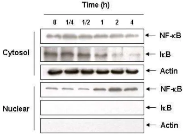
NF-κB의 translocation에 미치는 PMA의 영향
PMA를 시간별로 처리를 하였을 때 핵 안으로 translocation
된 NF-κB의 발현을 관찰한 결과, 처리시간 증가에 따라 핵 내에 존재하는 NF-κB의 발현이 증가하였으며 특히 PMA 처 리 후 2시간째에 강하게 발현됨을 확인할 수 있었다. 또한 PMA 처리 후 1시간 후부터 시간이 지남에 따라 세포질에 존 재하는 IκB의 발현이 서서히 감소하는 것을 확인할 수 있었 다. 따라서 PMA가 IκB의 인산화를 유발시킴으로써 NF-κB를 핵 안으로 translocation 시킨다는 것을 확인하였다(Fig. 5).
Fig. 4. Effect of fermented beans extracts on the PMA-induced PGE2production in U937 cells. The cells were pretreaed with 3 mg/ml of FS, FBA and FYA for 1 hr followed by incubation with PMA (40 nM) for 6 hr. The PGE2
accumulation in the medium was determined by an EIA kit as described in materials and methods. The data shown are means±SD of three independent experiments.
(A)
(B) (C)
Fig. 6. Effect of fermented beans extracts on NF-kB translocation in PMA-stimulated U937 cells. (A) The cells were pretreated with 3 mg/ml of FS, FBA and FYA for 1 hr before stimulation with PMA (40 nM) for 6 hr, and cytosol and nuclear proteins were prepared. The levels of IkB protein and NF-kB localization in cells was determined with anti-IKB and anti-NF-kB antibodies. (B and C) The levels of IkB in the cytosolic protein and NF-kB in nuclear proteins extracts normalized by control protein (actin).
Fig. 5. NF-kB translocation by PMA in U937 cells. The cells were treated with 40 nM of PMA for the indicated times, and cytosol and nuclear proteins were prepared. The NF-kB in nuclear proteins extracts and levels of IkB in the cyto- solic protein were determined by a Western blot analysis.
FS, FBA 및 FYA에 의한 NF-κB의 translocation 억제 PMA 처리에 의해 핵 안으로 translocation된 NF-κB의 발현 변화에 미치는 FS, FBA 및 FYA의 영향을 조사한 결과, PMA 에 의해 핵 안으로 translocation되었던 NF-κB가 FS, FBA 및 FYA의 처리에 의하여 translocation 정도가 현저하게 감소되 었으며, 세포질에서 PMA 처리에 의해 감소되었던 IκB의 발현 이 FS, FBA 및 FYA 처리에 의해 약간 증가하는 것을 확인할 수 있었다(Fig 6). 이상의 결과는 FS, FBA 및 FYA가 PMA에 의한 NF-κB의 활성 증가를 현저하게 억제하였음을 의미하는 것이다.
고 찰
Phorbol ester의 일종으로 tumor promoter인 PMA는 diac- ylglycerol과 같이 세포 내 단백질을 인산화 시키는 protein kinase C 활성인자로서 성장인자, 호르몬 및 세포의 성장과 분화를 조절하는 cytokine에 의한 기작을 연구하는데 사용되 며, cytokine 및 lipopolysaccharide와 마찬가지로 여러 세포들 에서 COX-2의 발현 및 PG의 생성을 촉진시키는 염증유발 인 자로 알려져 있다[2,12,19,23,26]. 본 연구에서는 U937 세포 모 델에서 FS, FBA 및 FYA의 항염증 효능을 확인하기 위하여 PMA 처리에 의해 증가되는 염증 유발 인자들의 발현에 미치 는 이들의 효과를 비교하였다. 이를 위하여 U937 세포의 증식 에 FS, FBA 및 FYA가 어떠한 영향을 미치는 지를 알아보기 위해 MTT assay에 의한 세포 증식 억제 여부를 조사한 결과, 6시간동안 최고 농도인 5 mg/ml 에서도 생존율이 80% 정도 였으며 세포사멸이 유도되지 않는 것을 확인함으로써 세포 증식에 FS, FBA 및 FYA가 직접적으로 큰 영향을 미치지 않았 음을 알 수 있었다(Fig. 1). 또한 U937 세포에서 PMA에 대한 COX-2의 발현을 증가 시킬 수 있는 조건을 동시에 설정하기 위하여, PMA를 다양한 농도별로 6시간 동안 처리 하였을 경 우, COX-1의 발현에는 큰 변화가 없었으나, COX-2는 40 nM 이상 처리군에서 강하게 발현이 증가되는 것을 확인하였다.
그리고 40 nM PMA에 의한 COX-2의 발현이 처리 6시간 후에 가장 발현이 높게 나타났다(Fig. 2).
현재까지 밝혀진 여러 가지 항염증 약물들의 작용기전 중
하나는 PG 합성을 억제하는 것이며, 이는 PG 합성에 관여하
는 효소인 COXs의 생성 및 발현 억제에 의한 것이다[6]. COXs
는 크게 COX-1 및 COX-2로 구분되는데, 특히 COX-2의 과발 현은 염증성 질환을 포함한 인체의 다양한 질병을 일으키는 요인으로 작용한다[1,2,6,10,19,23,29,34]. COX에 의해 합성되 는 PG는 모두 8 종류로 알려져 있으며, 다양한 조직에서 물질 대사를 조절하는데, 그 중 염증 반응을 유발하는 것으로 알려 진 PGE
2는 PG 합성효소에 의해 arachidonic acid로부터 PGH
2가 합성이 될 때 형성된다[1,8,9,23]. PGE
2는 혈관 확장, 기관지 확장, 위산억제, 염증, 발열 등의 작용을 하며 특히 염증성 질 환을 포함하는 다양한 생체반응에서 세포분열이나 증식에 영 향을 줌으로써 각종 질병의 유발과 진행에 관여하는 것으로 알려져 있다[1,2,8,19,26,29]. 본 연구에서는 U937 세포 모델에 서 PMA 처리 후 증가된 COX-2의 발현에 미치는 FS, FBA 및 FYA의 영향을 조사한 결과, 이들의 단독 처리군에서는 COX-1 뿐만 아니라 COX-2의 발현에 아무런 영향을 주지 않 았으나, PMA에 의해 과발현된 COX-2의 발현이 FS, FBA 및 FYA의 처리에 의해 매우 감소되었음을 전사 및 번역 수준에 서 확인하였다(Fig. 3). 아울러 PMA에 의해 생성이 증가된 PGE
2의 경우에도 FS, FBA 및 FYA를 처리하였을 때 현저히 억제되는 것을 확인함으로써, FS, FBA 및 FYA의 처리에 의한 COX-2의 발현 억제가 PGE
2의 생성을 억제시켰음을 추정할 수 있었다(Fig. 4).
한편 NF-κB는 DNA의 전사를 조절하는 단백질복합체로서, 자극이 없는 세포에서 NF-kB는 세포질에서 그들의 활성 저해 제로 알려져 있는 IκB와 결합되어 비활성화 상태로 존재한다.
비활성화된 NF-κB는 거의 모든 동물세포의 세포질에서 발견 되며 스트레스, cytokines, free radical, UV, oxidant, mitogens 그리고 항원 등과 같은 자극에 의해 활성화된다[7,20,21,22,32].
NF-κB의 활성은 세포가 자극을 받아 IκB 단백질이 IκB kinase (IKK)에 의해 인산화 되면서 시작되며, IκB와 분리된 NF-κB는 핵 안으로 이동이 용이해지게 된다. 핵 안으로 들어간 NF-κB 는 DNA의 promoter 및 enhancer들의 조절요소로 작용하는 데, 과다한 NF-κB의 활성 증가는 염증성 질환을 포함하여, 암, 자가면역질환, 폐혈성 쇼크, 바이러스성 감염 그리고 잘못된 면역발생 등과 연관되어진다[4,17,31,32]. 많은 연구에 따르면 콩에 함유된 다양한 천연물이 NF-κB의 활성을 억제할 수 있는 것으로 알려져 [3,24], 본 연구에서 사용한 콩 발효 산물들이 PMA에 의한 NF-κB의 활성에 어떠한 영향을 미치는지 조사하 였다. 이를 위하여 PMA를 단독 처리하였을 경우 세포질과 핵에서의 NF-κB와 IκB의 변화를 확인한 결과, PMA 처리시간 증가에 따라, 세포질에 존재하는 IκB의 발현양이 감소하였으 며, 핵으로의 NF-κB translocation이 증가하는 것을 확인하였 다(Fig. 5). 하지만 FS, FBA 및 FYA를 단독으로 처리한 경우에 는 NF-κB의 translocation 현상은 관찰되지 않았으며, PMA에 의한 NF-κB translocation을 현저하게 억제하였고, 세포질 상 에 존재하는 IκB는 발현양이 증가하는 것을 확인하였다(Fig.
6). 이는 FS, FBA 및 FYA가 U937 세포에서 PMA 처리에 의해
유발된 COX-2의 발현 감소 및 PGE
2의 생성 억제가 NF-κB의 활성 억제와 연관되어있음을 보여 주는 결과이다.
감사의 글
본 논문은 농림부 농림기술개발사업의 지원(307003-03- 1-HD140)에 의하여 이루어진 결과의 일부입니다.
References
1. Balzary, R. W. and T. M. Cocks. 2006. Lipopolysaccharide induces epithelium- and prostaglandin E(2)-dependent re- laxation of mouse isolated trachea through activation of cy- clooxygenase (COX)-1 and COX-2.
J. Pharmacol. Exp. Ther.
317, 806-812.
2. Brunelle, M., E. A. Sartin, L. G. Wolfe, J. Sirois, and M. Doré.
2006. Cyclooxygenase-2 expression in normal and neo- plastic canine mammary cell lines.
Vet. Pathol.
43, 656-666.3. Borrás, C., J. Gambini, M. C. Gómez-Cabrera, J. Sastre, F.
V. Pallardó, G. E. Mann, and J. Viña. 2006. Genistein, a soy isoflavone, up-regulates expression of antioxidant genes: in- volvement of estrogen receptors, ERK1/2, and NFkappaB.
FASEB J.
20, 2136-2138.4. Chen, F., V. Castranova, and X. Shi. 2001. New insights into the role of nuclear factor-kappaB in cell growth regulation.
Am. J. Pathol.
159, 387-397.5. Choi, U. K., M. H. Kim, N. H. Lee, Y. S. Jeong, O. J. Kwon, Y. C. Kim, and Y. H. Hwang. 2007. The characteristics of cheonggukjang, a fermented soybean product, by the de- gree of germination of raw soybeans.
Food Sci. Biotechnol.
16, 734-739.
6. Dempke, W., C. Rie, A. Grothey, and H. J. Schmoll. 2001.
Cyclooxygenase-2: a novel target for cancer chemotherapy?
J. Cancer Res. Clin. Oncol.
127, 411-417.7. Festa, M., A. Petrella, S. Alfano, and L. Parente. 2009. R-ro- scovitine sensitizes anaplastic thyroid carcinoma cells to TRAIL-induced apoptosis via regulation of IKK/NF- kappaB pathway.
Int. J. Cancer.
124, 2728-2736.8. FitzGerald, G. A. 2003. COX-2 and beyond: Approaches to prostaglandin inhibition in human disease.
Nat. Rev. Drug Discov.
2, 879-890.9. Grkovich, A., C. A. Johnson, M. W. Buczynski, and E. A Dennis. 2006. Lipopolysaccharide-induced cyclooxygenase-2 expression in human U937 macrophages is phosphatidic acid phosphohydrolase-1-dependent.
J. Biol. Chem.
281, 32978-32987.10. Habib, A., I. Shamseddeen, M. S. Nasrallah, T. A. Antoun, G. Nemer, J. Bertoglio, R. Badreddine, and K. F. Badr. 2007.
Modulation of COX-2 expression by statins in human mon- ocytic cells.
FASEB J.
21, 1665-1674.11. Jacoby, R. F., K. Seibert, C. E. Cole, G. Kelloff, and R. A.
Lubet. 2000. The cyclooxygenase-2 inhibitor celecoxib is a potent preventive and therapeutic agent in the min mouse model of adenomatous polyposis.
Cancer Res.
60, 5040-5054.12. Jiang, Y. J., B. Lu, P. C. Choy, and G. M. Hatch. 2003.
Regulation of cytosolic phospholipase A2, cyclooxygenase-1 and -2 expression by PMA, TNFalpha, LPS and M-CSF in human monocytes and macrophages.
Mol. Cell Biochem.
246, 31-38.13. Kim, H. B., H. S. Lee, S. J. Kim, H. J. Yoo, J. S. Hwang, G. Chen, and H. J. Youn. 2007. Ethanol extract of fermented soybean, Chungkookjang, inhibits the apoptosis of mouse spleen, and thymus cells.
J. Microbiol.
45, 256-261.14. Kim, J. I., C. Y. Jin, M. J. Seo, H. S. Lim, Y. C. Lee, W.
H. Joo, B. T. Choi, Y. K. Jeong, and Y. H. Choi. 2008.
Genistein-induced growth inhibition was associated with inhibition of cyclooxygenase-2 and telomerase activity in human cancer cells.
J. Life Sci.
18, 884-890.15. Kim, M. H., W. W. Kang, N. H. Lee, D. J. Kwon, O. J. Kwon, Y. S. Chung, Y. H. Hwang, and U. K. Choi. 2007. Changes in quality characteristics of cheonggukjang made with ger- minated soybean.
Kor. J. Food Sci. Technol.
39, 676-680.16. Kim, N. Y., E. J. Song, D. Y. Kwon, H. P. Kim, and M.
Y. Heo. Antioxidant and antigenotoxic activities of Korean fermented soybean.
Food Chem. Toxicol.
46, 1184-1189.17. Luo, S. F., C. C. Wang, C. S. Chein, L. D. Hsiao, and C.
M. Yang. 2003. Induction of cyclooxygenase-2 by lip- opolysaccharide in canine tracheal smooth muscle cells: in- volvement of p42/p44 and p38 mitogen-activated protein kinases and nuclear factor-kappaB pathways.
Cell Signal.
15, 497-509.18. Lee, J. O., M. H. Park, Y. H. Choi, Y. L. Ha, and C. H.
Ryu. 2007. New fermentation technique for complete diges- tion of soybean protein.
J. Microbiol. Biotechnol.
17, 1904-1907.19. Molina-Holgado, E., S. Ortiz, F. Molina-Holgado, and C.
Guaza. 2000. Induction of COX-2 and PGE(2) biosynthesis by IL-1beta is mediated by PKC and mitogen-activated pro- tein kinases in murine astrocytes.
Br. J. Pharmacol.
131, 152-159.20. Murakami, Y., T. Akahoshi, N. Aoki, M. Toyomoto, N.
Miyasaka, and H. Kohsaka. 2009. Intervention of an in- flammation amplifier, triggering receptor expressed on myeloid cells 1, for treatment of autoimmune arthritis.
Arthritis Rheum.
60, 1615-1623.21. Novak, U, B. G. Cocks, and J. A. Hamilton. 1991. A labile repressor acts through the NFkB-like binding sites of the human urokinase gene.
Nucleic Acids Res.
19, 3389-3393.22. Park, C., C. Y. Jin, B. T. Choi, W. H. Lee, and Y. H. Choi.
2008. Induction of apoptosis by HDAC inhibitor trichostatin A through activation of caspases and NF-kB in human pros- tate epithelial cells.
J. Life Sci.
18, 336-343.23. Park, C., J. O. Lee, C. H. Ryu, and Y. H. Choi. 2008.
Inhibition of phorbol 12-myristate-13-acetate induced cyclo- oxygenase-2 activity by three-step fermented soybeans.
J.
Life Sci.
18, 180-186.24. Park, K., K. Choi, H. Kim, K. Kim, M. H. Lee, J. H. Lee, and J. C. Kim. 2009. Isoflavone-deprived soy peptide sup- presses mammary tumorigenesis by inducing apoptosis.
Exp. Mol. Med.
41, 371-381.25. Phommalth, S., Y. S. Jeong, Y. H. Kim, K. H. Dhakal, and Y. H. Hwang. 2008. Effects of light treatment on isoflavone content of germinated soybean seeds.
J. Agric. Food Chem.
21, 10123-10128.
26. Renò, F., and M. Cannas. 2005. Effect of prostaglandin E2 on PMA-induced macrophage differentiation.
Prostaglandins Other Lipid Mediat.
75, 13-24.27. Shin, R., M. Suzuki, T. Mizutani, and N. Susa. 2007.
Improvement of experimentally induced hepatic and renal disorders in rats using lactic acid bacteria-fermented soy- bean extract (BiofermenticsTM).
Evid. Based Complement.
Alternat. Med.
1-7.28. Shon, M. Y., S. W. Lee, and S. H. Nam. 2007. Antioxidant and anticancer activities of glycine semen germinatum fer- mented with germinated black soybean and some bacteria.
Kor. J. Food Preserv.
14, 538-544.29. Stachowska, E., B. Dolegowska, V. Dziedziejko, M. Rybicka, M. Kaczmarczyk, J. Bober, M. Rac, B. Machalinski, and D.
Chlubek. 2009. Prostaglandin E2 (PGE2) and thromboxane A2 (TXA2) synthesis is regulated by conjugated linoleic acids (CLA) in human macrophages.
J. Physiol. Pharmacol.
60, 77-85.
30. Weng, S. X., M. H. Sui, S. Chen, J. A. Wang, G. Xu, J. Ma, J. Shan, and L. Fang. 2009. Parthenolide inhibits pro- liferation of vascular smooth muscle cells through induction of G0/G1 phase cell cycle arrest.
J. Zhejiang Univ. Sci. B.
10, 528-535.
31. Yamamoto, Y. and R. B. Gaynor. 2001. Therapeutic potential of inhibition of the NF-kappaB pathway in the treatment of inflammation and cancer.
J. Clin. Invest.
107, 135-142.32. Yamazaki, S, T. Muta, and K. Takesige. 2001. A novel IkappaB protein, IkappaB-zeta, induced by proinflammatory stimuli, negatively regulates nuclear factor-kappaB in the nuclei.
J. Biol. Chem.
276, 27657-27662.33. Yang, S. O., M. S. Kim, K. H. Liu, J. H. Auh, Y. S. Kim, D. Y. Kwon, and H. K. Choi. 2009. Classification of fer- mented soybean paste during fermentation by 1H nuclear magnetic resonance spectroscopy and principal component analysis.
Biosci. Biotechnol. Biochem.
73, 502-507.34. Yu, Y., J. Fan, Y. Hui, C. A. Rouzer, L. J. Marnett, A. J.
Klein-Szanto, G. A. FitzGerald, and C. D. Funk. 207.
Targeted cyclooxygenase gene (ptgs) exchange reveals dis- criminant isoform functionality.