ISSN 2288-1069 (Online)
http://dx.doi.org/10.12925/jkocs.2021.38.1.168
방사선 육종 차조기와 백출 복합물이 조골세포와 파골세포의 활성에 미치는 영향
심부용1,*
․지중구
2,✝1중부대학교 바이오융합학부, 강사
2중부대학교 한방보건제약학과, 교수
(2021년 2월 5일 접수: 2021년 2월 16일 수정: 2021년 2월 17일 채택)
Effects of Perilla frutescens var. crispa and Atractylodes macrocephala Koidzum i mixture on Osteoblast Differentiation and Osteoclast Formation
Boo-Yong Sim
1,*․Joong-Gu Ji2,✝1
Division of Integrated Biotechnology, Joongbu University
2
Department of Herbal Health & Pharmacy, Joongbu University
(Received February 5, 2021; Revised February 16, 2021; Accepted February 17, 2021)
요 약 : 본 연구는 방사선 육종 차조기와 백출 복합물의 조골세포 분화 활성 및 파골세포 형성 억제
를 조사하였다. 차조기와 백출 복합물은 MG-63 세포에서 ALP 활성 및 arlizarin red 염색을 확인하였 고 조골세포 형성의 영향은 RAW 264.7 세포에서 TRAP 활성과 TRAP 염색을 진행하였다. 세포 독성 시험에서 차조기와 백출 복합물은 50 ㎍/㎖ 농도 이하에서 안전한 것으로 확인되었다. ALP 활성 및 골 석회화 형성 능력은 대조군보다 활성이 낮았으나, 파골세포에서 TRAP 활성을 유의적으로 감소시켰으 며, 효과적으로 TRAP(+) 다핵세포를 억제하였다. 따라서 차조기와 백출 복합물은 골 흡수 억제 활성을 향상시켜 뼈 관련 질환의 예방 및 치료에 효과적인 것으로 보여진다.주제어 : 골 대사, 식·의약품 소재, 방사선 형질전환 차조기, 백출, 퇴행성관절염
Abstract : The effects of the Perilla frutescens var. crispa
andAtractylodes macrocephala Koidzumi
mixture on the activities of osteoblast differentiation and the restraint of osteoclast formation were investigated. thePerilla frutescens var. crispa
andAtractylodes macrocephala Koidzumi
mixture in the human osteoblast “MG-63” cell, was examined in relation to alkaline phosphatase (ALP) activity and alizarin red stains. In order to observe the effects of osteoclasts formation, we analyzed RAW 264.7 cell tartrate-resistant acid phosphatase (TRAP) activity and TRAP stains. In cytotoxicity testing, it was confirmed that apple extract is safe at a concentration of 50 ㎍/㎖ or less. The ALP activity and Bone calcification formation ability were thePerilla
✝
Corresponding author
(E-mail: [email protected])
frutescens var. crispa
andAtractylodes macrocephala Koidzumi
mixture had a lower activity than that of control group. However thePerilla frutescens var. crispa
andAtractylodes macrocephala Koidzumi
mixture significantly reduced activity of TRAP in the RAW 264.7 osteoclastic cell generation and effectively Inhibited the TRAP(+) multinuclear cells. Thus, our results demonstrate that thePerilla frutescens var. crispa
andAtractylodes macrocephala Koidzumi
mixture enhances the inhibitory activity of bone-resorption in RAW 264.7 cells. In conclusion, thePerilla frutescens var. crispa
andAtractylodes macrocephala Koidzumi
mixture seem to be effective in the prevention and treatment of bone related disorders.Keywords : bone metabolism, food and drug material, radiation mutant Perilla frutescens
var.crispa, Atractylodes macrocephala Koidzum
i,osteoarthritis
1. 서 론
손목과 어깨, 무릎 등 사용 빈도가 높은 부위 에서 염증, 기질 손상에 의해 발병되는 퇴행성 관절염(osteoarthritis)은 비만, 골밀도, 관절 변화 등과 같은 원인에 의해서도 발생하지만 주요 원 인은 노화에 의한 것으로, 전세계적인 고령화에 따라 알츠하이머, 치매 등과 같은 인지 능력 관 련 질환과 함께 발병율은 계속 증가하고 있는 추 세이다[1,2].
우리나라의 퇴행성 관절염의 현황을 살펴보면, 2013년도 대비 2018년에는 환자수는 약 15%, 진료비는 약 60% 정도 증가한 것으로 집계되어 퇴행성 관절염 치료와 예방 효과를 갖는 소재 및 식·의약품의 개발은 꾸준히 진행되고 있다[3,4].
다만, 퇴행성 관절염의 주된 연령대인 노령 환자 의 경우, 현재 치료에 활용되고 있는 약물(소염진 통제, 비스테로이드성 항염증제, 연골 보호제 등) 과 비약물(인공관절 치환술) 치료에 대해 미비한 효과나 회복, 재수술의 위험성 등을 이유로 권장 하지 않는 경우가 많은 실정이다[5,6]. 이로 인해 많은 사람들이 퇴행성관절염을 미연에 예방하고 자 운동 진행 여부와 별도로 인대, 근육 등을 강 화하는 건강기능식품 또는 영양 보조 식품 등을 섭취하고 있는 인구가 늘어나고 있고 이를 반영 하듯 관련 시장은 계속 확대되고 있다[7,8].
퇴행성 관절염의 예방과 개선이 진행되기 위해 인체에서는 골(뼈)과 관련된 세포가 중요 기전으 로 작용하게 되는데, 골세포와 골 형성에 관여하 는 조골세포(osteoblast)와 골 흡수에 관여하는 파 골세포(osteoclast) 등은 당 단백, 콜라겐과 같은 세포 외 기질과 함께 골 형성에 관여한다[9,10].
관절 가동 시 통증과 마찰음이 발생하는 퇴행성 관절염 환자의 방사선적 특징을 살펴보면, 골 끝 부위의 마모로 인해 골과 골 사이가 좁아져 연골 의 양이 적은 것을 확인할 수 있다. 이는 골의 형성이 파골세포와 조골세포가 각각 골 흡수 및 골 기질 형성, 무기질화 과정이 원활하지 않게 되어 과도한 골 손실이 발생한 것으로서 노령 환 자의 경우에는 파골세포의 활성이 조골세포 대비 상대적으로 높아 과도한 골 손실로 이어지므로 이를 극복하기 위한 소재개발을 위한 기초연구가 필요하다[11,12].
백출(
Atractylodes macrocephala Koidzum
i)은 국화과(Compositae)에 속하는 뿌리줄기로서 항 암, 항염증, 항산화 작용, 심혈관 작용, 당뇨, 간 보호 작용, 제산력 효능, 위산운동작용, 항알러지, 항우울 효과 등이 보고 되어있으며, 대표적인 지 표 물질은 atractylenolide I인 것으로 알려져 있 다[13-17].차조기(
Perilla frutescens
var.crispa
)는 꿀풀과 (Laminaceae)에 속하는 일년생의 초본으로 주로 약용작물과 향신채 등으로 활용되고 있으며, 대표 적 성분인 isoegomaketone은 항암을 비롯한 항염 증, apoptosis 등에 효과가 있는 것으로 보고된 바 있다[18-21]. 특히, 본 연구에서 사용된 방사 선 형질전환 차조기는 기존 차조기의 지표 성분 인 isoegomaketone 함량을 높인 것으로 이전 연 구에서 세포실험을 통해 활성산소 및 NF-κB, COX-2, 염증성 사이토카인 등의 생성을 유의적 으로 억제하는 결과를 보고하였으며, 백출과의 복 합물을 통해 collagen typeⅡ로 유도한 류마티스 성 관절염 동물모델을 통해 항관절염 효능을 제 시한 바 있다[22,23].Fig. 1. HPLC analysis of the isoegomakotone standard(A), atracylenolide Ⅰ standard(B), isoegomakotone contents of
Perilla frutescens
var.crispa
extracts(C), and atracylenolide Ⅰ contents ofAtractylodes macrocephala Koidzum
i extracts(D). Column; agilent eclipse plus C18 (250×4.6)mm, sample injection volume; 10 ㎕, flow rate; 1.0 ㎖/min, detector; PDA detector.따라서 본 연구에서는 앞선 연구 결과에 착안하 여 노령 환자의 골 강화용 소재개발을 진행하고 자 효능평가를 위해 MG-63 조골세포와 골수세 포 유래 파골세포를 활용하여 방사선 형질전환 차조기에 백출을 혼합한 추출물에 관한 분화 억 제 활성을 비교 분석하였다.
2. 재료 및 방법
2.1. 시료
방사선 형질전환 차조기(이하, 육종 차조기)는 SFC 바이오 (Korea)에서 충남 예산에 계약재배 를 통하여 재배한 후 수확하였으며, 건조물을 제 분하여 초임계추출에 사용하였으며, 백출은 동명 당제약 (Korea)에서 구매 후 제분하여 사용하였 다. 초임계 유체 추출은 추출(압력 400 bar, 온도 50℃), 분리(압력 40 bar, 온도 40℃), CO2 유량 (550 ㎖-12 min)의 조건에서 총 3시간 동안 진 행하였다. 추출 후 수분을 제거한 원료를 –4℃
에서 냉장 보관하며 실험에 이용하였다. 차조기와 백출 복합추출물은 각각의 추출물을 혼합하여 제 조하였으며, 차조기와 백출의 지표 물질인 isoegomaketone (IK), atracylenolide I의 함량이 각각 18-27 ㎎/g, 2.8-4.2 ㎎/g이 되도록 제조하 여 사용하였다(Fig. 1). 이후 제조된 혼합물을 – 18℃에서 냉동 보관하며 실험에 이용하였다.
2.2. 시약 및 기기
시약은 dulbecco's phosphate buffered saline (D-PBS : Welgene, Korea), Dulbecco's Modified Eagle's Medium (DMEM : Gibco BRL Co., U.S.A.), 우태아혈청 (fetal bovine serum, FBS : Invitrogen Co., U.S.A.), penicillin/
streptomycin (Gibco BRL Co., U.S.A.), cell viability assay kit (Daeillab sevice, Korea), β -glycerol phosphate (Sigma Co., U.S.A.), ascorbic Acid (Sigma Co., U.S.A.), dimethyl sulfoxide (DMSO : Sigma Co., U.S.A.), alizarin red (Sigma Co., U.S.A.), naphthol AS-BI phosphate (Sigma Co., U.S.A.), fast garnet GBC (Sigma Co., U.S.A.),등을 사용하였다. 또 한, 기기는 CO2 incubator (Forma scientific Co., U.S.A.), clean bench (Vision scientific Co., Korea), plate shaker (Lab-Line Co., U.S.A.), ELISA reader (Molecular Devices Co., U.S.A.) 등을 사용하였다.
2.3. 세포 배양
인간 유래 조골세포인 MG-63 세포를 RPMI 배지에 10%의 FBS와 1%의 penicillin 및 streptomycin을 첨가하여 37℃의 CO
배양기에 서 2~3일마다 계대배양하고, 분화유도를 위해 10 mM β-glycerol phosphate와 50 ㎎/㎖의 ascorbic acid를 첨가하여 분화유도 배지로 사용하였으며, 3일마다 배지를 교환하였다.
파골세포는 마우스 대식세포인 RAW 264.7 세 로를 DMEM 배지에 10%의 FBS와 1%의 penicillin 및 streptomycin을 첨가하여 37℃의 CO
배양기에서 2~3일마다 계대배양하고, 분화 유도를 위해 50 ng/㎖의 RANKL (receptor activator of nuclear factor kappa-B ligand)와 10 µM PD98059를 첨가하여 분화유도 배지로 사용하였으며, 3일마다 배지를 교환하였다.2.4. 세포 독성 측정
MG-63 세포를 96 well plate에 1×10⁵ cells/well로 분주하여 세포 배양기 (37℃, 5%
CO2)에서 24시간 동안 배양하였다. 배양 후 새 로운 배양액으로 교체하였으며, 시료를 5, 10, 25, 50, 100 ㎍/㎖ 농도로 처리한 후, 다시 24시 간 동안 세포 배양기에서 배양하였다. 배양 후 MTT 시약 5 ㎎/㎖를 분주하고 4시간 후 배양액 을 제거하고 침전물을 dimetyl sulfoxide (DMSO)에 용해하여 450 ㎚에서 흡광도를 측정 하여 대조군에 대한 세포 생존율을 백분율로 표 시하였다.
분화된 파골세포를 96 well plate에 5×10⁴ cells/well로 분주하여 세포 배양기 (37℃, 5%
CO2)에서 24시간 동안 배양하였다. 배양 후 새 로운 배양액으로 교체하였으며, 시료를 5, 10, 25, 50, 100 ㎍/㎖ 농도로 처리한 후, 다시 24시 간 동안 세포 배양기에서 배양하였다. 배양 후 MTT 시약을 10 ㎕씩 첨가하고 30분 후 450 ㎚ 에서 흡광도를 측정하여 대조군에 대한 세포 생 존율을 백분율로 표시하였다.
2.5. Alkaline phosphatase (ALP) 활성 측정 MG-63 세포를 96 well plate에 5×10
cells/㎖로 분주여 세포 배양기 (37℃, 5% CO2)에서 24시간 동안 배양하였다. 배양 후 분화유도 배지 로 교환하고 시료를 5, 10, 25, 50, 100 ㎍/㎖
농도로 처리하였다. 4일간 배양한 뒤 배양액을 제거하고 증류수로 세척한 다음 0.1% Triton X-100을 20 ㎕씩 첨가하여 37℃에서 30분간 용 해하였다. 용해한 세포의 상층액에 0.1 N glycine 과 100 mM의 p-nitrophenylphosphate (p-NPP) 를 첨가한 뒤 세포 배양기에서 30분간 반응시켰 다. 반응 후 0.1 N NaOH을 넣어 반응을 정지 시킨 다음, 405 nm에서 흡광도를 측정하여 대조 군에 대한 세포 생존율을 백분율로 표시하였다.
2.6. Alizarin Red S (ARS) 염색 관찰 MG-63 세포를 96 well plate에 5×10⁴ cells/well로 분주하여 세포 배양기 (37℃, 5%
CO2)에서 24시간 동안 배양하였다. 배양 후 석 회화 유도를 위해 분화유도 배지와 시료를 5, 10, 25, 50, 100 ㎍/㎖ 농도로 처리하여 3일마다 배지를 갈아주며 14일동안 배양하였다. 배양 후 배지를 제거하고 증류수로 세척한 뒤 fixative solution (citrate solution + acetone + formaldehyde)으로 1시간 고정시켰다. Alizarin red solution은 Alizarin red를 증류수에 녹여 40 mM로 농도를 맞추고 pH 4.2로 조정하였다, 고 정된 cell에 AR solution으로 실온에서 10분간 염색하고 증류수로 5번 세척한 뒤 PBS를 가하고 15분간 방치하였다. PBS를 제거한 뒤 현미경으로
석회화 형성 정도를 관찰하고 10%
cetylpyridinium chloride를 첨가한 10 mM sodium phosphate (pH 7.0) 용액을 200 ㎕씩 첨가하여 15분간 녹이고 얻은 흡광도를 570 nm 에서 측정하였다. 골 석회화 형성능은 대조군의 흡광도를 기준으로 계산하였다.
2.7. Tartrate resistant acid phosphatase (TRAP) 활성 측정
RAW 264.7 세포를 96 well plate에 5×10
cells/㎖로 분주여 세포 배양기 (37℃, 5% CO2) 에서 24시간 동안 배양하였다. 배양 후 배지를 제거하고 DMEM 배지에 분화 인자인 receptor activator of NF-κB ligand (RANKL) 50 ng/㎖, 10 uM PD98059와 시료를 5, 10, 25, 50, 100 ㎍/㎖ 농도로 처리하여 4일간 배양하였다.
이후 배지를 제거하고 PBS로 세척한 후 fixative solution (citrate solution+acetone+
formaldehyde)으로 세포를 고정하였고 기질 용액 으로는 1.36 ㎎/㎖ 4-nitrophenyl phosphate disodium salt, 10 mM tartrate를 포함하는 50 mM citrate buffer (pH 4.6)를 제조하여 고정한 세포에 분주하였다. 세포 배양기에서 30분간 반 응 후 효소 반응액을 0.1 N NaOH로 반응을 중 지시키고 405nm에서 흡광도를 측정하여 대조군 에 대한 활성도를 백분율로 표시하였다.
2.8. TRAP 염색 관찰
RAW 264.7 세포를 96 well plate에 5×10
cells/㎖로 분주여 세포 배양기 (37℃, 5% CO2) 에서 24시간 동안 배양하였다. 배양 후 배지를제거하고 DMEM 배지에 분화 인자인 receptor activator of NF-κB ligand (RANKL) 50 ng/
㎖, 10 uM PD98059와 시료를 농도별로 처리하 여 4일간 배양하였다. 이후 50 mM tartrate acid 를 포함하는 50 mM sodium acetate buffer에 naphthol AS-BI phosphate를 기질 용액으로 사 용하였고, 염색제로는 fast garnet GBC 용액을 사용하여 37℃에서 30분간 염색 후 현미경으로 관찰하였다.
2.9. 통계처리
모든 실험 결과는 평균값±표준편차(mean±
S.D)로 표시하였다. 그룹별 비교는 one-way analysis of variance (ANOVA) 방법을 이용하였 고, student’s t-test를 사용하여 통계적 유의성을 검증하였다(***
p
<0.001, **p
<0.01, *p
<0.05).3. 결과 및 고찰
3.1. 세포 독성
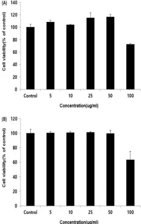
노령화에 의한 골 흡수(bone resorption)와 골 형성(bone formation)의 불균형은 퇴행성관절염, 골다공증과 같은 질환을 일으키게 된다[24]. 이러 한 불균형이 초래되는 기전은 활성화된 파골세포 와 활성이 저하된 조골세포에 의한 것으로 골 흡 수작용이 골 형성 능력보다 높아져 발생하기에 골 관련 질환 연구 시 조골세포와 파골세포가 주 로 활용된다[25,26]. 따라서 파골세포의 분화 억 제 또는 조골세포 활성 증가 등의 효능 검증을 통해 골의 재생과 퇴행을 방지하는 식·의약품 소 재 개발에는 최우선으로 독성평가를 진행하여 활 용이 가능한 농도 범위 설정은 필수적이다.
본 연구에서는 조골세포 및 파골세포를 활용한 연구에 차조기와 백출 복합물이 연구가 가능한 농도를 확인하기 위해 진행한 세포 독성 검사를 진행하였다. 그 결과, 차조기와 백출 복합물은 조 골세포와 파골세포에 5, 10, 25, 50, 100 (㎍/㎖) 농도로 처리하였을 때, 연구에 사용된 세포 모두 100 ㎍/㎖ 농도에서 대조군(100%) 대비 약 27~37% 정도의 독성이 나타났다(Fig. 2). 따라서 이후의 연구는 독성이 관찰되지 않은 50 ㎍/㎖
농도까지로 설정하여 진행하였다.
Fig. 2. Effects of
Perilla frutescens
var.crispa and Atractylodes macrocephala Koidzum
i mixture on cell viability in MG-63 osteoblastic cell(A) and RAW 264.7 ostoeclastic cell generation(B).Cell viability by MTT assay was performed in MG-63 cell and RAW 264.7 cell. The measured levels were expressed as percent of the control value (mean±standard deviation, three experiments).
3.2. Alkaline phosphatase (ALP) 활성 Alkaline phosphatase (ALP)는 대부분의 인체 조직에 존재하고 있으며, 골 생성 과정에서 활성 도가 높아지게 된다[27]. 이로 인해 일반적으로 ALP 발현은 조골세포의 분화에서 주요한 역할이 수행되며, 활성도 평가를 통해 조골세포의 분화 단계를 평가할 수 있는 결과로 사용되었다[27].
Concentration (㎍/㎖)
Control 5 10 25 50
Fig. 4. The morphology of cell at the end of cell cultures after alizarin red staining by
Perilla frutescens
var.crispa and Atractylodes macrocephala Koidzum
i mixture during differentiation 14 days.본 연구에서는 차조기와 백출 복합물이 조골세 포 분화에 영향을 미치는지를 확인하기 위해 ALP 활성을 측정하였다. 그 결과, 차조기와 백출 복합물은 조골세포에 5, 10, 25, 50 (㎍/㎖) 농도 로 처리하였을 때, 대조군(100%) 대비 ALP 활성 이 감소하였다(Fig. 3). 이와 같은 결과는 차조기 와 백출 복합물은 조골세포의 분화를 촉진하지 않는 것으로 보여지나 이를 구체적으로 확인하고 자 골 석회화 형성능 평가를 수행하였다.
Fig. 3. Effects of
Perilla frutescens
var.crispa and Atractylodes macrocephala Koidzum
i mixture on the Alkaline phosphatase activities of the MG-63 osteoblastic cell. Enzyme-linked immunosorbent assays was used to measure the ALP activity of MG-63 cell. The measured levels were expressed as percent of the control value (mean±standard deviation, three experiments).3.3. 골 석회화 형성 능력 관찰
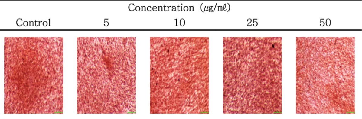
Alizarin은 식물성 염료로서 골 형성에 작용하 는 칼슘에 특이적으로 흡착이 높게 나타난다. 이 는 무기질화된 세포의 기질에 염색되어 석회화 정도에 따라 염색이 비례하므로 골 석회화 형성 능력을 판단하는 연구에 많이 사용된다[28].
본 연구에서는 차조기와 백출 복합물이 앞선 실험에서 조골세포 분화에 영향을 미치지 않았기 에 이를 구체화하고자 alizarin 염색을 진행하여 확인하였다. 그 결과, 차조기와 백출 복합물은 조 골세포에 5, 10, 25, 50 (㎍/㎖) 농도로 처리하여 14일이 지난 후 확인한 현미경 관찰 시 대조군 대비 alizarin이 염색된 범위가 많지 않았으며, 이 를 정량화한 결과에서도 대조군(100%) 대비 적 은 것으로 확인되었다(Fig. 4, 5). 이와 같은 결과 는 조골세포에 의한 석회화 과정에서 ALP의 정 확한 기능은 확실하지는 않지만, 현재까지는 유기 인산염을 가수분해하여 국소적으로 PO4 농도를 증가시켜 석회화를 촉진하는 것으로 보고되고 있 어 차조기와 백출 복합물은 조골세포 증식을 촉 진하여 염기성인 인산 분해효소가 골과 관련된 성장인자 또는 단백질에 자극을 주지 않아 퇴행 성 관절염 개선을 위한 골 생성에 관한 긍정적 효과는 기대하기가 어려울 것으로 판단된다. 이에 따라 파골세포에 대한 영향을 확인하고자 하였다.
Fig. 5. Quantification of mineralization on
Perilla frutescens
var.crispa and Atractylodes macrocephala Koidzum
i mixture in MG-63 osteoblastic cell during differentiation 14 days. Enzyme- linked immunosorbent assays was used to measure the ALP activity of MG-63 cell. The measured levels were expressed as percent of the control value (mean±standard deviation, three experiments).3.4. Tartrate resistant acid phosphatase (TRAP) 활성
파골세포는 분화가 진행되면서 단핵의 파골세 포를 형성하는데, 세포가 융합되면서 다핵의 성숙 한 파골세포를 형성하고 이는 골 표면에 부착하 여 골 흡수 역할을 수행하게 된다[29]. 이와 같은 파골세포는 TRAP과 칼시토닌(calcitonin) 수용체 를 가지게 되며, 산 생성이 활발해지는 특징을 보여 TRAP은 파골세포에 관한 표지 매개체로 널리 활용되고 있다[30,31].
본 연구에서는 차조기와 백출 복합물이 조골세 포에 효능이 나타나지 않았기에 파골세포에 관한 영향을 확인하고자 TRAP 활성을 측정하였다. 그 결과, 차조기와 백출 복합물은 파골세포에 5, 10, 25, 50 (㎍/㎖) 농도로 처리하였을 때, 대조군 (100%) 대비 25, 50 (㎍/㎖) 농도에서 유의적으 로(***
p
<0.001, **p
<0.01) 감소하였다(Fig. 6). 이 와 같은 결과는 차조기와 백출 복합물이 파골세 포의 증식 억제를 통해 연령대가 높아짐에 따라 발생하는 골 흡수 활성화에 의한 퇴화를 방어할 수 있는 원료임을 확인하였다. 이를 증명하고자 파골세포 염색을 진행하여 현미경 관찰을 통해 구체화하였다.Fig. 6. Effects of
Perilla frutescens
var.crispa and Atractylodes macrocephala Koidzum
i mixture on the tartrate resistant acid phosphatase activities of the RAW 264.7 osteoclastic cell generation. Enzyme-linked immuno- sorbent assays was used to measure the TRAP activity of RAW 264.7 cell.The measured levels were expressed as percent of the control value (mean±
standard deviation, three experiments).
***
p
<0.001, **p
<0.01 indicates a significant difference from the control group.3.5. Tartrate resistant acid phosphatase (TRAP) 염색 관찰
TRAP 염색을 진행할 경우, 핵 인자-kB 리간 드의 수용기 활성화제(RANKL)를 처리하지 않은 대조군은 파골세포로 유도하기 위해 사용된 RAW 264.7 세포가 원형 모양을 유지한 채 증식 하는데, TRAP 염색에서도 음성반응을 보여 연한 갈색이나 황토색을 보이게 된다[10]. 반면, RANKL을 처리한 세포에서는 진한 암갈색이나 적갈색으로 염색된 TRAP(+) 다핵세포가 관찰되 므로 파골세포 활성 억제를 증명하기 위한 시각 적 자료로 활용하게 된다[32].
본 연구에서는 차조기와 백출 복합물이 파골세 포의 TRAP 활성 억제 효능을 구체적으로 제시 하고자 TRAP 염색을 진행하여 확인하였다. 그 결과, RANKL을 처리한 대조군과 차조기와 백출 복합물을 처리한 실험군에서는 3개 이상의 핵을 갖는 TRAP(+) 다핵세포로 분화(Fig. 7-화살표) 가 유도된 것이 확인되었다(Fig. 7). 대조군과의 비교 시 실험군은 농도 의존적으로 분화가 TRAP 활성도 결과와 유사하게 적게 나타났으며, 25, 50 (㎍/㎖) 농도에서는 분화를 거의 억제하는
Concentration (㎍/㎖)
Control 5 10 25 50
Fig. 7. The morphology of cell at the end of cell cultures after tartrate resistant acid phosphatase staining by
Perilla frutescens
var.crispa and Atractylodes macrocephala Koidzum
i mixture during differentiation 30 min.것이 관찰되었다(Fig. 7). 이와 같은 결과는 차조 기와 백출 복합물이 파골세포 활성 억제를 통한 퇴행성관절염뿐만 아니라 골다공증과 같은 골 질 환의 예방과 개선을 위한 원료로서의 활용 가치 가 높다는 것을 보여주고 있다.
4. 결 론
본 연구는 방사선 육종 차조기와 백출의 초임 계 유체 추출 혼합물(이하, 차조기와 백출 복합 물)이 퇴행성관절염 질환의 식·의약품 소재로써 활용 가능성을 객관적으로 검증하고자 MG-63 조골세포와 RAW 264.7 파골세포의 분화에 미치 는 영향을 분석하였다.
차조기와 백출 복합물은 조골세포와 파골세포 의 독성평가를 통해 50 ㎍/㎖ 농도까지의 안전성 이 확인되었으며, 이를 바탕으로 진행한 조골세포 에서의 ALP 활성 분석과 alizarin red 염색을 통 한 평가에서는 조골세포 분화에는 효능이 확인되 지는 않았다. 반면, 파골세포에서의 TRAP 활성 은 25, 50 (㎍/㎖) 농도에서 대조군 대비 유의적 으로 감소시켰고 TRAP 염색 관찰에서도 농도 의존적으로 TRAP(+) 다핵세포의 분화를 억제하 여 25, 50 (㎍/㎖) 농도에서는 분화를 거의 억제 하는 것이 관찰되었다.
이상의 결과를 종합해 볼 때, 차조기와 백출 복합물은 안전성이 확보된 50 ㎍/㎖ 이하의 농도 에서 조골세포를 통한 골 석회화 능력보다는 파 골세포의 분화 억제를 통해 골 흡수를 억제할 수
있는 원료로써 인체의 노령화에서 나타나는 골 관련 기전에 효과적으로 사용될 수 있는 것으로 보여진다. 이에 차조기와 백출 복합물은 골 기능 강화와 골 관련 질환에 대한 예방과 개선에 효과 가 있을 것으로 판단되며, 이 같은 가설은 현재 진행되고 있는 퇴행성관절염을 유도한 동물 실험 을 통해 증명하고자 한다.
감사의 글
본 연구는 산업통상자원부와 한국산업기술진흥 원이 지원하는 광역협력권산업육성사업(2020- P0006184)으로 수행된 연구 결과입니다.
References
1. S. Glyn-Jones, A. J. R. Palmer, R.
Agricola, A. J. Price, T. L. Vincent, H.
Weinans, A. J. Carr, “Osteoarthritis”,
The Lancet
, Vol. 386, No. 9991, pp. 376-387, (2015).2. B. J. Park, H. J. Choi, B. Y. Sim, M. Y.
Yun, I. H. Yoo, D. H. Kim, “Effects of KV Pharmacopuncture on MIA-induced Osteoarthritis Rat”,
Journal of Physiology
& Pathology in Korean Medicine
, Vol. 31, No. 1, pp. 46-51, (2017).3. National Statistical Office(NSO), Statistics
of Senior Citizens, (2019).
4. National Health Insurance Service, Korean Statistical Information Service [Internet], Available From: http://kosis.kr/statHtml/
statHtml.do?orgId=350&tblId=DT_35001_A 074111&conn_path=I2. (accessed Dec., 18, 2019).
5. W. Zhang, G. Nuki, R. W. Moskowitz, S.
Abramson, R. D. Altman, N. K. Arden, M. Dougados, “OARSI recommendations for the management of hip and knee osteoarthritis: part III: Changes in evidence following systematic cumulative update of research published through January 2009”,
Osteoarthritis and cartilage
, Vol. 18, No. 4, pp. 476-499, (2010).6. A. Ghouri, P. G. Conaghan, “Prospects for Therapies in Osteoarthritis”,
Calcified Tissue International
, 1-12. (2020).7. S. Park, Y. S. Kim, D. Lee, Y. Kwon, J.
Park, S. Y. Lee, D. W. Nam, J. D. Lee, H. Kim, “Efficacy and safety of HT008 and glucosamine sulfate in the treatment of knee osteoarthritis: a randomized double-blind trial”,
The Korea Journal of Herbology
, Vol. 29, No. 4, pp. 45-52, (2014).8. B. Y. Sim, H. J. Choi, J. g. Ji, D. H. Kim,
“Effects of Yeonsan Ogye on monosodium iodoacetate-induced osteoarthritis in rats”,
The Korea Journal of Herbology
, Vol. 32, No. 2, pp. 41-47, (2017).9. H. N. Song, K. H. Leem, I. S. Kwun,
“Effect of water extract and distillate from the mixture of black goat meat and medicinal herb on osteoblast proliferation and osteoclast formation”,
Journal of Nutrition and Health
, Vol. 48, No. 2, pp.157-166, (2015).
10. H. S. Yoo, K. H. Chung, K. J. Lee, D. H.
Kim, J. H. An, “Effect of Gallus gallus var. domesticus (Yeonsan ogolgye) extracts on osteoblast differentiation and osteoclast formation”,
Microbiology and Biotechnology Letters
, Vol. 43, No. 4, pp. 322-329, (2015).11. A. M. Parfitt, “Osteonal and hemi‐osteonal remodeling: the spatial and temporal framework for signal traffic in adult human bone”,
Journal of cellular biochemistry
, Vol. 55, No. 3, pp. 273- 286, (1994).12. D. B. Solt, “The pathogenesis, oral manifestations, and implications for dentistry of metabolic bone disease”,
Current opinion in dentistry
, Vol. 1, No.6, pp. 783-791, (1991).
13. B. R. Yun, J. B. Weon, B. Lee, J. Lee, M.
R. Eom, J. M. Choong, "Quantitative Analysis of Atractylenolides I and III in Atractylodes japonica",
Kor. J. Pharmacogn
, Vol. 44, No. 1, pp. 53-59, (2013).14. J. H. Han, J. H. Kim, S. G. Kim, S. H.
Jung, D. H. Kim, G. E. Kim, W. K.
Whang, "Anti-oxidative Compounds from The Aerial Parts of Atractylodes macrocephala Koidzumi",
YAKHAK HOEJI
, Vol. 51, No. 2, pp.88-95, (2007).15. S. O. Lee, J. H. Seo, J. W. Lee, M. Y.
Yoo, "Inhibitory effects of the rhizome extract of Atractylodes japonica on the proliferation of human tumor cell lines",
Korean Journal of Pharmacognosy
, Vol.36, No. 3, pp.201-204. (2005).
16. S. H. le, M. H. Tran, J. S. Lee, Q. M.
Ngo, M. H. Woo, B. S. Min,
"Inflammatory Inhibitory Activity of Sesquiterpenoids from Atractylodes macrocephala Rhizomes",
Chem Pharm Bull
, Vol. 64, No. 5, pp.507-511, (2016).17. H. L. Huang, T. W. Lin, Y. L. Huang, R.
L. Huang, "Induction of apoptosis and differentiation by atractylenolide-1 isolated from Atractylodes macrocephala in human leukemia cells",
Bioorg Med Chem Lett
, Vol. 26, No. 8, pp. 1905-1909, (2016).18. Y. I. Lee, I. C. Shin, I. K. Lee, D. S.
Kim, “Variation of Leaf Pigment Contents in Progenies of Perilla Mutants Induced by Gamma Ray”,
Plant Breeding &
Biotechnology
, Vol. 31, No. 2, pp.110-113, (1999).
19. M. Hosoi, M. Ito, T. Yagura, R. P.
Adams, G. Honda, “cDNA isolation and functional expression of myrcene synthase from Perilla frutescens”,
Biological and Pharmaceutical Bulletin
, Vol. 27, No. 12, pp. 1979-1985. (2004).20. C. H. Jin, H. J. Lee, Y. D. Park, D. S.
Choi, D. S. Kim, S. Y. Kang, I. Y. Jeong,
“Isoegomaketone inhibits lipopolysaccharide- induced nitric oxide production in RAW 264.7 macrophages through the heme oxygenase-1 induction and inhibition of the interferon-β-STAT-1 pathway”,
Journal of agricultural and food chemistry
, Vol. 58, No. 2, pp. 860-867, (2009).21. B. O. Cho, C. H. Jin, Y. D. Park, H. W.
Ryu, M. W. Byun, K. I. Seo, I. Y. Jeong,
“Isoegomaketone induces apoptosis through caspase-dependent and caspase-independent pathways in human DLD1 cells”,
Bioscience, biotechnology, and biochemistry
, Vol. 75, No. 7, pp. 1306-1311, (2011).22. B. Y. Sim, J. H. Park, S. K. Kim, J. G. Ji,
“Effects of anti-inflammatory on Perilla frutescens var. crispa Induced by mutants with γ-Ray”.
Journal of the Korean Applied Science and Technology
, Vol. 36, No. 2, pp. 488-497, (2019).23. M. H. Park, C. J. Kim, J. Y. Lee, C. Y.
Keum, I. S. Kim, C. H. Jin, S. K. Kim,
“Anti-Arthritic Effect of Radiation Mutant Perilla frutescens var. crispa and Atractylodes macrophala koidz”,
Journal of the Korean Applied Science and Technology
, Vol. 37, No. 1, pp. 102-113, (2020).24. M. H. Jeon, M. H. Kim, “Effect of Hijikia fusiforme fractions on proliferation and differentiation in osteoblastic MC3T3-E1 cells”,
Journal of life science
, Vol. 21, No.2, pp. 300-308, (2011).
25. S. Boonen, P. Broos, J. Dequeker, R.
Bouillon, “The prevention or treatment of age‐related osteoporosis in the elderly by systemic recombinant growth factor therapy (rhIGF‐I or rhTGFβ): a
perspective”,
Journal of internal medicine
, Vol. 242, No. 4, pp. 285-290, (1997).26. S. H. Cho, K. G. Kim, S. R. Kim, J. A.
Lee, H. Moon, Y. Y. Hwang, “The effects of 17-β estradiol, medroxyprogesterone acetate and parathyroid hormone on the differentiation of osteoblast cell”,
Korean Journal of Obstetrics and Gynecology
, Vol.39, No. 1, pp. 1497-1506, (1996).
27. Y. Torii, K. Hitomi, Y. Yamahishi, N.
Tsukagoshi, “Demonstration of alkaline phosphatase participation in the mineralization of osteoblasts by antisense RNA approach”,
Cell biology international
, Vol. 20, No. 7, pp. 459-464, (1996).28. J. W. Lee, I. S. Lee, “Effects of Rubus coreanus Miquel extracts on the activity and differentiation of MC3T3-E1 osteoblastic cell”,
Journal of Life Science
, Vol. 14, No. 6, pp. 967-974, (2004).29. S. K. Mok, H. K. You, H. S. Shin, “The effects of prostaglandin and dibutyryl cAMP on osteoblastic cell activity and osteoclast generation”,
Journal of Periodontal and Implant Science
, Vol. 26, No. 2, pp. 448-468, (1996).30. M. Karst, G. Gorny, R. J. S. Galvin, M.
J. Oursler, “Roles of stromal cell RANKL, OPG, and M‐CSF expression in biphasic TGF‐β regulation of osteoclast differentiation”,
Journal of cellular physiology
, Vol. 200, No. 1, pp. 99-106, (2004).31. D. B. Solt, “The pathogenesis, oral manifestations, and implications for dentistry of metabolic bone disease”,
Current opinion in dentistry
, Vol. 1, No.6, pp. 783-791, (1991).
32. W. Sriarj, K. Aoki, K. Ohya, M.
Takahashi, Y. Takagi, H. Shimokawa,
“TGF-β in dentin matrix extract induces osteoclastogenesis in vitro”,