Ⅰ. 서 론
구강악안면 영역에서 골결손부의 재건에 다양한 외과적 술식이 사용되어 왔으며 최근에 일련의 연구를 통해 여러 가지 치료방법이 도입되고 있다.
현재까지 가장 이상적인 방법은 자가골 이식으로 알려져
있으나1, 이는 공여부의 제한이나 공여부의 반흔, 이식골편
의 흡수, 추가적인 환자의 통증, 감염 등의 이유로 그 사용 이 제한적이다2-4. 그 외에 동종골이나 이종골 이식은 공여 자나 공여 동물로부터의 질병의 전염과 감염, 그리고 자가
골 이식에 비해 골형성 효과가 낮은 단점이 있다5,6. 따라서 최근에는 platelet-rich fibrin (PRF)가 경조직의 치유 및 재생 을 촉진하는 것으로 보고되면서 골결손부에서의 치유목적 으로 사용되고 있다7. PRF란 정상치의 혈소판 수(150-400
×103/dL)보다 3-7배로 고농도 농축한 것을 말하며 platelet- rich plasma (PRP)와는 달리 제작과정이 단순하고 동일한 결과의 혈소판 농축을 얻을 수 있다8. PRF는 다량의 고농도 의 성장인자들, platelet-derived growth factor (PDGF), trans- forming growth factors (TGF)-β, insulin-like growth factor
(IGF)를 방출하여 골조직 치유를 유도한다9. 이 성장인자들
은 이식골 치유과정 시 angiogenesis, 화학주성(chemotaxis), 분열촉진(mitosis), stem cell 증식, 골편 간 결합력 제공, 피 브린 망을 통한 골전도율을 증가시키는 역할을 함으로써 초기 골재생을 촉진한다10.
Mark 등11은 혈소판 농축 혈장을 겔(gel)로 만들어 골이식
을 한 결과 방사선학적 골성숙도가 1.62-2.16배 우수하고 골구조에서의 밀도가 15-30% 개선되었다고 보고하였다.
김 성 곤
210-701 강원도 강릉시 강릉 대학로120 강릉원주대학교 치과병원 구강악안면외과 Seong-Gon Kim
Department of Oral and Maxillofacial Surgery, Dental hospital, Gangneung-Woonju National University
120 Daehangno, Gangneung, 210-701, Korea Tel: +82-33-640-2468 Fax: +82-33-641-2477 E-mail: [email protected]
가토의 두개 결손부에서의 실크 단백질과 platelet-rich fibrin (PRF)의 골형성 효과
송지영1∙권해용2∙권광준1∙박영욱1∙김성곤1
1강릉원주대학교 치과병원 구강악안면외과, 2농촌진흥청
The bone regenerative effect of silk fibroin mixed with platelet-rich fibrin (PRF) in the calvaria defect of rabbit
Ji-Young Song1, HaeYong Kweon2, Kwang Jun Kwon1, Young-Wook Park1, Seong-Gon Kim1
1Department of Oral and Maxillofacial Surgery, College of Dentistry, Gangneung-Wonju National University, Gangneung, Korea
2National Academy of Agricultural Science, RDA, Suwon, Korea
Introduction:This study evaluated the bone regenerative effect of silk fibroin mixed with platelet-rich fibrin (PRF) of a bone defect in rabbits.
Materials and Methods:Ten New Zealand white rabbits were used for this study and bilateral round shaped defects were formed in the parietal bone (diameter: 8.0 mm). The silk fibroin mixed with PRF was grafted into the right parietal bone (experimental group). The left side (control group) was grafted only PRF. The animals were sacrificed at 4 weeks and 8 weeks. A micro-computerized tomography (μCT) of each specimen was taken.
Subsequently, the specimens were decalcified and stained for histological analysis.
Results:The average value of plane film analysis was higher in the experimental group than in the control group at 4 weeks and 8weeks after surgery. However, the difference was not statistically significant.(P>0.05) The tissue mineral density (TMD) in the experimental group at 4 weeks after surgery was significantly higher than the control group.(P<0.05)
Conclusion:Silk fibroin can be used as a scaffold of PRF for rabbit calvarial defect repair.
Key words:Platelet-rich fibrin (PRF), Silk fibroin, Bone regeneration
[paper submitted 2010. 4. 13 / revised 2010. 5. 14 / accepted 2010. 5. 28]
Abstract (J Korean Assoc Oral Maxillofac Surg 2010;36:250-4)
Robiony 등12은 심하게 퇴축된 하악골에 자가골혈소판겔을 주입하고 하악골을 신장한 결과 우수한 골재생이 나타난 다고 보고하였다. Choukroun 등13은 PRF를 함유한 동결건 조이식골(freeze dried bone allograft, FDBA)를 이용한 상악 동 거상술 시에 control group에 비해 4개월 정도 골재생이 빠름을 보고하였다.
섬유상 단백질인 실크(silk)는 예로부터 비단이나 수술용 봉합재료로 사용되어 온 단백질이고 누에와 같은 유충에 서 만들어진다고 알려져 있다. 실크는 매우 강한 인장력을 보이며 수용액 상태에서 용해도가 매우 낮은 특성을 보인 다14. 이러한 실크 단백질은 상아질 막 단백질-1(dentin matrix protein-1)과 함께 수산화 인회석(hydroxyapatite)을 nucleation 시킬 수 있는 기능을 가지는 융합 단백질을 생물 학적으로 합성할 수 있다고 보고하였다15.
이에 본 연구는 골결손부에서의 골형성을 촉진시킬 수 있는 방법들 중 하나로 누에에서 추출한 실크 단백질에 혈 소판 농축 혈장을 혼합하여 가토의 골결손부에 직접 주입 하여 방사선상 골질변화 및 골밀도변화의 측정과 조직학 적 분석을 통한 실크 단백질 및 혈소판 농축 혈장의 골생성 에 대한 효과를 평가하고자 하였다.
Ⅱ. 연구대상 및 방법 1. 실험동물
본 실험에서는 가토(체중 2.0-2.5 kg, 뉴질랜드산) 10마리 를 실험동물로 사용하였으며 실온에서 고형사료와 물을 이용하여 일정 기간 사육하였다.
2. 실험재료
사용된 실크 단백질은 농촌진흥청(Suwon, Korea)에서 제 공받은 것을 사용하였다. PRF는 수술 직전 가토의 귀 정맥 에서 5 cc syringe을 이용하여 혈액 5 cc를 채취하고 3,000 rpm으로 12분간 원심분리 후 최종적으로 혈액이 3층으로 분리되면 중간층을 0.6-0.8 cc 정도 채취하였다.(Fig. 1)
3. 동물실험
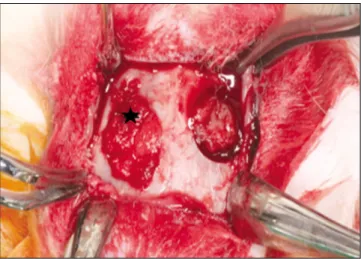
술전에 0.4 mL의 Ketamine 100 mg/mL (Ketara, Yuhan, Seoul, Korea)과 xylazine hydrochloride 10 mg/kg (Rompun, Bayer Korea, Seoul, Korea)을 가토의 대퇴부 근육에 주사하 여 전신마취를 시행하였다. 전두 부위에서 코 부위까지 제 모를 하고 povidone-iodine으로 소독한 후 1:100,000 에피네 프린을 함유한 2% 리도카인을 주사하였다. 양쪽의 귀를 잇 는 부위로부터 5 cm 전방 부위까지 두피의 정중앙 부위를 절개한 후 골막 하 박리를 이용하여 두개골 vault의 골막을 젖혀 측두골을 노출시킨다. 8 mm 직경의 trephine bur를 이
용하여 좌, 우측에 각각 2개의 직경 8 mm, 깊이 2 mm의 결 손부를 형성하였다. 실험군인 우측에는 실크 단백질에 PRF를 섞은 것을 결손부에 이식하고, 대조군인 좌측에 PRF 0.2 cc 정도를 이식한 후(Fig. 2), 골막과 근육을 3-0 silk 로 봉합하였다. 모든 실험동물은 술후 감염방지를 위해 gentamycin 1 mg/kg (Kookje, Seoul, Korea)을 하루 3번 3일 간 근육주사 하였다.
이후 실험동물을 두 그룹으로 나눠 각각 4주와 8주 후에 희생을 시켰다. 이때 측두골은 pericardium과 dura matter를 견고하게 유지하면서 머리뼈과 분리시켰으며 시편은 10%
formalin에 2일 동안 고정시키고 일반 방사선사진과 micro- computerized tomography (μCT) 촬영, 조직학적 분석을 시 행하였다.
Fig. 1. After centrifugation, the blood was separated in 3 layer. We used middle layer for grafting.
Fig. 2.The silk protein mixed with PRF was grafted into the right parietal bone (experimental group: asterisk) and the left side (control group) was grafted only PRF.
(PRF: platelet-rich fibrin)
★
4. 방사선학적 분석
술후 4주 및 8주에 가토를 희생하여 얻어진 시편은 eXplore Locus SP scanner (GE Medical Systems, London, Canada)를 이용하여 0.05 mm의 thickness로 촬영하였으며 이렇게 얻어진 이미지는 MicroView software (GE Medical Systems, London, Canada)를 이용하여 재구성하였다. 이후 software를 이용하여 defect 부위의 bone mineral content (BMC), bone mineral density (BMD), tissue mineral content (TMC), tissue mineral density (TMD)를 계산하였다. 또한 일 반 방사선사진을 분석하여 실험군과 대조군에서 방사선 불투과상의 비율을 계산하였다.
5. 조직학적 검사
술후 4주 및 8주에 가토를 희생하여, 조직괴를 형성 후 2 일간 10% 중성 포르말린에 고정하고, formic acid로 7일간 탈회한 후, 통상적인 방법에 의하여 탈수 및 파라핀 포매를 하였으며 , 4-6 μm의 표본을 poly-l-lysine을 도포한 슬라이 드에 부착하여 표본을 제작하였다. 조직절편에는 골이식 부위 및 정상 부위가 모두 포함되도록 제작하였으며 신생 골과 섬유조직의 형태 관찰과 변화를 알아보기 위해 hema- toxylin-eosin 염색을 시행하였다.
6. 분석방법
한 동물 안에 있는 샘플을 분석하기 위하여 SPSS (SPSS Inc., Chicago, IL, USA), paired t test를 이용하였다. 통계적 으로 유의한 범위는 P<0.05이다.
Ⅲ. 결 과 1. 방사선학적 분석결과
골결손부의 일반 방사선사진 분석에서 4주와 8주 모두 실험군에서 대조군보다 방사선 불투과상의 비율이 높은 것으로 나타났다. 하지만 통계분석에서는 4주와 8주 모두
대조군과 실험군에서 유의할만한 차이는 보이지 않았 다.(Fig. 3)
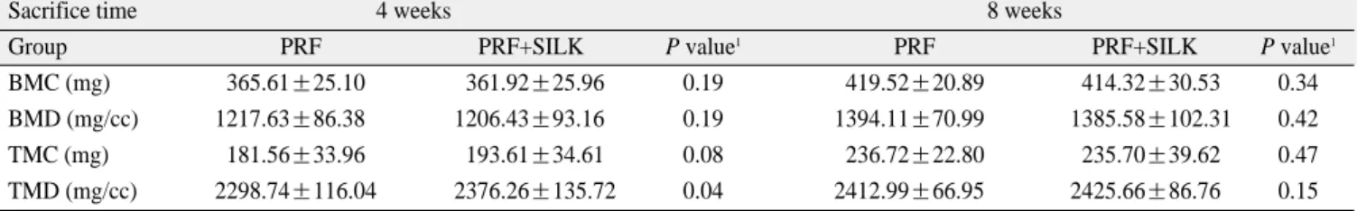
μCT를 통해 새로 형성된 골을 분석해 본 결과 4주의 TMC, TMD 값이 실험군에서 대조군보다 높게 관찰되었으 며, 통계분석에서는 TMD에서 실험군이 대조군보다 유의 할만한 증가를 나타냈다(P<0.05). 8주경에는 TMD 값이 실 험군에서 대조군보다 높게 측정되었으나 통계적으로 유의 할만한 차이를 나타내지는 않았다.(Table 1)
2. 조직학적 분석결과
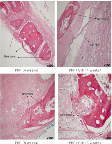
조직 절편을 hematoxylin-eosin 염색을 통하여 살펴본 결 과 4주와 8주경 모두 새로운 골의 형성을 보여주는 osteoid island의 형성을 관찰할 수 있었고(Fig. 4), 실험군에서는 macromolecule인 실크 단백질의 degradation을 위해 lym- phocyte나 numerous multinucleated giant cells (MNGC)에 의 해 매개되는 면역반응을 관찰할 수 있었다. 또한 대조군에 서는 남은 결손부에 dense fibrotic tissue가 발견되는 반면 실험군에서는 loose fiber가 주로 발견되었으며, silk의 degradation은 4주에 비해 8주에서 더 많이 진행된 것을 관 찰할 수 있었다.
Table 1.Microscopic computerized tomography analysis
Sacrifice time 4 weeks 8 weeks
Group PRF PRF+SILK P value1 PRF PRF+SILK P value1
BMC (mg) 365.61±25.10 361.92±25.96 0.19 419.52±20.89 414.32±30.53 0.34
BMD (mg/cc) 1217.63±86.38 1206.43±93.16 0.19 1394.11±70.99 1385.58±102.31 0.42
TMC (mg) 181.56±33.96 193.61±34.610.08 236.72±22.80 235.70±39.62 0.47
TMD (mg/cc) 2298.74±116.04 2376.26±135.72 0.04 2412.99±66.95 2425.66±86.76 0.15
(1: statistically significant P<0.05, BMC: bone mineral content, BMD: bone mineral density, TMC: tissue mineral content, TMD: tissue mineral density)
Fig. 3. Percent radiopacities within the PRF (test) and PRF+Silk (control) defects.
PRF Silk+PRF
4 weeks 8 weeks
80 70 60 50 40 30 20 10 0
percent of radiopacities
Ⅳ. 고 찰
구강악안면 영역에서 골결손부의 골이식은 흔한 술식이 다. 이를 위해서 많은 생체재료나 합성재료들이 많이 연구 개발되고 있다. 따라서 이번 논문에서 우리는 새로운 형태 의 골이식재인 silk fibroin을 가토의 생체 내에서 얻어진 혈 액을 바탕으로 제작된 PRF와 혼합하여 가토의 두개골에 이식할 때와 PRF 단독으로 이식할 때의 골형성 능력을 비 교하는 실험을 시행해 보았다. 이번 실험에 사용된 silk fibroin은 Bombyx mori에 의해 생산된 이후 조직 비계로써 폭넓게 연구되어지고 있으며16-18, 섬유소를 길게 늘인 fibrous protein으로 다양한 곤충과 거미로부터 얻어지며, 저렴하게 생산할 수 있고, 우수한 생체 적합성을 가지며 조 직 내에서 천천히 분해되며 가소성을 가진다19,20. 젤라틴과
hydroxyapatite 등과 혼합하여 다양한 형태, 예를 들면
fibroin 용액이나, 필름 또는 스폰지 형태로 적용할 수 있으 며 4가지 구조를 가지며 서로 다른 조합으로 구성되어 있 다: 1) elastic β-spirals 2) crystalline β-sheets rich in alanine 3) tight amino acid repeats forming α-helice 4) spacer regions. 다 른 동종 이식재나 이종 이식재와는 달리 교차감염의 위험
이 없으며 조작의 간편함이나 다른 기존의 재료와의 혼합 성이 우수하다. 또한 silk 단독으로의 골형성 인자의 부재 를 해결하기 위해 여러 가지 재료와 혼합하여 구조를 변경 시켰는데 silk fibroin 필름에 화학적으로 혼합된 arginin- glycin-aspartic acid (RGD) peptide는 이미 이전 연구에서 in vitro에서 osteoblast-like cell들이 RGD peptides (Arg-Gly- Asp)과 커플을 이루어 fibroin 필름에 부착하여 성장하면서 mineralized matrix를 생성하여 골형성을 유의하게 증가시 키는 것으로 보고되어 있으며21,22, 이외에 혼합된 bone mor- phogenetic protein 2 (BMP-2)와 nano-hydroxyapatite도 silk fibroin scaffold의 골형성을 유의하게 증가시키는 것으로 보고되었다17.
Silk fibroin protein에 혼합된 PRF는 환자로부터 얻어지는 재료로서 면역반응이나 염증반응과 관련된 우려가 적으 며, 혈소판이 풍부하고 여러 개의 성장인자를 가진 것으로 보고되었고 그중 platelet-derived growth factor (PDGF)는 정 상 골형성세포의 성장을 촉진한다고 알려져 있다9. 이번 실 험결과 일반 방사선 분석에서는 실험군이 모두 대조군에 비해 새로운 골의 형성이 높은 것으로 나타났으나 그 양에 서 유의할 만한 차이를 나타내지는 못했고, μCT 분석에서 도 만족할 만한 결과를 얻지는 못했다.
하지만 이번 실험결과에서 우리는 silk fibroin이 PRF의 scaffold로서 가능성을 확인할 수 있었다. 아마도 고분자 물 질인 silk fibroin의 흡수가 너무 느리게 진행되는 점이 신생 골의 형성에 장애가 된 것으로 추측된다. 실크 단백질의 생 체 내 흡수 속도는 실크 단백질의 분자량을 조절하거나 가 공방법을 달리함으로서 획득될 수 있다는 보고를23감안하 면 현재 형태의 실크 scaffold를 좀 더 개선할 수 있다면 좋 은 형태의 scaffold로서 PRF와 혼합하여 사용할 수 있다고 생각한다. 또한 최근 연구논문을 살펴봐도 silk와 PRF를 동 시에 임플란트 주변에 이식하거나, 가토의 두개골에서 silk 와 PRF의 혼합이식과 아무것도 이식하지 않은 것과 비교 했을 때 두 연구 모두에서 silk와 PRF의 혼합이식이 매우 유의성있는 이식재료라는 보고를24,25감안할 때 그 연구 가 치는 충분하다고 사료된다. 다만 이번 연구는 가토에서 실 행한 단기간의 연구이므로 사람에게 직접적으로 적용하기 는 어렵다. 이를 위해서는 좀 더 추가적인 연구가 필요하 다.
Ⅴ. 결 론
본 연구에서 인공적으로 형성한 가토의 두개골 결손부의 우측에는 실험군인 실크 단백질에 PRF를 섞은 것을 이식 하고, 좌측에는 대조군인 PRF를 이식한 후 4주, 8주 후 실 험동물을 희생하여 조직통계학적으로 분석하여 다음과 같 은 결론을 얻었다.
1. 일반 방사선사진 분석에서 4주와 8주 모두 실험군이 대조군보다 신생골 형성 비율이 높았지만 통계학적으
Fig. 4. Histologic section (H&E staining, original magnifica- tion ×100). Osteoid formation both in the experimental group and in the control group at 4 weeks and 8 weeks.
PRF (4 weeks) PRF+Silk (4 weeks)
PRF (8 weeks) PRF+Silk (8 weeks)
로 유의할만한 수준은 아니었다.(P>0.05)
2. μCT 분석에서 4주째 실험군의 TMD 수치가 대조군보 다 통계적으로 유의할 만하게 높았다.(P<0.05)
3. 조직학적 분석에서 실험군이 4주째보다 8주째 실크의 생체분해가 더 많이 일어났다.
이를 바탕으로 우리는 실크 단백질의 비계로서의 가능성 을 확인할 수 있었다. 실크의 분자량을 조절하거나 가공방 법을 달리하여 실크가 좀 더 빨리 생체 분해되게할 수 있다 면 좀 더 나은 실험결과를 보일 것이다. 추후 이에 대한 추 가적인 연구가 필요하겠다.
References
1. Buser D, Dula K, Hess D, Hirt HP, Belser UC. Localized ridge augmentation with autografts and barrier membranes.
Periodontol 2000 1999;19:151-63.
2. Laurie SW, Kaban LB, Mulliken JB, Murray JE. Donor-site mor- bidity after harvesting rib and iliac bone. Plast Reconstr Surg 1984;73:933-8.
3. Sommers BN, Eisenstein SM. Donor site pain from the ilium. A complication of lumbar spine fusion. J Bone Joint Surg Br 1989;
71:677-80.
4. Younger EM, Chapman MW. Morbidity at bone graft donor sites. J Orthop Trauma 1989;3:192-5.
5. Friedlaender GE, Horowitz MC. Immune responses to osteo- chondral allografts: nature and significance. Orthopedics 1992;
15:1171-5.
6. Carlson ER, Marx RE, Buck BE. The potential for HIV transmis- sion through allogeneic bone. A review of risk and safety. Oral Surg Oral Med Oral Pathol Oral Radiol Endod 1995;80:17-23.
7. Bhanot S, Alex JC. Current applications of platelet gels in facial plastic surgery. Facial Plast Surg 2002;18:27-33.
8. Marx RE. Platelet-rich plasma (PRP): what is PRP and what is not PRP? Implant Dent 2001;10:225-8.
9. Rodriguez A, Anastassov GE, Lee H, Buchbinder D, Wettan H.
Maxillary sinus augmentation with deproteinated bovine bone and platelet rich plasma with simultaneous insertion of en- dosseous implants. J Oral Maxillofac Surg 2003;61:157-63.
10. Tayapongsak P, O'Brien DA, Monteiro CB, Arceo-Diaz LY.
Autologous fibrin adhesive in mandibular reconstruction with particulate cancellous bone and marrow. J Oral Maxillofac Surg 1994;52:161-5.
11. Marx RE, Carlson ER, Eichstaedt RM, Schimmele SR, Strauss JE, Georgeff KR. Platelet-rich Plasma: growth factor enhance-
ment for bone grafts. Oral Surg Oral Med Oral Pathol Oral Radiol Endod 1998;85:638-46.
12. Robiony M, Polini F, Costa F, Politi M. Osteogenesis distraction and platelet-rich plasma for bone restoration of the severely at- rophic mandible: preliminary results. J Oral Maxillofac Surg 2002;60:630-5.
13. Choukroun J, Diss A, Simonpieri A, Girard MO, Schoeffler C, Dohan SL, et al. Platelet-rich fibrin (PRF): a second-generation platelet concentrate. Part V: histologic evaluations of PRF effects on bone allograft maturation in sinus lift. Oral Surg Oral Med Oral Pathol Oral Radiol Endod 2006;101:299-303.
14. Altman GH, Diaz F, Jakuba C, Calabro T, Horan RL, Chen J, et al. Silk-based biomaterials. Biomaterials 2003;24: 401-16.
15. Huang J, Wong C, George A, Kaplan DL. The effect of geneti- cally engineered spider silk-dentin matrix protein 1 chimeric pro- tein on hydroxyapatite nucleation. Biomaterials 2007;28:2358- 67.
16. Meinel L, Fajardo R, Hofmann S, Langer R, Chen J, Snyder B, et al. Silk implants for the healing of critical size bone defects.
Bone 2005;37:688-98.
17. Hirano Y, Mooney DJ. Peptide and protein presenting materials for tissue engineering. Adv Mater 2004;16:17-25.
18. Dal Pra I, Freddi G, Minic J, Chiarini A, Armato U. De novo en- gineering of reticular connective tissue in vivo by silk fibroin nonwoven materials. Biomaterials 2005;26:1987-99.
19. Stitzel J, Liu J, Lee SJ, Komura M, Berry J, Soker S, et al.
Controlled fabrication of a biological vascular substitute.
Biomaterials 2006;27:1088-94.
20. Chan CK, Kumar TS, Liao S, Murugan R, Ngiam M, Ramakrishnan S. Biomimetic nanocomposites for bone graft ap- plications. Nanomedicine (Lond) 2006;1:177-88.
21. Zhao J, Zhang Z, Wang S, Sun X, Zhang X, Chen J, et al.
Apatite-coated silk fibroin scaffolds to healing mandibular border defects in canines. Bone 2009;45:517-27.
22. Li C, Vepari C, Jin HJ, Kim HJ, Kaplan DL. Electrospun silk- BMP-2 scaffolds for bone tissue engineering. Biomaterials 2006;
27:3115-24.
23. Cao Y, Wang B. Biodegradation of silk biomaterials. Int J Mol Sci 2009;10:1514-24.
24. Jang ES, Park JW, Kweon H, Lee KG, Kang SW, Baek DH, et al. Restoration of peri-implant defects in immediate implant in- stallations by Choukroun platelet-rich fibrin and silk fibroin pow- der combination graft. Oral Surg Oral Med Oral Pathol Oral Radiol Endod 2010;109:831-6.
25. Kim JY, Choi JY, Jeong JH, Jang ES, Kim AS, Kim SG, et al.
Low molecular weight silk fibroin increases alkaline phosphatase and type I collagen expression in MG63 cells. BMB Rep 2010;
43:52-6.