Tuberc Respir Dis 2010;69:177-183
CopyrightⒸ2010. The Korean Academy of Tuberculosis and Respiratory Diseases. All rights reserved.
건강한 흡연자에서 폐기종의 유무에 따른 폐기능 변화
가톨릭대학교 의과대학 서울성모병원
1건강증진의학과,
2내과학교실 호흡기내과
심윤수1, 함은재1, 최규용1, 이숙영2, 김석찬2, 김영균2, 박성학2
Longitudinal Evaluation of Lung Function Associated with Em- physema in Healthy Smokers
Yun Su Sim, M.D.
1, Eunjae Ham, M.D.
1, Kyu Yong Choi, M.D.
1, Suk Young Lee, M.D.
2, Seok Chan Kim, M.D.
2, Young Kyoon Kim, M.D.
2, Sung Hak Park, M.D.
2Departments of
1Health Promotion Medicine,
2Respiratory and Critical Care Medicine, Seoul St. Mary’s Hospital, The Catholic University of Korea College of Medicine, Seoul, Korea
Background: Smoking reduces pulmonary function and induces various lung diseases. Recently, the rate of emphysema detection has increased due to lung cancer screening with low-dose chest computed tomography (CT).
The purpose of this study was to evaluate changes in lung function associated with emphysema in healthy smokers.
Methods: One hundred and ninety one healthy smokers, who had undergone a low-dose chest CT (LDCT) scan as part of lung cancer screening and had revisited the health center after a median 23.9 months’ time, were recruited into this study. The severity of emphysema was calculated by the direct observation of a radiologist and a pul- monologist indipendently. Longitudinal changes in lung function according to emphysema based on LDCT and type of smoker was analyzed.
Results: Of the participants in this study, 25% of healthy smokers had emphysema, which was mild in severity, in older patients (p=0.003) and in heavy smokers (p<0.001). FEV1/FVC and FEF25-75% were decreased in current smokers with emphysema (p=0.001 and p=0.009, respectively) and without emphysema (p=0.001 and p=0.042).
Although lung function was not decreased in ex-smokers without emphysema, FEV1/FVC and FEF25-75% were decreased in ex-smoker with emphysema (p=0.020 and p=0.010).
Conclusion: Upon examination with LDCT, the prevalence of emphysema was higher in healthy smokers was than in non-smokers. Lung function was diminished in smokers with emphysema, in spite of former smoker.
Key Words: Emphysema; Tomography, X-Ray Computed; Smoking; Spirometry
Address for correspondence: Young Kyoon Kim, M.D.
Department of Respiratory and Critical Care Medicine, Seoul St. Mary’s Hospital, 505, Banpo-dong, Seocho-gu, Seoul 137-701, Korea
Phone: 82-2-2258-2756, Fax: 82-2-2258-1856 E-mail: [email protected]
Received: May 13, 2010 Accepted: Aug. 26, 2010
서 론
흡연은 폐기능을 감소시키고 다양한 호흡기질환을 유 발한다1-4. 그 중에 만성폐쇄성폐질환은 흡연과 공해, 노령 인구의 증가로 유병률이 증가하고 있으나 특징적인 기도
폐쇄나 증상이 나타나기까지 상당한 기간이 걸리고 금연 외에 효과적인 치료가 어렵다5. 10년 동안 만성폐쇄성폐 질환 위험군을 추적관찰하였을 때 14%가 만성폐쇄성폐질 환자로 발전한다는 보고가 있어6 조기에 위험군을 발견하 는 것이 중요하다.
만성폐쇄성폐질환에서 흔히 보이는 폐기종은 말단 세 기관지 이하에서 영구적 확장과 폐포벽의 파괴가 동반되 면서 탄성반동압력(elastic recoil pressure)이 감소하게 되 어 호기 시에 비가역적인 기도폐쇄를 일으키게 되는 병리 조직학적 개념의 질환이지만 임상에서 진단은 병리조직 학적 소견에 의존하기보다는 폐기능 검사 및 방사선학적 소견을 이용한다7,8. 그러나, 폐기능 검사는 폐기종 초기에
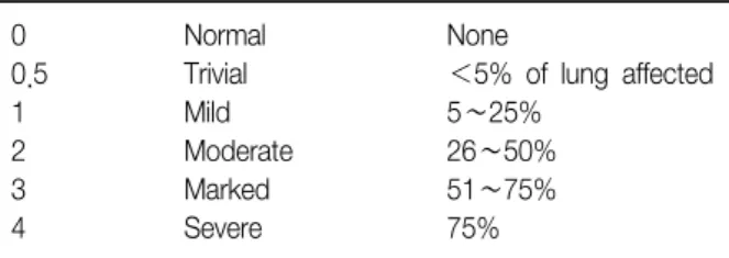
Table 1. Emphysema severity scale
0 Normal None
0.5 Trivial <5% of lung affected
1 Mild 5∼25%
2 Moderate 26∼50%
3 Marked 51∼75%
4 Severe 75%
는 정상 소견을 보이기도 하며, 단순흉부촬영은 폐혈관 음영이 감소하거나 폐의 과팽창이 나타날 때까지 진단이 어려울 수 있다9. 고해상도흉부전산화단층촬영(high reso- lution chest computer tomography, HRCT)은 비교적 초기 에 폐기종을 진단하며 직접 관찰하거나 소프트웨어로 감쇄 도를 계산하여 폐기종의 중등도를 평가할 수 있다10-12. 최근 폐암조기검진으로 건강한 흡연자에서 저선량흉부 전산화단층촬영(low dose chest computer tomography, LDCT)을 하게 되면서 무증상인 검진자에서 폐기종이 진 단되는 경우가 있다. 폐기종의 중등도와 폐기능의 상관관 계에 관한 연구에11-17 비해 폐기종을 가진 흡연자에서 폐 기능의 변화를 고찰한 연구는 드물다. 이에 본 연구에서 는 건강한 흡연자에서 나타나는 폐기종의 양상과 흡연 상 태에 따른 추적 폐기능의 변화를 알아보고자 하였다.
대상 및 방법
1. 연구 대상
2008년 10월부터 2009년 6월까지 서울성모병원 평생건 강증진센터에 방문하여 폐기능검사와 LDCT를 시행 받은 20세 이상의 성인 중에서 과거 본 병원에서 건강 검진을 받았으며 문진에 의해 만성폐쇄성폐질환과 기관지 천식 등의 폐질환 병력이 없고 기타 신체 장애가 존재하지 않을 경우를 건강하다고 정의하였으며 문진 및 검진 결과에서 종양이나 급성 혹은 만성 신부전과 심부전, 간 기능 부전 이 있는 경우는 제외하였다. 문진은 두 번의 방문에서 모 두 실시되었다. 대상자 274명 중에 남자가 239명이었으며 연령의 중앙값은 50세(사분위수, 43∼56세)이었고 191명 (70%)이 흡연자였다.
LDCT에서 결핵반흔, 기관지확장증, 흉막비후 등으로 한쪽 폐 면적의 1/4 이상이 손상되었거나 장경이 3 cm 이상인 결절이 있는 경우는 제외하였다.
2. 연구 방법
대상자들의 재방문 기간은 약 23.9개월(사분위수, 13.1
∼31.9개월)이었고, 현재 혹은 과거 흡연자 중에서 폐기종 의 유무에 따라 분류하였을 경우에 각 군 간에 재평가까지 의 기간은 차이가 없었다. 현재와 과거에 내원한 시기의 의무기록을 검토하여 성별과 나이, 과거력, 체질량지수와 혈액학적 지표를 조사하였다. 흡연력은 두 번째 방문을 기 준으로 평가하였고 첫 번째 방문 이후에 금연한 검진자는 8명이었으며 금연 기간은 모두 6개월 이상이었다. 폐기능
은 V max spectra 22 (Sensormedics, Yorba Linda, CA, USA)로 미국흉부학회의 폐기능검사 지침에 따라18 강제폐 활량(forced vital capacity, FVC), 1초간 강제호기량 (forced expiratory volume in 1 second, FEV1)과 폐활량 25∼75% 사이의 강제호기유량(forced expiratory flow be- tween 25 and 75% of vital capacity, FEF 25-75%)을 측정 하였다.
저선량 흉부전산화단층촬영에 사용된 기기는 SOMATOM Definition (Simens, Erlangen, Germany)로 두 번째 방문 에서 촬영하였다. 폐 실질 영상에 대해서 폐창(lung win- dow setting)에서 높이(window level) -650 HU, 너비 (window width) 1,500 HU를 적용하여 영상의학과 전문 의와 호흡기내과 전문의가 picture archiving and commu- nication system data base (PACS)를 통해 독립적으로 판 독한 후에 합의하였다.
폐기종 중등도 지수는 National Emphysema Treatment Trial에서 사용하였던 지수 체계를19 변형한 Hersh 등20의 논문을 참고하였다. 좌우 폐를 대동맥궁(aortic arch), 우 측 하폐정맥(right inferior pulmonary vein)을 기준으로 상(upper), 중(middle), 하(lower)로 각각 여섯 영역으로 나누어 0∼4점 사이의 점수를 합산하여 0점에서 최대 24 점이 되도록 하였다(Table 1).
가톨릭중앙의료원의 Institutional Review Board에서 허가를 얻었으며 후향적 연구의 특성으로 인해 동의서는 생략되었다.
3. 통계분석
통계 분석은 SPSS for window version 12.0 (SPSS Inc., Chicago, IL, USA) 프로그램을 이용하였다. 연속변수의 분석은 Mann-Whitney test를, 범주형 변수의 분석은 카이 제곱검정을 이용하였다. 흡연자에서 폐기능 변화는 Wil- coxon test로 분석하였다. p값이 0.05 미만일 때 통계적으 로 유의한 것으로 판단하였다.
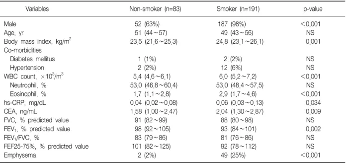
Table 2. Comparison of baseline characteristics and pulmonary function test in smoker and non-smoker
Variables Non-smoker (n=83) Smoker (n=191) p-value
Male 52 (63%) 187 (98%) <0.001
Age, yr 51 (44∼57) 49 (43∼56) NS
Body mass index, kg/m
223.5 (21.6∼25.3) 24.8 (23.1∼26.1) 0.001
Co-morbidities
Diabetes mellitus 1 (1%) 2 (2%) NS
Hypertension 2 (2%) 12 (6%) NS
WBC count, ×10
3/m
35.4 (4.6∼6.1) 6.0 (5.2∼7.2) <0.001
Neutrophil, % 53.0 (46.8∼60.4) 53.0 (48.4∼57.5) NS
Eosinophil, % 1.7 (1.1∼2.8) 2.9 (1.7∼4.6) <0.001
hs-CRP, mg/dL 0.04 (0.02∼0.08) 0.06 (0.03∼0.13) 0.034
CEA, ng/mL 1.58 (1.00∼2.47) 2.04 (1.30∼2.87) 0.009
FVC, % predicted value 91 (82∼99) 88 (80∼98) NS
FEV
1, % predicted value 98 (92∼105) 93 (84∼101) 0.002
FEV
1/FVC, % 83 (79∼86) 81 (76∼86) NS
FEF25-75%, % predicted value 101 (82∼125) 92 (78∼112) NS
Emphysema 2 (2%) 49 (25%) <0.001
The data are presented as number (%) or the median (interquartile range).
hs-CRP: high sensitive C-reactive protein; WBC: white blood cell; CEA: carcinoembryonic antigen; FEV
1: forced expiratory volume in 1 second; FVC: forced vital capacity; FEF25-75%: forced expiratory flow between 25 and 75% of vital capacity; NS: not significant.
Table 3. Emphysema severity score of 51 persons with emphysema in low dose chest CT
Right lung Left lung Upper zone 0.5 (0.5∼0.5) 0.5 (0∼0.5) Middle zone 0 (0∼0.5) 0 (0∼0.125)
Lower zone 0 0
Each lung 0.5 (0.5∼1) 0.5 (0∼0.625)
Total lungs 1.0 (0.5∼2)
The data are presented as the median (interquartile range).
결 과
1. 흡연자의 특성
191명의 흡연자가 비흡연자에 비해 체질량지수와 (p=0.001) 혈액 내 백혈구 수치(p<0.001), 호산구 분획 률(p<0.001), 고감도 C-반응성 단백질과(p=0.034) 태아 성암항원이(p=0.009) 유의하게 높았다. FEV1의 정상 예측 치에 대한 백분율은 흡연자가 낮았다(p=0.002) (Table 2).
흡연자와 비흡연자에서 체질량지수에 성별을 고려하여 분석하였을 때 유의한 차이를 보이지 않았고, FEV1은 연 령을 고려하여 분석하여도 흡연자에서 낮았다(p=0.001).
2. 폐기종 지수 및 특성
대상자 중에 폐기종을 가진 검진자는 51명(19%)이었으 며 49명이 흡연자였다(p=0.001). 폐기종은 주로 상엽에 존재하였으며 폐기종 지수의 중앙값은 1.0 (사분위수, 0.5
∼2)이었다(Table 3).
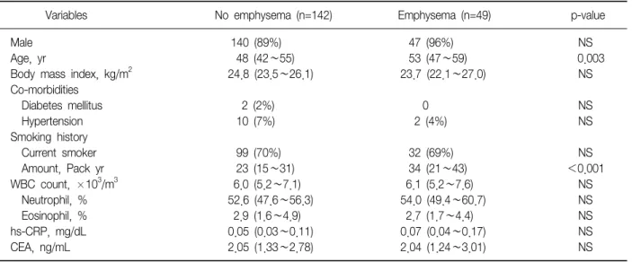
흡연자 중에 폐기종의 유무에 따라 비교하였을 때, 폐 기종을 가진 수진자에서 연령이 높고(p=0.003) 흡연력이 많았다(p<0.001) (Table 4).
3. 폐기종과 폐기능 변화
총 흡연자 191명 중에서 재방문에 측정한 폐기종군의 FEV1/FVC는 79% (사분위수, 74∼85%)로 첫 방문에 측정 한 83% (사분위수, 79∼86%)에 비해 감소하였으며(p<
0.001), FEF25-75% 예측치 백분율은 88% (사분위수, 69
∼103%)로 첫 방문시의 94% (사분위수, 79∼115%)에 비 해 감소하였다(p<0.001). 폐기종이 없는 검진자에서 재 방문 시에 측정한 FEV1/FVC는 81% (사분위수, 77∼86%) 로 첫 방문에 측정한 82% (사분위수, 78∼87%)에 비해 감 소하였으며(p=0.013), FEF25-75% 예측치 백분율은 94%
(사분위수, 79∼116%)로 첫 방문시의 99% (사분위수, 79
∼118%)에 비해 감소하였다(p=0.038) (Figure 1A).
현재 흡연자 131명에서는 재방문에 측정한 폐기종군의
Table 4. Comparison of baseline characteristics in smoker according to presence of emphysema
Variables No emphysema (n=142) Emphysema (n=49) p-value
Male 140 (89%) 47 (96%) NS
Age, yr 48 (42∼55) 53 (47∼59) 0.003
Body mass index, kg/m
224.8 (23.5∼26.1) 23.7 (22.1∼27.0) NS
Co-morbidities
Diabetes mellitus 2 (2%) 0 NS
Hypertension 10 (7%) 2 (4%) NS
Smoking history
Current smoker 99 (70%) 32 (69%) NS
Amount, Pack yr 23 (15∼31) 34 (21∼43) <0.001
WBC count, ×10
3/m
36.0 (5.2∼7.1) 6.1 (5.2∼7.6) NS
Neutrophil, % 52.6 (47.6∼56.3) 54.0 (49.4∼60.7) NS
Eosinophil, % 2.9 (1.6∼4.9) 2.7 (1.7∼4.4) NS
hs-CRP, mg/dL 0.05 (0.03∼0.11) 0.07 (0.04∼0.17) NS
CEA, ng/mL 2.05 (1.33∼2.78) 2.04 (1.24∼3.01) NS
The data are presented as number (%) or the median (interquartile range).
hs-CRP: high sensitive C-reactive protein; WBC: white blood cell; CEA: carcinoembryonic antigen; NS: not significant.
Figure 1. Change of lung function according to presence of emphysema in (A) smoker (n=191), (B) current smoker (n=131), and (C) ex-smoker (n=60) at initial and follow up visit. The middle line of bar shows median value with inter- quartile range (upper and lower lines). The data are pre- sented as % predicted value of FVC, FEV1, FEF25-75%, and FEV1/FVC. PE: pulmonary emphysema group; NPE:
no pulmonary emphysema group; FEV1: forced expiratory volume in 1 second; FVC: forced vital capacity;
FEF25-75%: forced expiratory flow between 25 and 75%
of vital capacity. *p<0.05.
FEV1/FVC는 80% (사분위수, 74∼87%)로 첫 방문에 측정 한 84% (사분위수, 79∼86%)에 비해 감소하였으며(p=
0.001), FEF25-75% 예측치 백분율은 90% (사분위수, 76
∼103%)로 첫 방문시의 96% (사분위수, 78∼118%)에 비 해 감소하였다(p=0.009). 폐기종이 없는 검진자에서 재방 문 시에 측정한 FEV1/FVC는 81% (사분위수, 76∼87%)로 첫 방문에 측정한 84% (사분위수, 78∼87%)에 비해 감소하 였으며(p=0.001), FEF25-75% 예측치 백분율은 91% (사분 위수, 78∼111%)로 첫 방문시의 97% (사분위수, 78∼
117%)에 비해 감소하였다(p=0.042) (Figure 1B).
과거흡연자 60명에서 폐기종이 없는 군에서 재방문에 측정한 폐기능은 첫 방문 시와 유의한 차이를 보이지 않았 고 폐기종군에서 재방문 시에 측정한 FEV1/FVC는 76%
(사분위수, 73∼84%)로 첫 방문에 측정한 81% (사분위수, 77∼85%)에 비해 감소하였으며(p=0.020), FEF25-75% 예 측치 백분율은 83% (사분위수, 60∼103%)로 첫 방문 시 의 91% (사분위수, 78∼113%)에 비해 감소하였다(p=
0.010) (Figure 1C).
현재 흡연자의 FEF25-75% 예측치 백분율의 1년간 감소 율은 폐기종군은 4.4% (사분위수, 0.6∼11.9%)이었고 폐 기종이 없는 대상자의 1.1% (사분위수, -2.8∼5.9%)보 다 컸으나 통계적으로 유의하지는 않았으며(p=0.056) 과거 흡연자 중에 폐기종군과 폐기종이 없는 대상자에서는 4.2%
(사분위수, -1.1∼16.9%)와 0.5% (사분위수, -4.3∼
5.5%)로 폐기종군에서 FEF25-75% 예측치 백분율의 1년 간 감소율이 더 컸다(p=0.044).
고 찰
흡연이 폐기능을 감소시키는 것은 잘 알려져 있으며3,4,21 본 연구에서는 건강한 흡연자를 대상으로 하여 정상 범위 의 폐기능을 보였지만 FEV1이 비흡연자에 비해 낮았다.
흡연이 폐기능의 감소를 가져오는 기전에 대해서는 기도 과민성의 증가나 폐포벽의 파괴로 인한 폐기종의 발생 등 다양하게 제시되고 있으며22,23 흡연자에서 호중구와 호산 구의 증가가 이러한 폐 손상이나 과민성과 관련되어 있다 는 연구가 있다24-26. 본 연구에서도 비흡연자에 비해 흡연 자에서 혈액 내에 호산구 분획률과 백혈구 수, 고감도 C 반응성 단백질이 증가되어 있어 전신적인 만성 염증상태 를 시사하였다.
본 연구에서 비흡연자에서는 2%에서 폐기종이 관찰되 었으나 흡연자의 약 25%에서 폐기종이 발견되었다. 건강
한 성인 흡연자가 대상이었던 Omori 등27의 연구에서 흡 연자의 약 26%, 비흡연자의 3%에서 폐기종이 발견되었고 Remy-Jardin 등28의 연구에서는 약 21%에서 폐기종이 관 찰되었던 것과 비슷한 경향을 보였으며, 60세 이상의 흡 연자가 대상인 Tylén 등17의 연구에서는 좀 더 많은 44%
에서 관찰되어 고령에서 더 많은 폐기종이 발견되는 것을 알 수 있었다. Satoh 등29이 부검자에서 실시한 연구에서 는 약 80%에서 폐기종이 발견되었으며 높은 연령과 관계 있었고, 본 연구에서도 폐기종이 있는 흡연자의 연령이 높 았다. 또한, 폐기종을 가진 흡연자의 흡연력이 높았던 것은 폐기종의 발생이 개인의 감수성에도 영향을 받으나 절대적 인 흡연량이 많을수록 폐실질의 파괴가 가속화 되어 폐기 종 발생을 일으키는 것과 관계가 있는 것을 시사한다.
본 연구는 건강한 흡연자를 대상으로 하였기 때문에 폐 기종은 중증도의 중위값은 1.0으로 경미하였고, 주로 상 엽에 분포하였다. 흡연자에서 폐기종의 분포를 본 Mair 등15의 연구에서 주로 폐 상엽과 중심부에 폐기종이 위치 했던 것과 일치하는 소견이며, 만성폐쇄성폐질환자에서 폐실질의 파괴는 경미한 경우에는 폐상부에 국한되며 질 병이 진행될수록 폐 전체에 걸쳐 나타나고 전형적으로 중 심 소엽성을 보이는 것으로 알려져 있다30.
본 연구에서 흡연자는 대부분 정상 범위의 폐기능 수치 를 보이고 있으나 추적한 폐기능을 분석할 경우에 현재 흡연자에서 폐기종의 유무에 관계 없이 재방문 시에 측정 한 FEF25-75%가 첫 방문에 측정한 값보다 유의하게 감소 하여 흡연에 의한 폐기능이 감소를 보여준 다른 연구3,4,21 와 일치하는 소견을 보였다. FEV1/FVC도 감소하는 경향 을 보였으며 흡연자와 폐기종을 가진 검진자가 연구 대상 이었기에 폐쇄성 폐기능 저하의 양상을 보이는 것으로 추 론해 볼 수 있으나 FVC와 FEV1 각각의 변화에 따라 다른 값을 가질 수 있기에 폐기능의 감소라고 해석하기 어렵다.
흡연에 의한 폐기능의 악화를 예방할 수 있는 가장 효 과적인 방법은 금연으로 알려져 있다3,4,21. Xu 등4은 9,191 명에서 3년 간격으로 6년 동안 폐기능 검사를 실시하였고, 비흡연자와 과거 흡연자의 FEV1 감소에는 차이가 없었으 나 현재 흡연자는 비흡연자와 과거 흡연자에 비하여 FEV1
감소가 더 심하다는 결과를 보여주었다. 본 연구에서는 과거 흡연자에서 폐기종이 없는 검진자는 재방문에 측정 한 폐기능 수치가 첫 방문에 측정한 값에 비해 유의한 감 소가 없어 비슷한 결과를 보였으나 폐기종군에서는 재방 문 시에 FEF25-75%의 감소를 보여, 폐기종을 가진 흡연자 는 금연 후에도 폐기종이 없는 흡연자에 비해 폐기능의
악화가 진행되는 것을 알 수 있었다. Remy-Jardin 등10도 폐기종을 가진 흡연자에서 더 빠른 폐기능 감소를 보고하 였으나 흡연상태에 따른 분류는 하지 않았다.
본 연구는 몇 가지 제한점을 지닌다. 먼저 LDCT가 HRCT에 비하여 폐기종을 발견하는데 민감도가 떨어져 3
∼5 mm 크기의 기종을 놓친다는 보고가 있다14. 그 외에 본 연구에서는 비교대상군의 연령과 성별의 분포가 일치 하지 않았다. 본 연구에서 흡연자에서 비흡연자에 비해 체질랑지수가 높았고, 이것은 흡연이 니코틴성 콜린 수용 체에 작용해 교감신경을 항진시켜 열을 발생하고 식욕부 진을 유발하여 체중 및 체질량지수가 낮아진다는 과거 연 구31와 다른 결과였으나, 비흡연군에 여성이 많았기에 성 별을 고려하여 이변량회기분석을 하였을 때에는 흡연자 와 비흡연자의 체질량지수에서 통계적으로 유의한 차이 를 보이지 않았다. 또한, 폐기종군에서 고령의 흡연자가 많은 것도 폐기능 악화에 영향을 줄 수 있으나 폐기능 감 소가 폐기종의 유무에 따라 뚜렷한 차이를 보였던 과거흡 연자 경우에 두 흡연자군 사이에 연령이 통계적으로 유의 한 차이가 없어 나이에 따른 영향은 배제될 수 있었다.
향후에는 흡연군과 연령군을 통제한 대상군에 대한 전향 적인 연구가 필요할 것으로 판단된다.
정상 범위의 폐기능을 가진 건강한 흡연자에서 폐암의 조기검진을 위해 실시한 LDCT에서 폐기종이 발견될 확률 은 상당히 높아서 증상 및 특징적인 폐기능 장애가 나타나 기 전에 잠재적인 만성폐쇄성폐질환자의 예측에 LDCT가 도움을 줄 수 있으며, 폐기종이 있는 흡연자는 금연에도 불구하고 폐기능의 감소가 폐기종이 없는 흡연자에 비하 여 분명하기에 밀접한 추적관찰이 필요하다.
감사의 글
This study was supported by a grant of the Korea Healthcare technology R&D Project, Ministry for Health, Welfare and Family Affairs, Republic of Korea (A040153).
참 고 문 헌
1. Buist AS, Nagy JM, Sexton GJ. The effect of smoking cessation on pulmonary function: a 30-month follow- up of two smoking cessation clinics. Am Rev Respir Dis 1979;120:953-7.
2. Heyneman LE, Ward S, Lynch DA, Remy-Jardin M,
Johkoh T, Müller NL. Respiratory bronchiolitis, respira- tory bronchiolitis-associated interstitial lung disease, and desquamative interstitial pneumonia: different enti- ties or part of the spectrum of the same disease proc- ess? AJR Am J Roentgenol 1999;173:1617-22.
3. Willemse BW, Postma DS, Timens W, ten Hacken NH.
The impact of smoking cessation on respiratory symp- toms, lung function, airway hyperresponsiveness and inflammation. Eur Respir J 2004;23:464-76.
4. Xu X, Dockery DW, Ware JH, Speizer FE, Ferris BG Jr. Effects of cigarette smoking on rate of loss of pul- monary function in adults: a longitudinal assessment.
Am Rev Respir Dis 1992;146(5 Pt 1):1345-8.
5. Murray CJ, Lopez AD. Alternative projections of mortal- ity and disability by cause 1990-2020: Global Burden of Disease Study. Lancet 1997;349:1498-504.
6. Cerveri I, Corsico A, Zoia MC. GOLD stage 0. Am J Respir Crit Care Med 2003;167:936.
7. The definition of emphysema. Report of a National Heart, Lung, and Blood Institute, Division of Lung Dis- eases workshop. Am Rev Respir Dis 1985;132:182-5.
8. Thurlbeck WM, Simon G. Radiographic appearance of the chest in emphysema. AJR Am J Roentgenol 1978;
130:429-40.
9. Nicklaus TM, Stowell DW, Christiansen WR, Renzetti AD Jr. The accuracy of the roentgenologic diagnosis of chronic pulmonary emphysema. Am Rev Respir Dis 1966;93:889-99.
10. Remy-Jardin M, Edme JL, Boulenguez C, Remy J, Mas- tora I, Sobaszek A. Longitudinal follow-up study of smoker's lung with thin-section CT in correlation with pulmonary function tests. Radiology 2002;222:261-70.
11. Lee JY, Lee KY, Choi EK, Kim SJ, Choi YH. Objective in vivo quantification of emphysema by thin-section CT: correlation with physiologic findings. Tuberc Respir Dis 1998;45:992-9.
12. Ahn JH, Park JM, Ko SH, Yoon JG, Kwon SS, Kim YK, et al. Correlation between high-resolution CT and pul- monary function tests in patients with emphysema.
Tuberc Respir Dis 1996;43:367-76.
13. Gupta PP, Yadav R, Verma M, Gupta KB, Agarwal D.
High-resolution computed tomography features in pa- tients with chronic obstructive pulmonary disease.
Singapore Med J 2009;50:193-200.
14. Horiuchi N, Fujita J, Suemitsu I, Yamasaki Y, Higa F, Tateyama M. Low-dose multislice CT and high-resolu- tion CT assessment of pulmonary emphysema in public school teachers. Lung 2007;185:25-30.
15. Mair G, Miller JJ, McAllister D, Maclay J, Connell M,
Murchison JT, et al. Computed tomographic emphyse- ma distribution: relationship to clinical features in a co- hort of smokers. Eur Respir J 2009;33:536-42.
16. Mastora I, Remy-Jardin M, Sobaszek A, Boulenguez C, Remy J, Edme JL. Thin-section CT finding in 250 volun- teers: assessment of the relationship of CT findings with smoking history and pulmonary function test results.
Radiology 2001;218:695-702.
17. Tylén U, Boijsen M, Ekberg-Jansson A, Bake B, Löfdahl CG. Emphysematous lesions and lung function in heal- thy smokers 60 years of age. Respir Med 2000;94:38-43.
18. American Thoracic Society. Standardization of spirome- try, 1994 update. Am J Respir Crit Care Med 1995;152:
1107-36.
19. National Emphysema Treatment Trial Research Group.
Patients at high risk of death after lung-volume-reduc- tion surgery. N Engl J Med 2001;345:1075-83.
20. Hersh CP, Jacobson FL, Gill R, Silverman EK. Comput- ed tomography phenotypes in severe, early-onset chro- nic obstructive pulmonary disease. COPD 2007;4:331-7.
21. Fletcher CM. Letter: Natural history of chronic bronchi- tis. Br Med J 1976;1:1592-3.
22. Auerbach O, Hammond EC, Garfinkel L, Benante C.
Relation of smoking and age to emphysema. Whole- lung section study. N Engl J Med 1972;286:853-7.
23. Rasmussen F, Siersted HC, Lambrechtsen J, Hansen HS, Hansen NC. Impact of airway lability, atopy, and tobac- co smoking on the development of asthma-like symp- toms in asymptomatic teenagers. Chest 2000;117:1330- 5.
24. Janoff A, Raju L, Dearing R. Levels of elastase activity in bronchoalveolar lavage fluids of healthy smokers
and nonsmokers. Am Rev Respir Dis 1983;127:540-4.
25. Totti N 3rd, McCusker KT, Campbell EJ, Griffin GL, Senior RM. Nicotine is chemotactic for neutrophils and enhances neutrophil responsiveness to chemotactic pep- tides. Science 1984;223:169-71.
26. Hällgren R, Samuelsson T, Venge P, Modig J. Eosino- phil activation in the lung is related to lung damage in adult respiratory distress syndrome. Am Rev Respir Dis 1987;135:639-42.
27. Omori H, Nakashima R, Otsuka N, Mishima Y, Tomigu- chi S, Narimatsu A, et al. Emphysema detected by lung cancer screening with low-dose spiral CT: prevalence, and correlation with smoking habits and pulmonary function in Japanese male subjects. Respirology 2006;
11:205-10.
28. Remy-Jardin M, Remy J, Boulenguez C, Sobaszek A, Edme JL, Furon D. Morphologic effects of cigarette smoking on airways and pulmonary parenchyma in healthy adult volunteers: CT evaluation and correlation with pulmonary function tests. Radiology 1993;186:107- 15.
29. Satoh K, Kobayashi T, Misao T, Hitani Y, Yamamoto Y, Nishiyama Y, et al. CT assessment of subtypes of pulmonary emphysema in smokers. Chest 2001;120:
725-9.
30. Repine JE, Bast A, Lankhorst I. Oxidative stress in chronic obstructive pulmonary disease. Oxidative Stress Study Group. Am J Respir Crit Care Med 1997;156(2 Pt 1):341-57.
31. Dallosso HM, James WP. The role of smoking in the regulation of energy balance. Int J Obes 1984;8:365-75.