⋅교신저자 : Jin-Young Moon, Gyeongju 780-714, South Korea College of Oriental Medicine, Dong-Guk University Tel. +82(54)770-2665, Fax. +82(54)770-2649.
E-mail: [email protected]
⋅투고 : 2011/05/18 심사 : 2011/06/13 채택 : 2011/06/16
The Effect of Pinus Densiflora Gnarl Extract for Pharmacopuncture on Human LDL Oxidation
Induced
by Free Radical and Metal Ion
Sun-Hee Leem
1⋅Kang-Pa Lee
2⋅Jin-Young Moon
11
Dept. of Meridian & Acupoint, College of Oriental Medicine, Dong-Guk University
2
Division of Bio Science, College of Science and Technology, Dong-Guk University
松節 약침액이 자유기와 금속 이온으로 유도된 인체 저밀도 지단백질의 산화 반응에 미치는 효과
임선희
1, 이강파
2, 문진영
11
동국대학교 한의과대학 경혈학교실,
2동국대학교 과학기술대학 생명과학과
Abstract
목적 : 이 연구는 관절 및 심혈관계 질환 치료에 사용되는 松節(
Pinus densifloraGnarl)을 약침용 시료로 조제하여 본 약물의 항산화 효능을 규명하고자 하였으며 이를 다양한 시스템에서 검토하였다.
방법 : FeCl
2-ascorbic acid system에서 흰쥐 간조직의 지질과산화 반응을 관찰하였고, Fenton reaction system에서 자 유기에 의한 plasmid DNA 분절을 유도하였다. 또한 deoxyribose assay를 통해 hydroxyl radical 소거능을 관찰하 였고, NBT reduction assay로 superoxide radical 소거능을 검토하였다. 또한 human low-density lipoprotein (LDL)의 산화를 유도하기 위해 CuSO
4와 AAPH를 사용하였으며 relative electrophoretic mobility (REM) assay 로 LDL 산화 억제 효능을 대조 항산화물질과 비교 검토하였다.
결과 : 송절 약침액은 자유기에 의한 간조직의 지질과산화(
p< 0.01)및 DNA 분절을 현저하게 억제하였으며, hydroxyl radical, superoxide radical (
p< 0.01), nitric oxide 및 peroxynitrite를 강하게 소거하였다. 또한 CuSO
4(IC
50= 9.2
± 0.2 μg/mL)와 AAPH (IC
50= 34.8 ± 5.1 μg/mL)에 의해 유도된 human LDL의 산화를 억제하였고, REM assay에 서도 산화 억제 효능을 재확인할 수 있었다.
결론 : 송절 약침액은 활성산소종 및 활성질소종를 소거하였고, 지질과산화를 억제하였으며, 특히 human LDL의 산화 적 손상을 방어하였다. 이에 본 약물은 자유기에 의한 심혈관의 산화적 손상을 효과적으로 보호할 것으로 판단된다.
Key words :
Pinus densiflora, lipid peroxidation, plasmid DNA, human low-density lipoprotein (LDL), REM assay
Ⅰ. Introduction
Red pine,
Pinus densiflora
Sieb. et Zucc., is widelydistributed in Japan, China and Korea. It has been reported that parts of the red pine, particularly the leaf and pollen, have anti-mutagenic, anti-tumor and anti-inflammatory effects1,2). The gnarl of
Pinus
densiflora
(PD), which is called as Song-Jeul in Korea, has been used as a functional food or health-promoting herbs in Asia for the treatmentof cardiovascular diseases (CD) such as paroxysmal cardiodynia3). However, no study has so far examined its possible mode of action. Oxidative stress caused by free radicals can cause pathogenesis in various diseases, including cancer, atherosclerosis and inflammation4,5). Reactive oxygen species (ROS) such as superoxide anion (O2·-), hydrogen peroxide (H2O2), hydroxyl radical (·OH) and singlet oxygen (1O2) are formed at inflammation sites and may play an important role in the tissue damage. ROS as well as reactive nitrogen species (RNS) such as peroxynitrite (ONOO-) and nitric oxide (·NO), participate in modifying phospholipids, mainly low -density lipoprotein (LDL)6,7). Several studies have demonstrated that oxidized-LDL is incorporated into monocytes and macrophages to form foam cells, which may promote the atherosclerotic process8,9). On the other hand, antioxidant agents such as α -tocopherol function as a protector against lipid peroxidation in biological systems by scavenging free radicals10), and play a useful role in minimizing the risk of atherosclerosis and myocardial infarction11,12). Hence, ROS-and RNS-induced oxidative stress plays an important role in the pathogenesis of CD.
Accordingly, there has been renewed interest in the role of natural antioxidants for the management of CD patients. In the present study, we evaluated the antioxidant activity of the PD pharmacopuncture solution in different types of lipid peroxidation and free radical generating systems.
Ⅱ. Materials and methods 1. Plant material
The gnarls of
Pinus densiflora
(PD) were obtained from a local market at Geochang, Gyeongsangnam-doin Korea. A voucher specimen was deposited in the herbarium of the College of Oriental Medicine, Dongguk University, Gyeongju, South Korea.
2. Chemicals
2,2-Diphenyl-1-picryl-hydrazyl (DPPH), (-)- epigallocatechin gallate (EGCG), butylated hydroxytoluene (BHT), dimethyl sulfoxide (DMSO), ethylene-diamine-tetraacetic acid (EDTA),L-ascorbic acid, α-tocopherol, deoxyribose, hydrogen peroxide (H2O2), nitro blue tetrazolium (NBT), trichloroacetic acid (TCA), 2-thiobarbituric acid (TBA), hypoxanthine, xanthine oxidase, bicinchoninic acid kit, DL-penicillamine, diethylenetriaminepentaacetic acid (DTPA), 4,5-diaminofluorescein (DAF-2), 3- morpholinosydnonimine hydrochloride (SIN-1), copper (II) sulfate (CuSO4), human LDL and gallic acid were purchased from Sigma Chemical Co. (St.
Louis, USA). Folin-Ciocalteu's phenol reagent was obtained from Merck (Germany), dihydrorhodamine 123 (DHR 123) from Molecular Probes (Eugene, USA) and peroxynitrite from Cayman Chemical Co. (Ann Arbor, USA). Blue/Orange 6X loading dye, agarose and ethidium bromide were obtained from Promega (Madison, USA). Ferric chloride (FeCl3), ferrous chloride (FeCl2) and 2,2'-azobis (2-methylpropionamidine) dihydrochloride (AAPH) were obtained from Wako Pure Chemical Industries Ltd. (Osaka, Japan).
3. Extraction procedure
For preparation of pharmacopuncture solution, 60 g of sample was finely cut and ground in an electric blender, and then extracted with boiling water 500 mL for 3 h. After filtration, the filtrate
was evaporated to 100 mL under vacuum condition, then supplemented with 99.9% ethanol to eliminate ethanol insoluble debris to the level of 75, 85 and 95% concentration followed by each step. Ethanol was removed by a rotary evaporater (EYELA N-1000, Japan). The extract was lyophilized, and the yield of the extract was 1.5% of the initial material.
4. Preparation of rat liver homogenate
Male Sprague-Dawley rats, 4-5 weeks old, were obtained from Orient Bio. (Seongnam, Korea).
They were housed in an air-conditioned room at a temperature of 24 ± 2 ℃ and humidity of 60 ± 5%. The animals were sacrificed and the hepatic tissue was homogenized in 0.15 M KCl buffer (pH 7.0). The rat liver homogenate was centrifuged for 20 min at 12000 rpm, and the amount of protein in the collected pellet was measured using a bicinchoninic protein kit. The rat liver homogenate was stored at -86 ℃.
5. FeCl 2 -ascorbic acid-stimulated lipid peroxidation in rat liver homogenate
The antioxidant activity of PD extract on lipid peroxidation in FeCl2-ascorbic acid system was determined by the method of Patro et al.13). Briefly, different concentrations of PD extract (0.001 - 0.5 mg/mL) were mixed with 0.5 mL of liver homogenate, 0.1 mL of Tris-HCl buffer (50 mM, ,pH 7.2), 0.05 mL of ascorbic acid (0.1 mM) and 0.05 mL of FeCl2 (4 mM). The mixture was incubated at 37
℃ for 1 h. And then 0.9 mL of distilled water and 2 mL of 0.6% TBA were added to the mixture, and was shaken vigorously and heated at 100 ℃ or 30 min. After cooling, 5 mL of n-butanol was
added and the mixture was shaken vigorously and centrifuged for 10 min at 1000 × g to obtain the n-butanol layer. The absorbance of the collected supernatant was measured spectrophotometrically at 532 nm using a spectrophotometer (UltroSpec 6300 Pro, Amersham, UK).
6. DNA nicking assay
The inhibitory effect of PD extract on DNA nicking was assessed by a slightly modified version of the method described by Lee et al.14). Briefly, supercoiled (SC) pBR322 plasmid DNA (2.0 μg) was mixed with varying concentrations of PD extract (10, 25 and 50 μg) in the presence of 300 mM H2O2, 50 μM ascorbic acid, and 80 μM FeCl3, and the final volume was brought up to 20 μL.
After incubation for 30 min at 37 ℃ 4 μL of Blue/Orange 6X loading dye was added and loaded onto 1% agarose gel in 1X TAE buffer in the presence of ethidium bromide. The gel was run at 100 V for 1 h, and photographed under UV illuminator.
7. Deoxyribose assay
The scavenging capacity of PD extract on hydroxyl radicals was measured through deoxyribose assay as described by Halliwell et al.15). To analyze the non-site-specific removal on the hydroxyl radicals, different concentrations (0.01 - 4 mg/mL) of PD extract were added to the reaction buffer (pH 7.4) containing 0.1 mM FeCl3, 0.1 mM EDTA, 1.5 mM H2O2, 2.5 mM deoxyribose, and 0.1 mM ascorbic acid. After incubation for 1 h at 37 ℃ 1 mL of 0.5% TBA and 1 mL of 2.8% TCA dissolved in 0.025 M NaOH were added to the mixture. The solution was heated in a water bath
at 80 ℃ for 30 min and maintained in ice to terminate the reaction, after which the absorbance was recorded from 532 nm. The chelating capacity of PD extract on the iron ions was determined by measuring the site-specific scavenging activity using the reaction buffer, as described but without EDTA.
8. DPPH radical scavenging assay
The scavenging activity of PD extract on DPPH radicals was determined according to the method of Kweon et al.16). Test samples were prepared in distilled water at different concentrations (5-500 μ g/mL), followed by adding l mL of DPPH ethanol solution (0.1 mM) and 450 μL of Tris-HCl buffer (50 mM, pH 7.4) to the 50 μL of the samples.
The mixture was shaken and incubated for 1 h at room temperature and the absorbance was measured at 517 nm. The IC50 value was defined as the concentration of test sample required to reduced 50% of the DPPH radical. In the experiment, ascorbic acid and BHT were used as positive controls.
9. NBT reduction assay
The scavenging activity of PD extracts on superoxide radical was estimated by the method of Gotoh and Niki17). The PD extracts of different concentrations (1-100 μg/mL) were added to the reaction solution containing 100 μL of EDTA (30 mM, pH 7.4), 10 μL of 30 mM hypoxanthine dissolved in 50 mM NaOH and 200 μL of NBT (1.42 mM). After standing for 3 min at room temperature, l00 μL of 0.5 U/mL xanthine oxidase was added and the mixture was supplemented with phosphate buffer (50 mM, pH 7.4) to the final volume of 3 mL. After incubation for 20 min at
room temperature, the absorbance was measured at 560 nm.
10. Nitric oxide scavenging assay
The scavenging activity of PD extracts on nitric oxide was determined according to the method of Sutherland et al.18). To make up DAF-2 solution, 1 mg of DAF-2 was dissolved in 0.55 mL of DMSO. The mixture was diluted with 50 mM phosphate buffer (1:400, v/v). The test sample (10 μL), at different concentrations, was mixed with 130 μL of 50 mM phosphate buffer (pH 7.4), 10 μL of 40 mM SIN-1 and 50 μL of DAF-2 solution. After the mixture was incubated at room temperature for 10 min, the fluorescent intensity of triazolofluorescein was measured using a fluorescence microplate reader (GENios-basic, TECAN, Austria) at excitation and emission wavelengths of 495 and 515 nm, respectively.
11. Peroxynitrite scavenging assay
The scavenging activity of PD extract on peroxynitrite was measured by a slightly modified version of the method reported by Crow19). Briefly, 10 μL of the PD extracts of different concentrations were mixed with 175.8 μL of rhodamine buffer (pH 7.4) containing 50 mM sodium phosphate dibasic, 50 mM sodium phosphate monobasic, 90 mM sodium chloride and 5 mM potassium chloride, followed by adding 4 μL of 5 mM DTPA and 0.2 μL of 5 mM DHR 123. Then the reaction was started by adding 10 μL of 10 μM peroxynitrite.
After the mixture was left standing at room temperature for 10 min, the fluorescent intensity was monitored at excitation and emission wavelengths
of 480 and 530 nm, respectively. The scavenging effect was expressed as the percentage inhibition of DHR 123 oxidation, and penicillamine was used as a positive control.
12. Copper and AAPH-mediated LDL oxidation
The inhibitory effect of PD extract on copper- and AAPH-mediated LDL oxidation was assessed by the method of Xu et al.20). For assay of copper-mediated LDL oxidation, the LDL (100 μg protein/mL) in PBS (pH 7.4) was mixed with PD extract. The oxidation was initiated by the addition of 10 μM CuSO4. After incubation at 37
℃ for 4 h, the level of LDL oxidation was measured by TBARS assay. Briefly, 1 mL of 20%
TCA was added to the mixture which was then centrifuged at 1500 × g for 10 min. The collected supernatant was mixed with 1 mL of 0.67% TBA in 0.05 M NaOH, and then heated at 90 ℃ or 45 min. After cooling, the absorbance was measured spectrophotometrically at 532 nm. In the case of AAPH-mediated LDL oxidation assay, the LDL (100 μg protein/mL) in PBS (pH 7.4) was mixed with PD extract, and then 4 mM AAPH was added to initiate the LDL oxidation. The mixture was incubated at 37 ℃ for 4 h, and 1 mM EDTA was added for termination of reaction. The level of LDL oxidation was measured by TBARS assay.
13. Relative electrophoretic mobility (REM) assay
The REM of LDL was estimated by agarose gel electrophoresis21). The LDL (120 μg protein/mL) in PBS (pH 7.4) was incubated with PD extract and 10 μM CuSO4 for 12 h at 37 ℃. After incubation, roughly 3 μg of LDL protein was
processed using 0.7% agarose gel for 1 h at a constant voltage of 85 V in TAE buffer containing 40 mM Tris, 40 mM acetic acid, and 1mM EDTA.
The gel was stained with Coomassie brilliant blue R-250. The inhibition percentage was by the following equation: % inhibition = [(migration distance ofoxidative LDL - migration distance of sample LDL)/(migration distance of oxidative LDL - migration distance of native LDL)] × 100.
14. Determination of total phenolics
For quantitative analysis of total phenolic compounds, we used Folin-Ciocalteu reagent, as reported by Amakura et al.22). The sample aliquots (40 μL) were mixed with 200 μL of Folin-Ciocalteu reagent and 1160 μL of distilled water. The mixture was incubated for 3 min at room temperature after which 600 μL of 20% sodium carbonate was added.
The absorbance at 765 nm was measured after standing for 2 h. The amount of total phenolic compounds was expressed as gallic acid equivalents by reference to the gallic acid standard calibration curve (mg of gallic acid/g of sample).
15. Statistical analysis
Data are expressed as mean ± standard deviation (SD) of three separate experiments and analyzed by SPSS (version 12.0 for Windows, SPSS Inc.).
Statistical significance was confirmed by Student's
t
-test. Values ofp
< 0.05 were considered statistically significant.Ⅲ. Results
1. Inhibitory effect on lipid peroxidation of rat liver
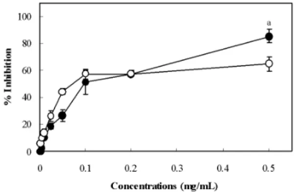
Initially, we evaluated the antioxidant activity of PD extract on FeCl2-ascorbic acid-induced lipid peroxidation of rat liver homogenate. Fig. 1 shows the antioxidant effect of PD extract compared with α-tocopherol, used as a control antioxidants.
In the FeCl2-ascorbic system, PD extract and α -tocopherol showed a dose-dependent inhibition of lipid peroxidation. At the 0.5 mg/mL concentration, the percentage inhibition of PD extract and α -tocopherol on lipid peroxidation was 85.5% (
p
<0.01) and 65.1%, respectively. However, the PD extract (IC50 = 0.113 ± 0.011 mg/mL) and α -tocopherol (IC50 = 0.113 ± 0.003 mg/mL) exhibited almost equal IC50values under the same conditions.
Fig. 1. Inhibitory effect of PD extract on FeCl
2- ascorbic acid-induced lipid peroxidation.
The concentration of the tested PD extract (● and α -tocopherol (○ ranged from 0.001 to 0.5 mg/mL. The line illustrates the percentage inhibition, and each value represents the mean ± SD of three separate experiments.
a
The level of significance for differencesbetween the experimental and α-tocopherol values was set at
p< 0.01.
2. Inhibitory effect on oxidative DNA damage
The protective effect of PD extract on hydroxyl radical-induced DNA nicking was evaluated by incubating SCpBR322 plasmid DNA in presence of H2O2 and FeCl3 for 30 min at 37 ℃ As shown in Fig. 2, addition of Fenton's reagents to the pBR322 plasmid increased the conversion of DNA from SC form to an open circular (OC) form, indicating increased DNA nicking by hydroxyl radicals (lane 2). However, PD extract (10, 25 and 50 μg) revealed a dose-dependent and prominent inhibition of DNA nicking (lanes 5, 6 and 7), and this inhibitory effect was similar to that of 5 U of catalase (lane 3) and 100 mM EDTA (lane 4), indicating their protective role against hydroxyl radical-induced DNA damage in the Fenton reaction system.
Fig. 2. Inhibitory effect of PD extract on pBR 322 plasmid DNA nicking.
Agarose gel electrophoresis showed the open circular (OC) and supercoiled (SC) forms of plasmid DNA induced by hydroxyl radicals. SC pBR 322 plasmid DNA (500 ng) was added to the Fenton’s reagent in the absence (lane 2) or presence (lanes 5-7) of PD extract (10, 25, and 50 μg) for 30 min at 37 ℃ Lane 1 shows native plasmid DNA. Lanes 3 and 4 show plasmid DNA with Fenton’s reagent containing 5 U of catalase and 100 mM EDTA, respectively.
3. Inhibitory effect on deoxyribose degradation
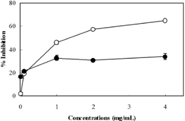
The activity of PD extract on hydroxyl radicals generated by Fenton reaction was tested using deoxyribose assay, and the results are shown in Fig. 3. In the non-site-specific and site-specific assays,
PD extract inhibited the hydroxyl radical-induced deoxyribose degradation in a concentration-dependent manner. In the site-specific assay, PD extract at a concentration of 4 mg/mL decreased the generation of hydroxyl radicals by 65.0%. On the other hand, PD extract weakly scavenged hydroxyl radicals by 33.9% in the non-site-specific assay at the same concentration.
Fig. 3. Antihydroxyl radical effect of PD extract in the deoxyribose assay system.
The non-site-specific (● and site-specific (○ activities of PD extract on hydroxyl radical formation were measured,and the results are expressed as the percentage of inhibition. The concentration of the tested PD extract ranged from 0.01 to 4.0 mg/mL. Each value represents the mean of three separate experiments.
4. Free radical scavenging activity
Fig. 4 shows the scavenging property of PD extract against DPPH radical compared to that of reference antioxidants. In the DPPH radical scavenging assay, ascorbic acid exhibited the highest activity (IC50 = 5.4 ± 0.4 μg/mL). On the other hand, PD extract showed good scavenging activity (IC50 = 20.4 ± 3.0 μg/mL) at a level slightly lower than that of BHT (IC50 = 16.5 ± 2.4 μg/mL). In the superoxide scavenging test(Fig. 5), PD extract showed the highest activity (IC50 = 6.0 ± 0.6 μ
g/mL), followed by EGCG (IC50 = 14.1 ± 1.3 μ g/mL), and caffeic acid (IC50 = 18.3 ± 2.0 μg/mL).
At concentrations of 10 - 100 mg/mL, differences between PD extract and reference compounds were statistically significant (
p
< 0.01). Table 1 shows the scavenging activities of PD extract on RNS, including nitric oxide and peroxynitrite. The PD extract effectively scavenged nitric oxide (IC50 = 5.7 ± 1.3 μg/mL), but the scavenging effect was lower than that of the well known antioxidant, resveratrol (IC50 = 1.7 ± 0.1 μg/mL). Moreover, the peroxynitrite scavenging capacity of PD extract (IC50 = 3.0 ± 0.4 μg/mL) was comparable to that of the peroxynitrite scavenger, penicillamine (IC50= 1.3 ± 0.1 μg/mL).
Fig. 4. Scavenging activity of PD extract on DPPH radical.
The PD extract (●, ascorbic acid (○ and BHT (◊
were incubated with 0.1 mM DPPH in ethanol for 1 h at room temperature. The absorbance of each sample was measured at 517 nm. The radical scavenging activity was expressed by the percentage inhibition. Each value represents the mean ± SD of three separate experiments.
Different letters above the line indicate significant difference
(
p< 0.05).
aPD extract vs. ascorbic acid.
bPD extract
vs. BHT.
Fig. 5. Scavenging activity of PD extract on superoxide anions.
Superoxide anions generated by hypoxanthine-xanthine oxidase system, and the scavenging activity of PD extract (●, caffeic acid (○ and EGCG (◊, were measured by monitoring the NBT reduction. The radical scavenging activity was expressed by the percentage inhibition. Each value represents the mean of three separate experiments.
Different letters above the line indicate significant difference (
p< 0.01).
aPD extract vs. caffeic acid.
bPD extract vs.
EGCG.
Test sample IC50 (μg/mL) Nitric Oxide Peroxynitrite PD extract 5.7 ± 1.3 3.0 ± 0.4
Resveratrol* 1.7 ± 0.1 ―
Penicillamine* ― 1.3 ± 0.1
The results are expressed as IC
50values, and each value represents the mean ± SD of three separate experiments.
*Used as a positive control.
Table 1. Scavenging Activity of PD Extract on Nitric Oxide and Peroxynitrite
5. Inhibitory effect on human LDL oxidation
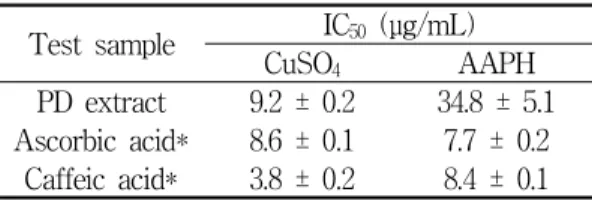
Table 2 shows the inhibitory effect of PD extract and commercial reference antioxidants on human LDL oxidation. In the copper-mediated LDL oxidation model system, caffeic acid (IC50 = 3.8 ± 0.2 μg/mL) revealed the highest antioxidant property of all the test samples, and PD extract showed a similar activity (IC50 = 9.2 ± 0.2 μg/mL) to that of ascorbic acid (IC50 = 8.6 ± 0.1μg/mL). In the
AAPH-mediated LDL oxidation model system, PD extract exhibited a relatively low antioxidant ability (IC50 = 34.8 ± 5.1 μg/mL) compared to that of ascorbic acid (IC50 = 7.7 ± 0.2 μg/mL) and caffeic acid (IC50 = 8.4 ± 0.1 μg/mL).
Test sample IC50 (μg/mL)
CuSO4 AAPH
PD extract 9.2 ± 0.2 34.8 ± 5.1 Ascorbic acid* 8.6 ± 0.1 7.7 ± 0.2 Caffeic acid* 3.8 ± 0.2 8.4 ± 0.1
The level of LDL oxidation was measured by TBARS assay. The results are expressed as IC
50values, and each value represents the mean ± SD of three separate experiments. *Used as a positive control.
Table 2. Effect of PD Extract on Copper- and AAPH-Mediated LDL Oxidation
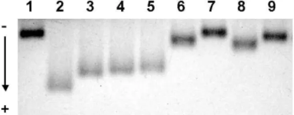
6. Effect on REM of oxidized human LDL
Fig. 6 shows the inhibitory effect of PD extract on REM of oxidized LDL. Incubation with 10 μM CuSO4 alone enhanced REM of LDL (lane 2), compared to that of native LDL (lane 1). In contrast, incubation in 10 μM EDTA (lane 7) and 10 μg/mL of BHT (lane 9) protected the copper-mediated LDL oxidation by 100% and 94.4%, respectively. At a concentration of 25 μg/mL, the percentage inhibition of PD extract (lane 6) was 83.3%, which was greater than that of 20 μ g/mL of ascorbic acid (72.2%, lane 8).
Fig. 6. Effect of PD extract on the relative electrophoretic mobility of LDL.
Human LDL (120 μg protein/mL) was added to PD extract and reference antioxidants including EDTA, ascorbic acid, and BHT. The LDL oxidation was initiated by adding 10 μM CuSO
4. After incubation for 12 h at 37
℃ about 3 μg of LDL protein was loaded on 0.7% agarose gel for 1 h, and the gelwas stained with Coomassie brilliant blue R-250. Lane 1, native LDL; lane 2, oxidized LDL; lanes 3-6, PD extract (10, 15, 20 and 25 μ g/mL, respectively); lane 7, EDTA (10 μM); lane 8, ascorbic acid (20 μg/mL); and lane 9, BHT (10 μ g/mL).
7. Content of total phenolics
The content of total phenolics of PD extract was determined by the Folin-Ciocalteu method using gallic acid as the standard, and compared with that of the dry plant material. As shown in Table 3, the amount of total phenolic compounds of PD extract was 132.0 ± 7.0 mg/g, which was about twice that of the dry plant material (61.4 ± 18.4 mg/g).
Test sample Total Phenolics (mg/g) Dry Plant Material 61.4 ± 18.4
PD Extract 132.0 ± 7.0
Amount of total phenolics was determined by the Folin -Ciocalteu method, and expressed as gallic acid equivalents per g of extract and dry plant material. Values are mean ± SD of three different experiments in triplicate.
Table 3. Total Phenolic Contents in PD Extract and Dry Plant Material
Ⅳ. Discussion
Generally, the hydroxyl radical is well known to be the most reactive free radical, and its reactions play a critical role in oxidative damage via the formation of the peroxy radical13,14). To evaluate the scavenging property of PD extract on the hydroxyl radical in the present study, we used the FeCl2-ascorbic acid-induced lipid peroxidation system.
In this assay system, ascorbic acid can exert a pro-oxidant action, thereby generating hydroxyl radicals which play a key role in initiating the lipid peroxidation16). In rat liver homogenate, the FeCl2- ascorbic acid-induced lipid peroxidation was markedly inhibited by PD extract, and this inhibitory effect was similar to that of α-tocopherol(Fig. 1). To further test the effect of PD extract on hydroxyl radical, we used the DNA nicking assay system.
The hydroxyl radicals generated by the Fenton reaction have been reported to stimulate DNA nicking23), and this oxidative DNA damage was inhibited by EDTA and catalase, which chelates iron ions and inactivates H2O224). In this assay, the hydroxyl radical enhanced the DNA oxidative damage. However, PD extract markedly inhibited hydroxyl radical-induced DNA nicking, and this inhibitory activity was similar to that of EDTA and catalase(Fig. 2).
To confirm the capacity of PD extract to remove hydroxyl radicals in both site-specific and non-site -specific cases, we used the deoxyribose assay system. The site-specific assay exhibited a relatively higher inhibitory effect on hydroxyl radicals than the non-site-specific assay. This result suggested that inhibitory action of PD extract on hydroxyl radical-induced deoxyribose degradation may be related to the chelacapacity of the metal ions
rather than the direct removal effect of hydroxyl radicals(Fig. 3). Antioxidants transform the purple colored DPPH radical (DPPH·) into a colorless compound (DPPH-H) by donating hydrogen. To evaluate the direct scavenging activity of PD extract on free radical, we used the DPPH radical scavenging assay. The PD extract revealed a potent free radicals scavenging capacity, which was similar to that of ascorbic acid(Fig. 4). These results indicated that PD extract has an excellent hydrogen donating action. Superoxide anion, also called the superoxide radical, can be formed during normal metabolism, in the autoxidation of organic compounds, in phagocytosis, and in enzymatic reactions25). Moreover, superoxide anions are the precursors of hydrogen peroxide and hydroxyl radicals via the Fenton reaction26). Recently, vascular cell studies have shown that superoxide radicals generated by NAD(P)H oxidase and xanthine oxidase play a critical role in the early stages of atherosclerosis27,28,29). In particular, xanthine oxidase generates superoxide anions by catalyzing hypoxanthine to uric acid, and these superoxide radicals convert NBT to diformazan30). To test the scavenging activity of PD extract on superoxide anions, we used a hypoxanthine -xanthine oxidase system and measured the level of superoxide anion-induced NBT reduction(Fig. 5).
In this assay, the PD extract displayed significant (
p
< 0.01) scavenging activity in the superoxide radical generating system, suggesting that this extract may be useful in preventing oxidative damage by superoxide radicals. These results confirmed that PD extract is an efficient scavenger of ROS, including hydroxyl radical and superoxide anions, and acts as an iron chelating agent. To further investigate the antioxidant ability of PD extract, we examined the RNS scavenging activity usingperoxynitrite and nitric oxide generating systems, respectively. The peroxynitrite is the product of the reaction between superoxide and nitric oxide in vivo. It has been reported that vascular endothelial cells produce both superoxide and nitric oxide to generate peroxynitrite within the vascular wall31), which can cause LDL modification and endothelial cell damage32). The peroxynitrite-modified LDL can be recognized and taken up by the macrophages via the scavenger receptor, leading to the formation of foam cells33,34,35), which may be a critical event in the development and progression of atherosclerosis.
Although peroxynitrite is much more reactive than superoxide and hydrogen peroxide, endogenous protectors against peroxynitrite are lacking in the biological system. In the current study, the level of DHR 123 oxidation by native peroxynitrite was measured and compared with penicillamine to determine the scavenging activity of PD extract on peroxynitrite. In this assay, the scavenging activity of PD extract on peroxynitrite was similar to that of penicillamine, which is a well known peroxynitrite scavenger. In addition, we measured the fluorescence intensity of triazolofluorescein caused by the reaction of DAF-2 with nitric oxide generated from SIN-1 in order to evaluate the scavenging property of the PD extract on nitric oxide. In this test, the PD extract displayed a strong scavenging property on nitric oxide, indicating that PD extract is a potent scavenger of RNS, including nitric oxide and peroxynitrite(Table 1).
These data suggest that the PD extract can act as a versatile natural antioxidant in the ROS and RNS generating model system, and may be useful in preventing the oxidative modification of LDL.
According to the results of ROS and RNS scavenging tests, we assessed the possible antioxidant
activity of PD extract against LDL oxidation using two different of human LDL oxidation tools:
copper- and AAPH (a peroxy free radicals generator) -mediated oxidation model systems. From the results of both tests, PD extract was more effective in inhibiting the copper-mediated LDL oxidation.
(Table 2), indicating that it may be effective to prevent the human LDL oxidation via metal ions chelating actions rather than free radical scavenging actions. To confirm the inhibitory activity of PD extract on copper-mediated LDL oxidation, we used REM assay, which is another model of LDL oxidation and has been used as a specific system to evaluate the anti-LDL oxidation effect of natural plants and herbs36). From the REM assay data, PD extract effectively suppressed the electrophoretic mobility during exposure of LDL to copper-induced oxidation(Fig. 6). According to the results of antioxidant tests on lipidperoxide, DNA damage, ROS, RNS and human LDL, the total phenolic amount in PD extract was determined by Folin -Ciocalteu assay. The antioxidant properties of natural plant materials have been reported to be correlated with their total phenolic content. As shown in Table 3, the amount of total phenolic compounds of PD pharmacopuncture solution was twice that of the dry plant material, suggesting that the potent antioxidative and antiradical capacity of PD pharmacopuncture solution was probably due to the high level of phenolic compounds.
Ⅴ. Conclusions
Our studies have confirmed the effectiveness of
Pinus densiflora
gnarl pharmacopuncture solution in preventing lipid peroxidation of rat liver homogenate and oxidative damage of supercoiled plasmid DNA.In addition,
Pinus densiflora
gnarl was also effective in scavenging on ROS and RNS, and protecting against copper- and AAPH-induced human LDL oxidation, and it has a high level of total phenolics.The present results suggest new information that
Pinus densiflora
gnarl is a potent free radical scavenger and metal ion chelator for the prevention of the oxidative diseases including atherosclerosis and myocardial infarction. Therefore, additional researches will be needed to isolate and identify the antioxidant compounds.Acknowledgements
This work was supported by the research program of Dongguk University.
References
1. Kwak CS, Moon SC, Lee MS. Antioxidant, antimutagenic, and antitumor effects of pine needles (
Pinus densiflora
). Nutrition and cancer.2006 ; 56 : 162-71.
2. Choi EM. Antinociceptive and antiinflammatory activities of pine (
Pinus densiflora
) pollen extract.Phytotherapy research. 2007 ; 21 : 471-5.
3. Hur J. Dong-eui-bo-gam. Seoul : Bupin Publishing.
2005 : 522-3.
4. Minuz P, Fava C, Lechi A. Lipid peroxidation, isoprostanes and vascular damage. Pharmacological reports. 2006 ; 58 : 57-68.
5. Dandona P, Ghanim H, Brooks DP. Antioxidant activity of carvedilol in cardiovascular disease.
Journal of hypertension. 2007 ; 25 : 731-41.
6. Trostchansky A, Batthyany C, Botti H, Radi R, Denicola A, Rubbo H. Formation of lipid-protein adducts in low-density lipoprotein by fluxes of
peroxynitrite and its inhibition by nitric oxide.
Archives of biochemistry and biophysics. 2001 ; 395 : 225-32.
7. Asmis R, Begley JG, Jelk J, Everson WV. Lipoprotein aggregation protects human monocyte-derived macrophages from OxLDL-induced cytotoxicity.
Journal of lipid research. 2005 ; 46 : 1124-32.
8. de Vries HE, Buchner B, van Berkel TJ, Kuiper J. Specific interaction of oxidized low-density lipoprotein with macrophage-derived foam cells isolated from rabbit atherosclerotic lesions.
Arteriosclerosis, thrombosis, and vascular biology.
1999 ; 19 : 638-45.
9. Tabata T, Mine S, Kawahara C, Okada Y, Tanaka Y. Monocyte chemoattractant protein-1 induces scavenger receptor expression and monocyte differentiation into foam cells. Biochemical and biophysical research communications. 2003 ; 305 : 380-5.
10. Hafeman DG, Hoekstra WG. Lipid peroxidation in vivo during vitamin E and selenium deficiency in the rat as monitored by ethane evolution.
The Journal of nutrition. 1977 ; 107 : 666-72.
11. Halliwell B. Free radicals and antioxidants: a personal view. Nutrition reviews. 1994 ; 52 : 253-65.
12. Suarna C, Wu BJ, Choy K, Mori T, Croft K, Cynshi O, Stocker R. Protective effect of vitamin E supplements on experimental atherosclerosis is modest and depends on preexisting vitamin E deficiency. Free radical biology & medicine.
2006 ; 41 : 722-30.
13. Patro BS, Bauri AK, Mishra S, Chattopadhyay S. Antioxidant activity of Myristica malabarica extracts and their constituents. Journal of agricultural and food chemistry. 2005 ; 53 : 6912-8.
14. Lee JC, Kim HR, Kim J, Jang YS. Antioxidant property of an ethanol extract of the stem of
Opuntia ficus-indica
var. Saboten. Journal of agricultural and food chemistry. 2002 ; 50 : 6490-6.15. Halliwell B, Gutteridge JM, Aruoma OI. The deoxyribose method: a simple "test-tube" assay for determination of rate constants for reactions of hydroxyl radicals. Analytical biochemistry.
1987 ; 165 : 215-9.
16. Kweon MH, Hwang HJ, Sung HC. Identification and antioxidant activity of novel chlorogenic acid derivatives from bamboo (
Phyllostachys edulis
). Journal of agricultural and food chemistry.2001 ; 49 : 4646-55.
17. Gotoh N, Niki E. Rates of interactions of superoxide with vitamin E, vitamin C and related compounds as measured by chemiluminescence.
Biochimica et biophysica acta. 1992 ; 1115 : 201-7.
18. Sutherland H, Khundkar R, Zolle O, McArdle A, Simpson AW, Jarvis JC, Salmons S. A fluorescence-based method for measuring nitric oxide in extracts of skeletal muscle. Nitric Oxide.
2001 ; 5 : 475-81.
19. Crow JP. Dichlorodihydrofluorescein and dihydrorhodamine 123 are sensitive indicators of peroxynitrite in vitro: implications for intracellular measurement of reactive nitrogen and oxygen species. Nitric Oxide. 1997 ; 1 : 145-57.
20. Xu MZ, Lee WS, Han JM, Oh HW, Park DS, Tian GR, Jeong TS, Park HY. Antioxidant and anti-inflammatory activities of N-acetyldopamine dimers from
Periostracum Cicadae
. Bioorganic& medicinal chemistry. 2006 ; 14 : 7826-34.
21. Reid VC, Mitchinson MJ. Toxicity of oxidised
low density lipoprotein towards mouse peritoneal macrophages in vitro. Atherosclerosis. 1993 ; 98 : 17-24.
22. Amakura Y, Umino Y, Tsuji S, Tonogai Y.
Influence of jam processing on the radical scavenging activity and phenolic content in berries. Journal of agricultural and food chemistry.
2000 ; 48 : 6292-7.
23. Lindqvist C, Nordstrom T. Generation of hydroxyl radicals by the antiviral compound phosphonoformic acid (foscarnet). Pharmacology & toxicology.
2001 ; 89 : 49-55.
24. Badisa VL, Latinwo LM, Odewumi CO, Ikediobi CO, Badisa RB, Ayuk-Takem LT, Nwoga J, West J. Mechanism of DNA damage by cadmium and interplay of antioxidant enzymes and agents.
Environmental toxicology. 2007 ; 22 : 144-51.
25. Nageswara RM, Aleksandr V, Marschall SR.
Oxidative stress and vascular disease. Arteriosclerosis, thrombosis, and vascular biology. 2005 ; 25 : 29-38.
26. Kujala TS, Loponen JM, Klika KD, Pihlaja K. Phenolics and betacyanins in red beetroot (Beta vulgaris) root: distribution and effect of cold storage on the content of total phenolics and three individual compound. Journal of agricultural and food chemistry. 2000 ; 48 : 5338-42.
27. Warnholtz A, Nickenig G, Schulz E, Macharzina R, Brasen JH, Skatchkov M, Heitzer T, Stasch JP, Griendling KK, Harrison DG, Bohm M, Meinertz T, Munzel T. Increased NADH-oxidase -mediated superoxide production in the early stages of atherosclerosis: evidence for involvement of the renin-angiotensin system. Circulation.
1999 ; 99 : 2027-33.
28. Wassmann S, Wassmann K, Nickenig G.
Modulation of oxidant and antioxidant enzyme expression and function in vascular cells.
Hypertension. 2004 ; 44 : 381-6.
29. Madamanchi NR, Vendrov A, Runge MS.
Oxidative stress and vascular Disease.
Arteriosclerosis, thrombosis, and vascular biology.
2005 ; 25 : 29-38.
30. Madamanchi NR, Hakim ZS, Runge MS.
Oxidative stress in atherogenesis and arterial thrombosis: the disconnect between cellular studies and clinical outcomes. Thrombosis and Haemostasis. 2004 ; 3 : 254-67.
31. Hogg N, Darley-Usmar VM, Wilson MT, Moncada S. Production of hydroxyl radicals from the simultaneous generation of superoxide and nitric oxide. The Biochemical journal.
1992 ; 281 : 419-24.
32. Steinbrecher UP, Parthasarathy S, Leake DS, Witztum JL, Steinberg D. Modification of low density lipoprotein by endothelial cells involves lipid peroxidation and degradation of low density lipoprotein phospholipids. Proceedings of the National Academy of Sciences of the United States of America. 1984 ; 81 : 3883-7.
33. Goldstein JL, Ho YK, Basu SK, Brown MS.
Binding site on macrophages mediated uptake and degradation of acetylated low density lipoprotein, producing massive cholesterol deposition. Proceedings of the National Academy of Sciences of the United States of America.
1979 ; 26 : 333-7.
34. Liu SX, Zhou M, Chen Y, Wen WY, Sun MJ.
Lipoperoxidative injury to macrophages by oxidatively modified low density lipoprotein may play an important role in foam cell formation.
Atherosclerosis. 1996 ; 121 : 55-61.
35. Holvoet P, Collen D. beta-VLDL hypercholesterolemia
relative to LDL hypercholesterolemia is associated with higher levels of oxidized lipoproteins and a more rapid progression of coronary atherosclerosis in rabbits. Arteriosclerosis, thrombosis, and vascular biology. 1997 ; 17 : 2376-82.
36. Yoon MA, Jeong TS, Park DS, Xu MZ, Oh
HW, Song KB, Lee WS, Park HY. Antioxidant effects of quinoline alkaloids and 2,4-di-tert- butylphenol isolated from