Article Info
Received October 3, 2019 Revised November 29, 2019 Accepted December 26, 2019 Corresponding Author Tae-lim Yoon
E-mail: [email protected] https://orcid.org/0000-0002-1718-2205
Key Words
Anterior fascia of neck Hyoid bone
Motion therapy Range of motion
Background: Neck pain can be caused by any structure in the neck, such as intervertebral discs, ligaments, muscles, facet joints, dura mater, and nerve roots. The hyoid bone is a struc- ture that is also related to head and neck posture, neck movement and pain, but there are no studies on hyoid deviation, neck pain, and range of motion (ROM).
Objects: The purpose of this study was to investigate the effect of fascia relaxation and mobilization of the hyoid bone on the ROM, pain, and lateral deviation of the hyoid bone.
Methods: Twenty-five patients with neck pain identified by the lateral motion test (10 males [35.13 ± 7.67 years, 172.69 ± 3.90 cm, 78.77 ± 6.96 kg] and 15 females [35.13 ± 10.05 years, 161.11 ± 4.09 cm, 52.59 ± 2.98 kg]) was chosen randomly. Baseline values for pain, neck ROM, and lateral deviation in the hyoid bone were recorded using a visual analogue scale (VAS), goniometer, and tape measure. Then, each patient was treated with hyoid fascia relaxation and mobilization, and all results were recorded after intervention. Comparison of the results before and after intervention was analyzed using paird t-test (p < 0.05).
Results: Right rotation, extension, VAS, and rotational asymmetry statistically significant dif- ferences (p < 0.05). Right rotation and extension increased ROM, rotational asymmetry ratio and VAS decreased. However, there was no significant difference in flexion, left rotation, center point (p > 0.05).
Conclusion: Fascia relaxation and hyoid mobilization could improve the ROM of cervical extension, asymmetry of the cervical rotation and neck pain.
Copyright ⓒ Korean Research Society of Physical Therapy
This is an Open Access article distributed under the terms of the Creative Commons Attribution Non-Commercial License (http://creativecommons.org/licenses/by-nc/4.0) which permits unrestricted non-commercial use, distribution, and reproduction in any medium, provided the original work is properly cited.
INTRODUCTION
Neck pain is a major issue faced by modern society [1]. The one-year prevalence of neck pain in the general population was 29% in men, 40% in women, and 54% in men in occupa- tions and 76% in women [2,3]. Neck pain can originate in any part of the neck, such as intervertebral discs, ligaments, mus- cles, facet joints, and dural, while nerve roots, tumors, traumas (such as fractures or whips), infections, inflammatory disorders (such as rheumatoid arthritis), and congenital disorders can all be potential causes; however, in most cases, it is difficult to detect the root cause of neck pain. In addition, neck pain is assumed to be a disease caused by various factors, which are work-related or non-work-related and can be divided into physical, psychosocial, and individual risk factor subgroups [1].
Among the various interventions for treating such neck pain, manual therapy is widely used. Several studies have reported that fascia-relaxation is a form of manual treatment that ap- plies low-intensity, long-time stretching to the fascia, with subjects experiencing appropriate length, pain reduction, and increased function from pain relief in pain-sensitive struc- tures, such as nerves and blood vessels [4,5]. In a study of the relationship between cervical vertebral mobilization and neck pain, cervical posteroanterior oscillatory mobilization was performed. Moreover, significant improvements in neck pain have been reported since this intervention [6]. For instance, in a study applying myofascial release (MFR) to mechanical neck pain symptoms, MFR was applied to the upper trapezius [7], and compared the effects of McKenzie movement, Kine- sio taping and MFR on anterior head posture. In the study, it
Physical Therapy Korea
PTK https://doi.org/10.12674/ptk.2020.27.1.70 pISSN: 1225-8962 eISSN: 2287-982X Phys Ther Korea. 2020;27(1):70-77
Original Article
The Effect of Fascia Relaxation and Mobilization of the Hyoid on the Range of Motion, Pain, and Deviation of the Hyoid in Neck Pain
Byung-jin Lee
1, BHSc, PT, Tae-lim Yoon
2, PhD, PT
1
Department of Physical Therapy, The Graduate School, Cheongju Uneversity,
2Department of Physical Therapy, College of Health &
Medical Science, Cheongju University, Cheongju, Korea
was applied to levator scapula, neck extensor and scapula [8].
however, most of these interventions can only be applied to the back of the neck.
The hyoid bone is a structure that is also involved in neck movement and pain, and its interventions must be addressed.
Head and neck posture controls the balance of gravity and functional movement with the tone of the head, neck, and shoulder muscles. The hyoids attached to these structures are connected to the styloid process of the temporal bone, with the temporal lobe at the top of the thyroid cartilage. The thyroid cartilage membrane is connected to the border and attached closely to the cervical spine through the cervical fascia [9-11].
The suprahyoid muscle attachment point also moves with C1 during axial rotation. Hyoid bone-related muscles are associ- ated with neck pain because they depend on the hyoid bone to determine the curvature of the cervical spine, to balance the craniovertebral joint, and to stabilize the upper cervical spine [12].
Therefore, previous studies have suggested treatment with hyoid bone. Specifically, DeStefano [13] introduced hyoid mo- bilization in Greenman’s Principles of Manual Medicine fourth edition, and fascia pain and hyoid location studies suggest that restoration of the hyoid bone and fascia pain reduction oc- cur simultaneously [14]. The sternohyoid contracts to stabilize the hyoid bone. Nevertheless, there is a lack of studies on the effect of the hyoid bone on neck pain when it is mediated by mobilization or fascia relaxation. In the neck pain, the inclu- sion of hyoid bone kinematics would be helpful in the diagno- sis and treatment of neck disorders, such as muscle strength of the cervical spine and loading of discs [15].
The purpose of this study is to investigate the effect of mo- bilization and MFR of the hyoid bone on the range of motion (ROM), pain, and lateral deviation of the hyoid bone in pa- tients with neck pain with lateral deviation of the hyoid bone.
This study hypothesized that applying fascia relaxation and mobilization of the hyoid bone in patients with neck pain from lateral deviation of the hyoid bone would improve their ROM, pain, and lateral deviation of the hyoid bone.
MATERIALS AND METHODS
1. Subjects
The subject sample size was based on a pilot study of five subjects. Comparison of left and right rotation before and after
intervention by G*Power was estimated using an effect size of 1.60, a power of 0.8, and a significance level of α = 0.05. The actual power was 0.96, and the required sample size was esti- mated to be 20. This study used a random sampling of patients who visited a rehabilitation medicine clinic in Cheongju. As a result, 30 subjects initially participated; however, five subjects dropped out, so 25 patients were ultimately studied.
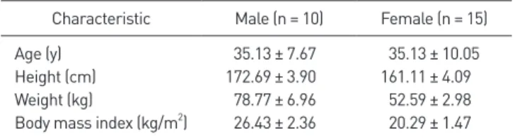
Participants were selected from Cheongju Allbarun Reha- bilitation Medicine, Korea. The inclusion criteria for this study included: 1) patients with neck pain in the rotation of the cervical spine. Patients’ pain levels were expressed in visual analogue scale (VAS) at the time of doctor’s consultation; and 2) patients with deviation in the lateral motion test. The lat- eral motion test was used as a screen test to examine patients with left and right deviation of the hyoid bone [16]. The cor- nua were palpated, and lateral motions were compared with both index fingers. If the hyoid bone moves similarly in both directions, there is no deviation, and if more motion occurs on either the right or left side, it is evaluated that the hyoid bone is displaced in the more moving direction. The exclusion criteria for this study included: 1) severe medical conditions (e.g., cancer, rheumatoid arthritis, ankylosing spondylitis, or other related autoimmune diseases); 2) symptoms of cervical myelopathy (e.g., disagreement of hands, arms and legs, active gait disturbances) and bladder incontinence (e.g., neuromus- cular sensory changes, muscle weakness, or reflexes); and 3) a history of pregnancy or postpartum, intervertebral disc-related diseases, neck pain from traumatic events or cervical spine surgery [17]. A total of 25 subjects who met the selection crite- ria and exclusion criteria (excluding 5 patients with interverte- bral disease) were selected. The general measured characteris- tics of the participants in this study were gender, age, weight, and height, as shown in Table 1. The subjects provided their voluntary consent after the purpose and method of the study was fully explained before the experiment to fully understand the contents of the experiment. All methods and procedures were approved by the Institutional Review Board (IRB) of the
Table 1. General characteristics of the subjects (N = 25)
Characteristic Male (n = 10) Female (n = 15)
Age (y) 35.13 ± 7.67 35.13 ± 10.05
Height (cm) 172.69 ± 3.90 161.11 ± 4.09
Weight (kg) 78.77 ± 6.96 52.59 ± 2.98
Body mass index (kg/m
2) 26.43 ± 2.36 20.29 ± 1.47
Values are presented as mean ± standard deviation.
Cheongju University Institutional Ethics Committee (Human Research) Committee: approval number (IRB no. 1041107- 201812-HR-032-01).
2. Procedure
Before the measurement, the subjects were accustomed to the process. The baseline measurement, flexion, extension, right rotation, and left rotation movement range of the cervical spine were recorded, and then a VAS was used to measure sub- jective neck pain in patients with cervical spine rotation. Then, the left and right deviations of the hyoid bone were recorded by measuring the center point. After baseline measurement, the intervention was performed with hyoid fascia relaxation and mobilization. Immediately after the intervention, the same therapist measured the flexion, height, right turn, left turn range and center point to record the left and right deviations of the hyoid bone. VAS was recorded as pain during rotation of the cervical spine. To determine the asymmetry of the left and right rotations, the difference before and after the intervention was summed to obtain the mean and standard deviation. The right to left rotation ratio was also calculated. Five subjects were excluded prior to pre-intervention evaluation due to cer- vical disc (cervical spine surgery) (Figure 1).
3. Outcome Measures
Hyoid bones were measured using a tape measure (total length: 150 cm) spaced 1 mm apart. In order to establish a baseline before identifying the left and right deviations in the hyoid bone, the measurer palpated the midpoint of the jaw and the jugular notch to mark the neck centerline connecting the two points and confirmed that the centerline coincided with the midline of the body. The examiner stood facing the subject and palpated a thyroid scar and the hyoid bone located 1 cm above. Both index fingers moved outward along the hy- oid body and palpated greater cornua on both sides of the hy- oid bone. After marking both greater cornua, the distance be- tween the two points was measured to indicate the midpoint.
The vertical distance from the midpoint of the hyoid bone to the centerline of the neck was measured using a tape measure (Figure 2). The right side was marked with ‘+’ and the left side was marked with ‘ –’ to record the direction and distance of the deviation (intra-rater reliability, 0.899; inter-rater reliability, 0.635).
4. Intervention 1) Mobilization
The patient 1) laid in an anatomical position, while the therapist sat to the side and fixed the head with one hand. The patient then 2) palpated the thyroid scar with the other hand, and palpated the hyoid bone, which is located 1cm above.
Next the patient 3) palpated the greater cornua on both sides of the hyoid bone with the thumb and index finger. When grabing hypoid bone, care should be taken not to disturb the breathing and compress the carotid arteries. 4) Mobilize left
ROM
VAS Center point test
ROM
VAS Center point test
Pre-measured (n = 25)
Selection of subject (lateral motion test [n = 30])
Excluded (n = 5)
Fascia relaxation and mobilization of hyoid bone (n = 25)
Post-measured (n = 25)
Analysis
Figure 1. Flow chart of patient recruitment and follow-up assessments.
VAS, visual analogue scale; ROM, range of motion.
Figure 2. Methods of measuring the center point.and right at a speed that allows you to move from the left end to the right end in one second, with a range and intensity of some resistance. Repeat 4 times for 30 seconds to relax the muscles around the hyoid bone (Figure 3) [13].
2) Myofascial relaxation
The patient 1) laid in an anatomical position, and the thera- pist sat on the side and held the head in one hand. The patient then 2) palpated the thyroid cartilage notch with the other hand, and palpated the hyoid bone 1 cm above. Next, the pa- tient 3) moved outward along the hyoid body with his thumb and index finger to palpate the greater cornua on both sides of the hyoid bone. This was repeated gently 30 seconds to two minutes to change the length of the fascia (Figure 4) [18].
5. Data Analysis
The results obtained by this study were compared and ana- lyzed using IBM SPSS for Windows ver. 23.0 (IBM Corp., Ar- monk, NY, USA). The normality of the quantitative variables was assessed using the Shapiro–Wilk test, and all data was normally distributed. A paired t-test was used to compare the
values of flexion, extension, left/right rotational range, VAS, and the center point ratios of the cervical spine according to hyoid relaxation and before and after mobilization. Statistical significance was determined at the 0.05 level.
RESULTS
The results revealed, in regard to differences in the cervical spine range before and after intervention, There was a signifi- cant difference in the extension of the study subjects (p = 0.001) and right rotation (p = 0.013) but no statistically significant difference in flexion (p = 0.696) and left rotation (p = 0.354) (Table 2). Additionally, the VAS evaluation (p = 0.001) showed a significant difference, but the center point (p = 0.278) showed no statistically significant difference (Table 2). Lastly, rotational asymmetry after intervention (p = 0.001) exhibited a significant difference (Figure 5).
DISCUSSION
The purpose of this study was to investigate the effect of
Figure 4. Hyoid myofascial release.
Figure 3. Hyoid mobilization.
Table 2. ROM, pain, center point, rotational asymmetry before and after intervention (N = 25)
Variable Pre-intervention Post-intervention p-value Mean difference (95% CI)
Flexion (°) 55.76 ± 11.79 55.04 ± 11.19 0.696 0.72 (–3.04 to 4.48)
Extension (°) 57.56 ± 10.287 66.60 ± 8.793 0.001* –9.04 (–14.06 to –4.02)
Right rotation (°) 60.00 ± 12.11 64.24 ± 11.54 0.013* –4.24 (–7.51 to –0.97)
Left rotation (°) 63.64 ± 9.55 65.48 ± 10.87 0.354 –1.84 (–5.85 to 2.17)
VAS 3.96 ± 1.098 2.28 ± 1.208 0.001* 1.68 (1.16 to 2.20)
Center point 0.98 ± 1.917 0.66 ± 2.139 0.278 0.32 (–0.28 to 0.92)
Rotational Asy (°) 9.72 ± 4.971 4.44 ± 4.84 0.001* 5.28 (2.37 to 8.19)
Values are presented as mean ± standard deviation. ROM, range of motion; CI, confidence interval; VAS, visual analogue scale; Asy, asymmetry. *p < 0.05.
fascia relaxation and mobilization of the hyoid bone on neck pain, neck range and rotational asymmetry, and hyoid bone deviation in patients experiencing neck pain. After the base- line measurement, the intervention of hyoid fascia relaxation and mobilization was performed. As a result, the extension and right rotation of the cervical spine increased, and the rota- tional asymmetry and VAS of the neck decreased significantly;
however, there was no significant change in the lateral devia- tions of the hyoid bone before and after the intervention.
After intervention, the cervical extension and right rotation range increased. A study of three-dimensional (3D) electro- magnetic and motion-tracking devices (Fastrak; Polhemius, Colchester, VT, USA) found that, in patients with mechanical neck pain, there was a significant reduction in the ROM in right rotation and extension of the neck [19]. Moreover, one of the many hypotheses about right rotation and ROM reduc- tion in mechanical neck disorders patients argued that this is the result of predominant, hands-on right rotation restriction [20]. Patients with poor posture-related neck pain will also experience a reduced cervical extension range. A previous study reported that as pain increased, the neck flexion and ex- tension range decreased [21]. These previous studies indicate that subjects with neck pain experienced a general decrease in ROM, and the present study also showed that the overall increase in the cervical ROM after MFR in various studies after intervention was also significant in terms of functional out- comes. Clinical significance has been shown to produce effects [7,22]. When mobilization was applied to the cervical spine, the extension of the neck and right rotation increased; thus, performing hyoid fascia relaxation and mobilization is recom-
mended.
After the intervention of hyoid fascia relaxation and mobili- zation, VAS decreased. Several previous studies have reported that mobilization has an effect on pain relief. In previous stud- ies, pain was reduced in severe movements after joint mobi- lization [23], and in a study of patients with acute neck pain, the average pain value decreased from 10 to 2, while the most severe pain level decreased from 10 to 2.5 after mobilization [24]. In addition, there was a significant change in pain and movement disorders when MFR was applied to patients with chronic lumbar disc herniation, which was different from the present study [25]. The myofascial relaxation technique is an effective passive technique that releases the damaged areas of sliding-fascial mobility and improves pain awareness in the short-term in patients with nonspecific neck pain or low back pain (p < 0.001) [26]. MFR applied to the neck and upper limbs was effective in reducing the pain intensity of the neck [7]. Ad- ditionally, a systematic review and meta-analysis of random- ized controlled trials (RCTs), MFR application lowered pain lev- els and increased joint coverage [27]. Therefore, hyoid fascia relaxation and mobilization can be useful in reducing cervical spine pain and improving movement disorders.
There was no significant change in the lateral deviations of hyoid before and after measurement. Although neck move- ment and the role of the hyoid bone and hyoid muscle are interrelated and play an important role, there has been no study on hyoid lateral deviations after hyoid fascia relaxation and mobilization intervention. A previous study suggests that physiotherapist techniques can change the relationship be- tween the head, neck, and hyoid bone [10]. Moreover, a study of young adults showed no effect on the relationship between head posture, front and rear, and up and down position of the hyoid bone; however, in a recent study, 3D cephalometric analysis revealed that the position of the hyoid bone changed significantly from the spine to the lower jaw after treatment for myofascial pain [14]. Contrary to expectation, however, no significant changes could be identified in this study, which may be due to the short duration of the intervention of the hyoid muscles, since the actual length change was difficult.
Another possibility may be that the width of the lateral devia- tion of the hyoid bone due to hyoid fascia relaxation and mo- bilization was less than the minimal detectable change in the measurement method or due to manual measurement errors.
Therefore, although there was no statistical difference in study,
Asymmetry()
Before 25
20
15
10
5
After Intervention
0
Figure 5. Differences in rotational asymmetry before and after interven-