The Reliability of Thickness Measurement of the Deep Fiber of the Lumbar Multifidus Using Ultrasonography
Doh-heon Jung, B.H.Sc., P.T.
Su-jung Kim, M.Sc., P.T.
Dept. of Rehabilitation Therapy, The Graduate School, Yonsei University Chung-hwi Yi, Ph.D., P.T.
Heon-seock Cynn, Ph.D., P.T.
Dept. of Physical Therapy, College of Health Science, Yonsei University Institute of Health Science, Yonsei University
Houng-sik Choi, Ph.D., P.T.
Dept. of Physical Therapy, Hanseo University
Abstract
1)The reliability of the thickness measurement of the lumbar multifidus (LM) using real-time ultra- sonography (US) was determined in only the superficial fiber of the lumbar multifidus (SM). However, previous studies have not examined the reliability of the deep fiber of the LM (DM). The purpose of this study was to determine the intrarater and the interrater reliability of the thickness measurements of DM using US. Eleven healthy males participated in the study. The thickness of the DM was measured with an US in the prone position. Reliability was examined using intraclass correlation coefficients (ICC), standard error of the measurement (SEM), and the Bland and Altman plot. ICC(3,1) was used to calculate the inter- rater reliability of the thickness measurement of DM using the values from both the first and second test sessions. Additionally, ICC(3,1) was used to calculate the intrarater reliability of the measurements over two days using the measurements obtained in test session 1 and test session 2. The results of this study were as follows: 1) the ICC(3,1) value for interrater reliability was .94 in the first test session, and .93 in the second test session. 2) the ICC(3,1) values for intrarater reliability of the measurements over two days was .90 in both the first examiner and the second examiner. The intrarater reliability and interrater reli- ability of the DM measurements, obtained via the US protocol used in this research was excellent.
Therefore, we conclude that the thickness measurement of the DM obtaioned from the US protocol used in this research would be useful for clinician assessment of the thickness of the DM.
Key Words: Deep fiber of lumbar multifidus; Muscle thickness; Reliability; Ultrasound imaging.
Introduction
The lumbar multifidus (LM) is a deep paraspinal muscle that has plays an important role in lumbar sta- bility (Freeman et al, 2010; Lonnemann et al, 2008).
Though the LM consists of multiple layers (Lonnemann et al, 2008; MacDonald et al, 2006), it can be classified into two muscle fibers: the superficial fiber of the lum- bar multifidus (SM) and the deep segmental fiber of lumbar multifidus (DM) (MacDonald et al, 2006).
Biomechanical differences between the SM and the DM, based on anatomical differences, have been demonstrated (MacDonald et al, 2006). Macintosh and Bogduk (1986) also described that the SM crosses the multiple spinal level between the second and the fifth vertebrae, where- as the DM crosses only two spinal levels. Accordingly, the SM extends and rotates the lumbar spine with erec- tor spinae; however, the DM stabilizes the lumbar spine (MacDonald et al, 2006). McGill (1991) defined that DM length does not changes during spinal motion, and DM Corresponding author: Houng-sik Choi [email protected]
Characteristics Mean±SD
Age (yrs) 25.9±4.7
Height (㎝) 174.5±3.2
Weight (㎏) 72.8±10.9
Table 1. General characteristics of subjects (N=11) has a greater percentage of slow twitch (Type I) muscle
fiber than does the SM, consequently contributing to lumbar stability (Sirca and Kostevc, 1985).
The muscle activity of the LM has been detected by fine wire electromyography, and its muscle size has been measured by computerized tomography, magnetic resonance imaging, and ultrasonography (US) (Danneels et al, 2000; Freeman et al, 2010; Vasseljen et al, 2006). Compared to other diagnostic equipments, real-time ultrasound imaging is noninvasive and cost-effective. Many researchers and physical thera- pists have used ultrasound imaging of the lumbar multifidus for clinical assessment, research and visual biofeedback (Hides et al, 1998; Koppenhaver et al, 2009b; Stokes et al, 2005; Van et al, 2006).
Several studies assessed the reliability of ultra- sound imaging measurement of the LM using intra- class correlation coefficients (ICC) (Hides et al, 1995;
Koppenhaver et al, 2009a; Pressler et al, 2006;
Strokes et al, 2005; Wallwork et al, 2007). The reli- ability of cross-sectional area (CSA) measurement of the LM was high (ICC=.72~1.00) (Hides et al, 1995;
Pressler et al, 2006; Strokes et al, 2005). However, in the findings of previous studies, the CSA measure- ments using US, were conducted on the entire thick- ness of the LM, without differentiating the SM from the DM. Recently, studies measured the SM thickness from the distance between the zygapophyseal joint (L2/3-L4/5) and the superior border of the LM (Koppenhaver et al, 2009a; Wallwork et al, 2007). The measurement of muscle thickness was also highly re- liable showing an ICC range from .87 to .97. Though these recent studies on the SM thickness measure- ment provided valuable information, the thickness measurement of the DM, which represents the deep stabilizer of lumbar region as compared to the SM, has not been measured using real-time US.
Therefore, the purpose of this study was to exam- ine the intrarater and interrater reliability of the DM thickness measurement in the parasagittal view of ultrasound imaging.
Methods
Participants
Eleven healthy male volunteers were recruited for this study. All subjects were free from low back pain, previous lumbar injury or surgery, spinal de- formity, or neuromuscular or joint diseases in the lumbar and lower extremities for six months prior to enrollment in this study. Subjects who had partici- pated in any regular training programs involving the back muscles within the previous three months were excluded. Table 1 summarizes the mean age, height, and weight of the subjects.
Examiners
Two physical therapists separately assessed the thickness of the DM separately. Before testing, to re- duce measurement error, both examiners in the present research underwent a total of 14 hours of hands-on training according to recommended protocol including precise location of anatomical landmarks, probe place- ment and pressure application, and the cursor marking point (Stokes et al, 2005).
Procedures2)
Because muscle contractions during measurements influenced muscle thickness (Wallwork et al, 2007), to avoid this confounding influence, the DM thickness was measured in a relaxed state of the muscle of healthy male subjects (Koppenhaver et al, 2009b). A real-time ultrasound image of the DM was generated at 25 ㎐ computerized US,1) with a 50 ㎜ 7.5-㎒ liner array probe. The subjects were placed in a prone po- sition on an examination plinth. An inclinometer was
1) SonoAce X4, Medison Co Ltd, Seoul, South Korea.
Figure 1. Image of the lumbar multifidus.
(L4/5:4/5 zygapophyseal joint, L5/S1:
L5/S1 zygapophyseal joint).
Figure 2. A thickness measure of the DM (L4/5: L4/5 zygapophyseal joint, L5/S1:
L5/S1 zygapophyseal joint).
used over the lumbo-sacral junction and a pillow was placed to flatten the lumbar curve to less than 10°
(Kiesel et al, 2007). The examiner palpated the L4, L5 spinous processes, and then marked a dot over the L4 spinous process. The probe was placed longitudinally over the lumbar spine with the mid-point over the L4 spinous process for the thickness of the DM. It was moved laterally and angled slightly medially until the L4/L5 zygapophyseal joint could be found (Kiesel et al, 2007; Richardson et al, 2004). The scan point is directly over the multifidus during the relaxed state of the muscle (Figure 1). An on-screen caliper was used to obtain a thickness of the DM between the L4/5 and the L5/S1 zygapophyseal joint. The linear distance of the DM was measured from the lowest margin of lamina to the inferior boarder of the SM (Figure 2).
This measurement protocol is based on the suggestion by Richardson (2004) who indicated that DM is ar- ranged between two zygapophyseal joints in the par- asagittal view of an ultrasound image.
Examiner 1 removed the previously test setting and then left the room. The subject was instructed to rest for 10 minutes. Examiner 2 entered the room and re- peated the same procedure for the DM measurement to finish test session 1. On the next day, test session 2 was conducted. The subjects were retested by both examiners using the same measurement protocol uti- lized in test session 1.
Statistical Analysis
For the interrater reliability of the DM thickness measurement with an US, the ICC(3,1) was calculated in both test session 1 and test session 2. The intrarater reliability, ICC(3,1) covering two days was calculated by each examiner using the measurements obtained in test session 1 and test session 2. Additionally, the Bland and Altman plot was used to provide a visual representation of the degree of agreement.
Each ICC was interpreted according to the following modification of the criteria proposed by Portney and Watkins (2000): ICC<.50, poor; ICC=.50~.75, moderate;
ICC .75<r<.90, good; and ICC>.90, excellent.
Furthermore, to examine the consistency of the meas- urement, the standard error of the measurement (SEM) was calculated [SEM=standard deviation×(1-ICC)1/2].
Results
Descriptive statistics of the measurements of the DM are shown in Table 2. Interrater reliability was excellent;
[ICC(3,1)=.93~.94; 95% CI (test session 1)=.78~.98, 95%
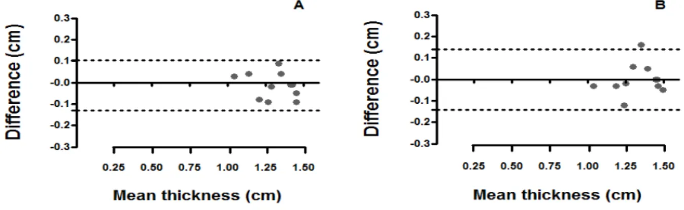
CI (test session 2)=.77~.98; p<.01]. Intrarater reliability was excellent; [ICC(3,1)=.90; 95% CI (examiner 1)=.6 8~.97, 95% CI (examiner 2)=.67~.97; p<.01]. All ICCs and SEMs are listed in Table 3. A Bland and Altman plot for reliability is shown in Figure 3, 4.
Test session 1 Test session 2
Examiner 1 Examiner 2 Examiner 1 Examiner 2
Mean (㎝) 1.300 1.329 1.315 1.330
SD .129 .154 .145 .141
Range 1.060~1.430 1.020~1.470 1.030~1.500 1.040~1.520
Table 2. Comparison of the thickness measurement of the deep fiber of lumbar multifidus between examiners (N=11)
Interrater reliability Intrarater reliability Test session 1 Test session 2 Examiner 1 Examiner 2
ICCa .94 .93 .90 .90
95% confidence interval .78~.98 .77~.98 .68~.97 .67~97
SEMb (㎝) .01 .01 .02 .02
p <.01 <.01 <.01 <.01
aIntraclass correlation coefficients, bStandard error of the measurement.
Table 3. Reliability of the thickness measurement of the deep fiber of lumbar multifidus (N=11)
Figure 3. Bland and Altman plot showing interrater reliability. A: test session 1, B: test session 2 (dashed line: mean difference A: .02, B: .01, dotted line: mean difference±2SD).
Figure 4. Bland and Altman plot showing intrarater reliability. A: examiner 1, B: examiner 2 (dashed line: mean difference A: .01, B: .0009, dotted line: mean difference±2SD).
Discussion
The purpose of this study was to establish the intrarater and the interrater reliability for the thick- ness measurement of the DM in a parasagittal view using an US. The results of this study showed ex- cellent reliability and a small SEM, indicating that an US protocol used in this research can reliably measure the thickness of DM in a resting condition.
To the best of our knowledge, this is the first study to demonstrate that an US can provide reli- able measurements of the thickness of the DM in healthy male subjects. This study has also proven that noninvasive US can be used to as an easy, simple, and cost-effective measurement for clinical for evaluation and diagnosis.
This study is novel because no previous studies re- ported the method of measuring the DM thickness or the reliability of the DM thickness measurement using an US in vivo. Even the anatomical, biomechanical, and functional importance of the DM was emphasized from the literature, and the muscle activity was meas- ured using a fine wire electrode to verify the reduced muscle activity in symptomatic low back patients (Hides, 2008) and to identify the effects of lumbar sta- bilization intervention on the CSA and the thickness of the LM as a whole (Koppenhaver, 2009b). In recent studies, only the SM thickness was measured; how- ever, the DM thickness was not determined (Freeman, 2010; Koppenhaver, 2009b).
However, in this study, the DM thickness measure- ment was conducted by measuring the linear distance from the lowest margin of the lamina to the inferior boarder of the SM between the L4/5 and the L5/S1 zygapophyseal joint; this is based on the suggestion by Richardson (2004) who indicated the DM is arranged between two zygapophyseal joints in the parasagittal view of the ultrasound image. It was found that the LM was separated by 3~4 distinct layers, morphologi- cally and anatomically (Lonnemann et al, 2008).
However, standardized guidelines that identify the DM
from these layers were not suggested. Therefore, the authors of this study suggest that the DM thickness measurement method, using an US, can be considered as an alternative method, substituting invasive fine wire electromyography.
Furthermore, using this DM measurement proto- col, the effect of the lumbar stabilization exercise, especially for deep segmental lumbar stabilization can be examined. Given that recent studies have noted that the CSA of the LM show differences between males and females, and healthy subjects and chronic LBP based on age (Hides et al, 2008;
Stokes et al, 2005), it is recommended that the ef- fects of exercise should be found in the local DM, compared with the SM, using the DM thickness measurement protocol used in this study.
There are limitations in this study. First, the DM thickness measurement was conducted only at a resting condition, thus the degree of DM thickness change during muscle contraction cannot be identified in the present study. Second, this study recruited only eleven subjects, so reliability studies with a large sample group need to be conducted. Third, even though excellent reliability was established for the DM thickness measurement using an US in this study, further study is needed to obtain validity of the DM thickness using a gold standard.
Conclusion
The purpose of this study was to compare the in- trarater and the interrater reliability for the thickness measurement of the DM using US in a parasagittal section. Intrarater reliability of the DM measurements over two days was excellent. Additionally, the inter- rater reliability of the DM measurements was also excellent. Therefore, we conclude that the thickness measurement of the DM, using US protocol in this research, was useful for clinical diagnosis as well as clinical evaluation.
This article was received October 1, 2010, and was accepted November 5, 2010.
References
Danneels LA, Vanderstraeten GG, Cambier DC, et al.
CT imaging of trunk muscles in chronic low back pain patients and healthy control subjects.
Eur Spine J. 2000;9(4):266-272.
Freeman MD, Woodham MA, Woodham AW. The role of the lumbar multifidus in chronic low back pain: A review. PM R. 2010;2(2):142-146.
Hides JA, Richardson CA, Jull GA. Magnetic reso- nance imaging and ultrasonography of the lum- bar multifidus muscle. Comparison of two dif- ferent modalities. Spine. 1995;20(1):54-58.
Hides JA, Richardson CA, Jull GA. Use of real-time ultrasound imaging for feedback in rehabilitation.
Man Ther. 1998;3(3):125-131.
Hides J, Gilmore C, Stanton W, et al. Multifidus size and symmetry among chronic LBP and healthy asymptomatic subjects. Man Ther. 2008;13(1):43-49.
Kiesel KB, Uhl TL, Underwood FB, et al.
Measurement of lumbar multifidus muscle con- traction with rehabilitative ultrasound imaging.
Man Ther. 2007;12(2):161-166.
Koppenhaver SL, Hebert JJ, Fritz JM, et al. Reliability of rehabilitative ultrasound imaging of the trans- versus abdominis and lumbar multifidus muscles.
Arch Phys Med Rehabil. 2009a;90(1):87-94.
Koppenhaver SL, Hebert JJ, Parent EC, et al.
Rehabilitative ultrasound imaging is a valid measure of trunk muscle size and activation during most isometric sub-maximal contractions:
A systematic review. Aust J Physiother.
2009b;55(3):153-169.
Lonnemann ME, Paris SV, Gorniak GC. A morpho- logical comparison of the human lumbar multi- fidus by chemical dissection. J Man Manip Ther. 2008;16(4):E84-E92.
MacDonald DA, Moseley GL, Hodges PW. The lum- bar multifidus: Does the evidence support clin- ical beliefs? Man Ther. 2006;11(4):254-263.
Macintosh JE, Bogduk N. The biomechanics of the lumbar multifidus. Clin Biomech.
1986;1(4):205-213.
McGill SM. Kinetic potential of the lumbar trunk mus- culature about three orthogonal orthopaedic axes in extreme postures. Spine. 1991;16(7):809-815.
Portney LG, Watkins MP. Foundations of Clinical Research: Applications to practice. 2nd ed.
Upper Saddle River, NJ, Prentice Hall Inc., 2000.
Pressler JF, Heiss DG, Buford JA, et al.
Between-day repeatability and symmetry of multifidus cross-sectional area measured using ultrasound imaging. J Orthop Sports Phys Ther.
2006;36(1):10-18.
Richardson CA, Hodges PW, Hides JA. Therapeutic Exercise for Lumbopelvic Stabilization: A motor control approach for the treatment and pre- vention of low back pain. 2nd ed. Philadelphia, PA, Churchill Livingstone, 2004.
Sirca A, Kostevc V. The fibre type composition of thoracic and lumbar paravertebral muscles in man. J Anat. 1985;141:131-137.
Stokes M, Rankin G, Newham DJ. Ultrasound imaging of lumbar multifidus muscle: Normal reference ranges for measurements and practical guidance on the technique. Man Ther. 2005;10(2):116-126.
Van K, Hides JA, Richardson CA. The use of real-time ultrasound imaging for biofeedback of lumbar multifidus muscle contraction in healthy subjects. J Orthop Sports Phys Ther. 2006;36(12):920-925.
Vasseljen O, Dahl HH, Mork PJ, et al. Muscle activ- ity onset in the lumbar multifidus muscle re- corded simultaneously by ultrasound imaging and intramuscular electromyography. Clin Biomech.
2006;21(9):905-913.
Wallwork TL, Hides JA, Stanton WR. Intrarater and interrater reliability of assessment of lumbar multifidus muscle thickness using rehabilitative ultrasound imaging. J Orthop Sports Phys Ther.
2007;37(10):608-612.