Comparison of Clinical and Structural Outcomes of Open and Arthroscopic Repair for Massive Rotator Cuff Tear
Nam Su Cho, Sang Won Cha, Hee Seok Shim, Hyung Suk Juh, Yong Girl Rhee
Shoulder & Elbow Clinic, Department of Orthopaedic Surgery, College of Medicine, Kyung Hee University, Seoul, Korea
Background: Management of massive rotator cuff tears can be challenging because of the less satisfactory results and a higher retear rate regardless of the use of open or arthroscopic repair technique.
Methods: We retrospectively analyzed 102 cases of massive rotator cuff tear treated with either open or arthroscopic repair. Open repair was performed in 38 patients; and arthroscopic repair, in 64 patients. The mean age at the time of surgery was 59.7 years in the open group and 57.6 years in the arthroscopic group.
Results: The Constant score increased from the preoperative mean of 55.9 to 73.2 at the last follow-up in the open repair group and from 53.8 to 67.6 in the arthroscopic repair group (p<0.001 and <0.001, respectively). The University of California at Los Angeles (UCLA) score increased from a preoperative mean of 17.7 to 30.8 at the last follow-up in the open group and from 17.5 to 28.7 in the arthroscopic group (p<0.001 and <0.001, respectively). No statistically significant difference in the Constant and UCLA scores was ob- served between the two groups at the last follow-up (p=0.128 and 0.087, respectively). Retear was found in 14 patients (36.8%) in the open group and 39 patients (60.9%) in the arthroscopic group (p=0.024).
Conclusions: Open and arthroscopic repairs of massive rotator cuff tears may provide satisfactory clinical results with no significant dif- ference. However, a significantly lower retear rate was observed for the open repair group compared with the arthroscopic repair group.
(Clin Shoulder Elbow 2016;19(2):60-66)
Key Words: Shoulder; Rotator cuff; Tendon injuries; Massive; Open; Arthroscopy
Clinics in Shoulder and Elbow Clinics in Shoulder and Elbow Vol. 19, No. 2, June, 2016
http://dx.doi.org/10.5397/cise.2016.19.2.60
Received May 4, 2015. Revised July 15, 2015. Accepted August 16, 2015.
Correspondence to: Yong Girl Rhee
Department of Orthopaedic Surgery, College of Medicine, Kyung Hee University, 23 Kyungheedae-ro, Dongdaemun-gu, Seoul 02774, Korea Tel: +82-2-958-8370, Fax: +82-2-964-3865, E-mail: [email protected]
Financial support: This research was supported by the Sports Scientification of Convergent R&D Program through the National Research Foundation of Korea (NRF) funded by the Ministry of Science, ICT & Future Planning (NRF-2014M3C1B1033319). Conflict of interests: None.
Introduction
Recently, arthroscopic repair has been widely accepted with evolving instrumental development and wide surgical experi- ence for treatment of rotator cuff tears.1,2) Most symptomatic large to massive rotator cuff tears can be managed successfully using an arthroscopic approach and some favorable outcomes have been reported.1-3)
Many studies1-3) have reported good clinical outcomes of ar- throscopic repair for large to massive rotator cuff tears; however retear of the repaired tendon remains a significant clinical issue.
Previous studies have reported retear rates ranging from 47%
to 94% after arthroscopic repair of large to massive rotator cuff
tears at 1- and 2-year follow-up.4,5) Although not synonymous with clinical failure, a retear is associated with poorer clinical outcomes than repairs that achieve structural healing. Many studies have reported that a healed rotator cuff resulted in a su- perior clinical outcome.5-7)
However, tendon retraction, adhesions, and poor tissue qual- ity, which are common in large to massive rotator cuff tears, make repair one of the most technically complex procedures in the shoulder.8) In cases involving less mobile and severely retracted large to massive tears, complete repair of a torn cuff to the native footprint may be difficult using only arthroscopic technique. Arthroscopic repair of severely retracted large to mas- sive rotator cuff tears has a less satisfactory result with a high rate
of retears.9,10) Therefore, open technique can be more effective for these kinds of tears. Some surgeons prefer an open approach for management of severely retracted large and massive tears.
However, relatively few studies have evaluated the clinical and structural outcomes of open and arthroscopic repairs for massive rotator cuff tears.
The purpose of this study was to evaluate and compare the clinical and structural outcomes of open and arthroscopic re- pairs for massive rotator cuff tears and to correlate the clinical outcomes of the respective repair techniques with respect to rotator cuff integrity. We hypothesized that there would be no significant differences in postoperative clinical outcomes and in the retear rate between the open and arthroscopic repair group.
Methods
This study was retrospective in nature and final approval of exemption by the Institutional Review Board was obtained (IRB Approval No.: KMC IRB 1404-04).
Patient Selection
A total of 102 patients (102 shoulders) who underwent sur- gical repair of a massive rotator cuff tear and routine follow- up magnetic resonance imaging (MRI) at least 6 months after surgery from February 2006 to January 2010 were enrolled in this study. Patients with partial or small, medium, and large- sized rotator cuff tears, acromioclavicular arthritis requiring distal clavicle resection, advanced glenohumeral arthritis, or rotator cuff tears and a worker’s compensation claim were excluded from the study. Patients who had undergone revision procedures were also excluded. According to the repair methods, 38 shoul- ders were treated using the open technique; and 64 shoulders, using the arthroscopic technique. The mean age of patients at the time of the operation was 59.7 years (range, 44–79 years) in the open repair group and 57.6 years (range, 40–75 years) in the arthroscopic repair group. The mean postoperative follow-
up periods were 26.2 months (range, 18–40 months) and 28.1 months (range, 19–38 months), respectively. According to the classification of DeOrio and Cofield,11) the extent of the tear was determined intraoperatively under direct arthroscopic visualiza- tion after debridement of the degenerated tendon edges. The tear size was measured in the anteroposterior dimension using a calibrated probe introduced through the posterior portal while viewing from the lateral portal. No significant differences in de- mographic data were found between the two groups (Table 1).
Preoperative and Postoperative Evaluations
We performed a retrospective analysis of the prospectively collected patient data. All patients underwent a physical exami- nation 1 day before the operation. Postoperative evaluation was performed regularly on an outpatient basis (at 3 weeks, 6 weeks, 3 months, 6 months, 9 months, and 12 months postoperatively and at the last follow-up visit), and the results of the last follow- up examination were analyzed. Preoperative and postoperative subjective pain scores were measured using the visual analog scale (VAS). For shoulder range of motion (ROM), forward flex- ion, external rotation at the side, and internal rotation to the posterior were assessed before and after the operation. Quan- titative measurement of muscle strength of the rotator cuff was performed using a portable, handheld Nottingham Mecmesin Myometer (Mecmesin Co., Nottingham, UK). Elevation strength was tested with the patient in the seated position, with the arm flexed to 90o in the scapular plane. External and internal rota- tions were tested with the shoulder in a neutral position and the elbow in 90o flexion. The Constant score12) and shoulder rating scale of the University of California at Los Angeles (UCLA)13) were used for clinical assessment.
Operative Techniques
Open repair tended to be performed in more severe cases with less mobile and severely retracted massive tears where a sufficient repair is difficult using arthroscopic technique. In some cases, arthroscopic repair was attempted, and then abandoned for an open approach. And, in some cases, we proceeded di- rectly with open surgery without an attempt at arthroscopic re- pair based on preoperative radiologic findings.
1) Open repair
All procedures were performed by the senior author with the patient in a 30o beach chair position under general anesthesia. A superolateral approach with a 6 to 8 cm incision extended from just lateral to the coracoid process over the anterolateral corner of the acromion was used for the open procedures. Once the raphe which demarcates the anatomic division between anterior and middle deltoid was identified, the muscle was split between its anterior and lateral portions, parallel to its muscle fibers. Blunt dissectors were then used to separate the muscle fibers parallel to their orientation beginning at the anterolateral corner of the Table 1. Patient Demographics
Variable Open repair
group Arthroscopic repair group
No. of patient 38 64
Sex (male/female) 23/15 38/26
Right/left 25/13 33/31
Mean age (yr) 59.7 (44–79) 57.6 (40–75)
Dominant/nondominant 24/14 36/28
Mean follow-up period (mo) 26.2 (18–40) 28.1 (19–38) Mean postoperative MRI time (mo) 6.4 (6–12) 7.1 (6–12) Values are presented as number only or median (range).
MRI: magnetic resonance imaging.
acromion and extending the muscle-splitting incision inferiorly for approximately 5 cm. Once adequate exposure had been obtained, the size and location of the rotator cuff tear and the degree of degeneration of the biceps tendon were evaluated, which was followed by a subacromial decompression with or without acromioplasty, as indicated. Traction sutures were ap- plied to the rotator cuff edges to improve visualization of the tear. After debridement of the footprint, the tear was repaired by Mason-Allen sutures to the greater tuberosity using transosseous tunnels. Tenotomy or tenodesis was performed in cases where pathology of the long head of the biceps tendon was detected.
Mason-Allen sutures with No. 2 Ethibond sutures (Ethicon, Somer- ville, NJ, USA) were used to repair the elevated anterior deltoid muscle to the anterior acromion through bone tunnels. The split in the deltoid was sutured using No. 1 Vicryl sutures (Ethicon).
2) Arthroscopic repair
All operations were performed by the same surgeon who performed the open repairs with the patient in a beach chair po- sition with the back of the bed flexed to 70o. A posterior viewing portal and an anterior working portal were used in assessment of the glenohumeral joint. After performance of diagnostic arthros- copy through the posterior portal, the arthroscope was inserted through the posterior portal to the subacromial space, and a lat- eral portal was created. After performance of subacromial bur- sectomy through this portal, the pattern of the rotator cuff tear in the subacromial space was observed. If severe fibrillation was observed inferior to the acromion, except when the acromion was seen as being flat on the preoperative radiographs, the pa- tient was young, or the rotator cuff tear was caused by a definite trauma, acromioplasty was performed based on the plain radio- graphs and arthroscopic findings. For subacromial viewing and rotator cuff repair, addition of a portal providing a Grand Can- yon view14) near the posterolateral corner of the acromion was established. Using the Banana Suture Lasso (Arthrex, Naples, FL, USA) through the ‘Three Sister’ portals (anterior subclavian portal, modified Neviaser portal, and posterior infraspinous por- tal),15) one end of each fiber wire pair was shuttled through the torn cuff edge, and standard arthroscopic rotator cuff repair was performed.
Postoperative Rehabilitation
All patients followed a standard postoperative rehabilitation program. From the day of the operation, passive exercises in- cluding pendulum exercise, passive forward flexion, and exter- nal rotation exercises were performed in a tolerable range. Ac- tive exercises were not allowed until 6 weeks postoperatively or until regaining full passive ROM. Active-assisted exercises were started at 6 weeks postoperatively and muscle strengthening exercises were gradually introduced thereafter. Return to heavy demand activity or manual labor was delayed until 6 months.
Assessment of Tendon Healing
For assessment of tendon healing, anatomical evaluation of the cuff repair was performed using MRI as the imaging modal- ity, as it provides multiplanar imaging of the postoperative shoul- der. All 102 patients underwent routine MRI at least 6 months after surgery for assessment of tendon integrity. All studies were obtained using a 1.5-T scanner (Signa; GE Medical Systems, Mil- waukee, WI, USA) using routine pulse sequences. The images were reviewed by an experienced senior radiologist who was informed that the patients had undergone surgery for rotator cuff repair but was blinded to the size and location of the tear that had been repaired. Continuity and retear of the tendon were as- sessed on MRIs according to established criteria.16) A diagnosis of full-thickness retear (i.e., anatomic failure of healing) was made when a fluid-equivalent signal was found or when the supraspi- natus, infraspinatus, or subscapularis tendon was not visible on at least one T2- or proton density-weighted image,.
Statistical Analysis
An independent t-test was used for comparison of the UCLA, Constant, and VAS scores and the deltoid muscle thickness be- tween the open and arthroscopic repair groups. A paired t-test was used for comparison of the preoperative and postopera- tive UCLA, Constant, and VAS scores between the two groups.
Significance was set at α=0.05, with 95% confidence intervals.
The statistical package for the social sciences (SPSS) ver. 17.0 software package (SPSS Inc., Chicago, IL, USA) was used for sta- tistical analyses.
Results
PainIn the open repair group the subjective VAS at rest decreased from the preoperative mean of 1.8 ± 0.7 to 0.2 ± 0.1 at the last follow-up examination (p<0.001). The mean VAS score during motion declined to 1.7 ± 1.1 from the preoperative value of 5.6
± 1.7 (p<0.001). In the arthroscopic repair group, the mean VAS scores at rest and during motion, respectively, improved from the preoperative values of 1.1 ± 0.3 and 5.7 ± 2.0 to 0.3
± 0.1 and 2.2 ± 1.7 at the last follow-up examination (p<0.001 and <0.001, respectively). At the last follow-up, the two groups did not show statistically significant differences in VAS scores at rest and during motion (p=0.194 and 0.145, respectively) (Table 2).
Range of Motion
In the open repair group, the mean active ROM for forward flexion changed from 156.6o (range, 120o–170o) preoperatively to 161.9o (range, 150o–180o) at the last follow-up examina- tion; external rotation at the side, from 53.6o (range, 15o–80o) to 50.9o (range, 30o–80o); and internal rotation to the posterior,
from T10.9 (range, T5–L1) to T10.0 (range, T3–T12). In the arthroscopic group, the mean preoperative ROM for forward flexion, external rotation at the side, and internal rotation to the posterior were measured at 143.6o (range, 130o–170o), 55.5o (range, 20o–80o), and T11.3 (range, T5–L4), respectively. At the last follow-up examination, the results were 160.3o (range, 140o–180o), 55.3o (range, 20o–90o), and T10.6 (range, T3–L3), respectively. Compared with the preoperative measurements, the postoperative values for all motions did not differ significant- ly between the two groups. In the evaluation of ROM at the last follow-up examination, no statistically significant difference was observed between the two groups (p=0.702, 0.333, and 0.321, respectively) (Table 2).
Muscle Strength
In the open repair group, the muscle strength for forward flexion, external rotation, and internal rotation increased from the preoperative mean of 5.8, 7.1, and 8.6 kg, respectively, to 5.1, 6.7, and 9.3 kg, respectively, at the last follow-up examina- tion, but no statistically significant differences were observed when compared with the preoperative strengths (p=0.246, 0.372, and 0.445, respectively). In the arthroscopic repair group, the preoperative muscle strength was 5.6 kg during forward flex- ion, 7.0 kg during external rotation, and 8.2 kg during internal rotation. At the last follow-up examination, the muscle strength had changed to 4.2, 6.0, and 8.5 kg, respectively, but the dif- ferences were not statistically significant compared with the
preoperative results (p=0.221, 0.814, and 0.981, respectively).
No statistically significant differences in the muscle strengths at the last follow-up examination were observed between the two groups (p=0.063, 0.234, and 0.250, respectively) (Table 2).
Clinical Assessment
In the open repair group the Constant scores increased from the preoperative mean of 55.9 to 73.2 at the last follow- up examination (p<0.001). The corresponding figures in the arthroscopic group improved from 53.8 to 67.6 (p<0.001). The preoperative UCLA score was 17.7 in the open repair group and 17.5 in the arthroscopic group. The UCLA score at the last follow-up examination was 30.8 in the open repair group and 28.7 in the arthroscopic group (Table 2).
The open repair group had 10 excellent (26.3%), 24 good (63.2%), and 4 fair results (10.5%). The arthroscopic repair group had 12 excellent (18.8%), 42 good (65.6%), 7 fair (10.9%), and 3 poor results (4.7%). A statistically significant improvement in the Constant and UCLA scores was observed in both groups, however the difference in the scores between the two groups was not statistically significant (p=0.128 and 0.087, respectively).
Structural Results
In assessment of the repair integrity in both groups on the postoperative MRIs, 14 retears (36.8%) were observed in the open repair group. In the arthroscopic repair group, retears were observed in 39 shoulders (60.9%). The retear rate was signifi- cantly lower in the open repair group than in the arthroscopic repair group (p=0.024) (Table 3).
Functional Outcomes in the Complete Healing and Retear Groups
In the healing group the Constant score increased from the preoperative mean of 54.9 to 70.8 at the last follow-up ex- amination (p<0.001). The corresponding figures for the retear group improved from 53.4 to 68.5 (p<0.001). The UCLA score at the last follow-up was 29.8 in the healing group and 29.1 in the retear group. No significant difference in functional outcome was observed between the healing and retear groups (p=0.526 Table 2. Comparison of Postoperative Results between the Open and Ar-
throscopic Repair Groups
Variable Open repair
group (n=38) Arthroscopic repair group (n=64) p-value
VAS (pain at rest) 0.2 ± 0.1 0.3 ± 0.1 0.194
VAS (pain during motions) 1.7 ± 1.1 2.2 ± 1.7 0.145 ROM
FF (o) 161.9 (150–180) 160.3 (140–180) 0.702
ERs (o) 50.9 (30–80) 55.3 (20–90) 0.333
IRp (level) T10.0 (T3–T12) T10.6 (T3–L3) 0.321 Muscle strength (kg)
FF 5.1 4.2 0.063
ER 6.7 6.0 0.234
IR 9.3 8.5 0.250
Constant score 73.2 ± 12.8 67.6 ± 17.6 0.128
UCLA score 30.8 ± 3.2 28.7 ± 6.4 0.087
Values are presented as mean ± standard deviation, median (range), or mean only.
VAS: visual analog scale, ROM: range of motion, FF: forward flexion, ERs:
external rotation at the side, IRp: internal rotation to the posterior, ER: exter- nal rotation, IR: internal rotation, UCLA: the University of California at Los Angeles.
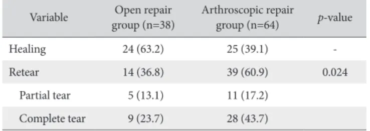
Table 3. Comparison of Anatomic Results between the Open and Ar- throscopic Repair Groups
Variable Open repair
group (n=38) Arthroscopic repair
group (n=64) p-value
Healing 24 (63.2) 25 (39.1) -
Retear 14 (36.8) 39 (60.9) 0.024
Partial tear 5 (13.1) 11 (17.2) Complete tear 9 (23.7) 28 (43.7) Values are presented as number (%).
and 0.654, respectively) (Table 4).
Discussion
A retear after surgical repair of a massive rotator cuff tear is a common complication. Association of several factors including patient age, preoperative tear size, degree of muscular atrophy, degree of fatty infiltration of the cuff muscle, surgical technique, and inappropriate rehabilitation with tendon retears has been demonstrated.17-19) In general, when successful repair of a mas- sive rotator cuff tear is achieved, excellent clinical results may be attained and joint degeneration may be halted or at least mark- edly decelerated.20,21) However, with respect to less mobile and severely retracted massive rotator cuff tears, literature comparing and analyzing clinical results and repair integrity between open and arthroscopic repairs is lacking.
Trappey and Gartsman22) insisted that a low-tension environ- ment is critical for rotator cuff healing. Other biomechanical studies have demonstrated that the elements for successful repair of a rotator cuff tear are strong fixation,23,24) a high interface pres- sure and a wide interface area between the tendon and bone,25) and minimization of stress concentration inside the tendon.26) In cases of arthroscopic repair of a massive rotator cuff tear, ef- fective anatomical repair is difficult because the repair construct is under inevitably undue tension even after sufficient release.
Therefore, the retear rate of massive rotator cuff tears is generally higher than that of smaller rotator cuff tears. Recent studies have reported postoperative healing rates after arthroscopic repair of
massive rotator cuff tears of 47% to 94%.4,17,27) In this study, the retear rate was 60.9% after arthroscopic repair of massive rotator cuff tears, which is within a similar range with that reported in other relevant literature; however, the retear rate was 36.8% for open repair, which was a statistically significant lower retear rate than that for arthroscopic repair.
There are several explanations for the favorable clinical and anatomical results of open rotator cuff repair in this study. First, the authors used the deltoid splitting technique for the approach to repair of the torn rotator cuff and minimized postoperative dehiscence of the deltoid using the Mason–Allen stitch to reat- tach and strongly fix the coracoacromial ligament and the del- toid detached from the anterior acromion. Second, for sufficient release of a less mobile and severely retracted torn cuff, a small Darrach retractor was used for release in the anterior, superior, and posterior directions to minimize tension and repair at the anatomical footprint. In addition, the Mason–Allen and conven- tional transosseous techniques were used for firm fixation.
Liu and Baker28) initially reported no significant clinical differ- ences in evaluation of patients with intact or torn rotator cuffs us- ing arthrography 2 years after surgery. In contrast to these results, Gazielly et al.17) and Harryman et al.6) reported superior clinical outcomes in patients with intact repairs regardless of initial tear size. Zumstein et al.29) reported significantly increased Constant scores and abduction strength with preserved repair integrity after massive rotator cuff repairs. Galatz et al.5) reported excel- lent early clinical results despite loss of repair integrity, which appeared to deteriorate over time. Although structural failure of repair of massive tears can and does occur, it does not necessar- ily imply a poor clinical outcome.5,6) In our study, despite struc- tural failures, excellent pain relief and improvement in the ability to perform activities of daily living were demonstrated during the
>24-month follow-up period. This result, combined with those of multiple studies documenting frequent progression of rota- tor cuff tears over time, suggests that early improvement despite retear may be attributable to partial restoration of shoulder force couples that deteriorate with the described natural history of tear progression.30) Other possible explanations for the clinical improvement despite loss of repair integrity include the potential beneficial effects of bursectomy and the postoperative rehabilita- tion protocols associated with rotator cuff repair surgery.29)
Our study had a few limitations. First, as it was retrospec- tive in nature, our study had limitations similar to those of other retrospective studies. Second, the two techniques were not ran- domized, thus selection bias could be an issue. However, open repair tended to be performed in more severe cases. Neverthe- less, if the open repair group showed more favorable results, open repair may be considered to have stronger advantages.
The strength of this study was that reliable and validated MRIs were used to measure the postoperative structural outcome. An- other strength is that this study is a rare and noteworthy piece of Table 4. Comparison of Postoperative Results between the Complete Healing
and Retear Groups
Variable Complete healing
group (n=49) Retear group
(n=53) p-value VAS (pain during motions) 2.2 ± 1.1 1.8 ± 1.7 0.633 ROM
FF (°) 162.0 159.6 0.554
ERs (°) 53.3 51.9 0.688
IRp (level) T10.0 T10.6 0.321
Muscle strength (kg)
FF 4.6 4.4 0.628
ER 6.2 6.1 0.926
IR 7.8 7.7 0.878
Constant score 70.8 ± 14.3 68.5 ± 15.6 0.526
UCLA score 29.8 ± 3.7 29.1 ± 4.4 0.654
Values are presented as mean ± standard deviation or mean only.
VAS: visual analog scale, ROM: range of motion, FF: forward flexion, ERs:
external rotation at the side, IRp: internal rotation to the posterior, ER: exter- nal rotation, IR: internal rotation, UCLA: the University of California at Los Angeles.
literature comparing and analyzing clinical results and repair in- tegrity after open and arthroscopic repairs of less mobile and se- verely retracted massive rotator cuff tears. The growing number of advantages of recent arthroscopic repair has led to decreasing attention to open repair; however this study showed that open repair results in more-favorable structural outcomes for massive rotator cuff tears; therefore, in cases of a less mobile and se- verely retracted tendon, open repair is recommended instead of stressing arthroscopic repair.
Conclusion
Open and arthroscopic repairs of massive rotator cuff tears provided satisfactory clinical results with no significant difference.
However, a significantly lower retear rate was observed for the open repair group compared with the arthroscopic repair group.
Open repair may be recommended for appropriately selected patients with less mobile and severely retracted massive rotator cuff tears.
References
1. Gartsman GM, Khan M, Hammerman SM. Arthroscopic repair of full-thickness tears of the rotator cuff. J Bone Joint Surg Am.
1998;80(6):832-40.
2. Tauro JC. Arthroscopic rotator cuff repair: analysis of technique and results at 2- and 3-year follow-up. Arthroscopy. 1998;14(1):
45-51.
3. Burkhart SS, Danaceau SM, Pearce CE Jr. Arthroscopic rota- tor cuff repair: analysis of results by tear size and by repair technique-margin convergence versus direct tendon-to-bone repair. Arthroscopy. 2001;17(9):905-12.
4. Bishop J, Klepps S, Lo IK, Bird J, Gladstone JN, Flatow EL. Cuff integrity after arthroscopic versus open rotator cuff repair: a prospective study. J Shoulder Elbow Surg. 2006;15(3):290-9.
5. Galatz LM, Ball CM, Teefey SA, Middleton WD, Yamaguchi K.
The outcome and repair integrity of completely arthroscopi- cally repaired large and massive rotator cuff tears. J Bone Joint Surg Am. 2004;86(2):219-24.
6. Harryman DT 2nd, Mack LA, Wang KY, Jackins SE, Richardson ML, Matsen FA 3rd. Repairs of the rotator cuff. Correlation of functional results with integrity of the cuff. J Bone Joint Surg Am. 1991;73(7):982-9.
7. Nho SJ, Adler RS, Tomlinson DP, et al. Arthroscopic rotator cuff repair: prospective evaluation with sequential ultrasonography.
Am J Sports Med. 2009;37(10):1938-45.
8. Denard PJ, Jiwani AZ, Lädermann A, Burkhart SS. Long-term outcome of arthroscopic massive rotator cuff repair: the impor- tance of double-row fixation. Arthroscopy. 2012;28(7):909-15.
9. Verma NN, Dunn W, Adler RS, et al. All-arthroscopic versus mini-open rotator cuff repair: a retrospective review with
minimum 2-year follow-up. Arthroscopy. 2006;22(6):587-94.
10. Yoo JC, Ahn JH, Koh KH, Lim KS. Rotator cuff integrity after arthroscopic repair for large tears with less-than-optimal foot- print coverage. Arthroscopy. 2009;25(10):1093-100.
11. DeOrio JK, Cofield RH. Results of a second attempt at surgi- cal repair of a failed initial rotator-cuff repair. J Bone Joint Surg Am. 1984;66(4):563-7.
12. Constant CR, Murley AH. A clinical method of functional as- sessment of the shoulder. Clin Orthop Relat Res. 1987;(214):
160-4.
13. Ellman H, Hanker G, Bayer M. Repair of the rotator cuff. End- result study of factors influencing reconstruction. J Bone Joint Surg Am. 1986;68(8):1136-44.
14. Lo IK, Burkhart SS. Arthroscopic repair of massive, contracted, immobile rotator cuff tears using single and double inter- val slides: technique and preliminary results. Arthroscopy.
2004;20(1):22-33.
15. Rhee YG, Vishvanathan T, Thailoo BKBR, Rojpornpradit T, Lim CT. The” 3 Sister Portals” for arthroscopic repair of massive ro- tator cuff tears. Tech Shoulder Elb Surg. 2007;8(2):53-7.
16. Gusmer PB, Potter HG, Donovan WD, O’Brien SJ. MR imag- ing of the shoulder after rotator cuff repair. AJR Am J Roent- genol. 1997;168(2):559-63.
17. Gazielly DF, Gleyze P, Montagnon C. Functional and anatomi- cal results after rotator cuff repair. Clin Orthop Relat Res.
1994;(304):43-53.
18. Gerber C, Fuchs B, Hodler J. The results of repair of massive tears of the rotator cuff. J Bone Joint Surg Am. 2000;82(4):505- 15.
19. Goutallier D, Postel JM, Gleyze P, Leguilloux P, Van Driessche S.
Influence of cuff muscle fatty degeneration on anatomic and functional outcomes after simple suture of full-thickness tears.
J Shoulder Elbow Surg. 2003;12(6):550-4.
20. Bedi A, Dines J, Warren RF, Dines DM. Massive tears of the rotator cuff. J Bone Joint Surg Am. 2010;92(9):1894-908.
21. Bigliani LU, Cordasco FA, McLlveen SJ, Musso ES. Operative repair of massive rotator cuff tears: Long-term results. J Shoul- der Elbow Surg. 1992;1(3):120-30.
22. Trappey GJ 4th, Gartsman GM. A systematic review of the clinical outcomes of single row versus double row rotator cuff repairs. J Shoulder Elbow Surg. 2011;20(2 Suppl):S14-9.
23. Burkhead WZ Jr, Skedros JG, O’Rourke PJ, Pierce WA, Pitts TC. A novel double-row rotator cuff repair exceeds strengths of conventional repairs. Clin Orthop Relat Res. 2007;461:106-13.
24. Kim DH, Elattrache NS, Tibone JE, et al. Biomechanical com- parison of a single-row versus double-row suture anchor tech- nique for rotator cuff repair. Am J Sports Med. 2006;34(3):407- 14.
25. Park MC, Cadet ER, Levine WN, Bigliani LU, Ahmad CS. Ten- don-to-bone pressure distributions at a repaired rotator cuff footprint using transosseous suture and suture anchor fixation
techniques. Am J Sports Med. 2005;33(8):1154-9.
26. Sano H, Yamashita T, Wakabayashi I, Itoi E. Stress distribu- tion in the supraspinatus tendon after tendon repair: suture anchors versus transosseous suture fixation. Am J Sports Med.
2007;35(4):542-6.
27. Mellado JM, Calmet J, Olona M, et al. Surgically repaired mas- sive rotator cuff tears: MRI of tendon integrity, muscle fatty de- generation, and muscle atrophy correlated with intraoperative and clinical findings. AJR Am J Roentgenol. 2005;184(5):1456- 63.
28. Liu SH, Baker CL. Arthroscopically assisted rotator cuff repair:
correlation of functional results with integrity of the cuff. Ar- throscopy. 1994;10(1):54-60.
29. Zumstein MA, Jost B, Hempel J, Hodler J, Gerber C. The clini- cal and structural long-term results of open repair of massive tears of the rotator cuff. J Bone Joint Surg Am. 2008;90(11):
2423-31.
30. Maman E, Harris C, White L, Tomlinson G, Shashank M, Boyn- ton E. Outcome of nonoperative treatment of symptomatic rotator cuff tears monitored by magnetic resonance imaging. J Bone Joint Surg Am. 2009;91(8):1898-906.