ISSN 0378-6471 (Print)⋅ISSN 2092-9374 (Online)
http://dx.doi.org/10.3341/jkos.2016.57.6.924
Original Article
백내장 수술 후 맥락막 두께 변화와 굴절력 오차
The Variation of Choroidal Thickness and Refractive Error after Cataract Surgery
노현철⋅박철용⋅오종현
Hyun Cheol Roh, MD, Choul Yong Park, MD, PhD, Jong-Hyun Oh, MD, PhD
동국대학교 일산병원 안과
Department of Ophthalmology, Dongguk University Ilsan Hospital, Goyang, Korea
Purpose: To evaluate the effect of cataract surgery on subfoveal choroidal thickness (SCT) and investigate the relationship be- tween the variation of SCT and refractive error.
Methods: We retrospectively reviewed the medical records of 47 patients (47 eyes) who underwent uneventful phacoemulsifica- tion cataract surgery from March 2012 to February 2014. SCTs were measured using spectral-domain optical coherence tomog- raphy performed before surgery and at 1 month, 3 months and 6 months postoperatively. We investigated the differences in tar- get refraction (TR) and postoperative spherical equivalent (SE), intraocular pressure (IOP) and central macular thickness (CMT) at all follow-ups.
Results: Compared with preoperative measurements, SCT showed a significant increase of 5.9 ± 13.3 μm at postoperative 1 month and 7.6 ± 18.1 μm at postoperative 3 months (p = 0.004 and p = 0.006, respectively), but no significant differences at post- operative 6 months (p = 0.104). The correlation between the variation of SCT and the differences in postoperative SE and TR were not significant at 1 month and 6 months, but were positively significant at 3 months (r = 0.310, p = 0.034). The variation of SCT showed no significant correlations with the postoperative change in IOP and CMT.
Conclusions: SCT significantly increased up to 3 months after cataract surgery. The variation of SCT may affect the post- operative refractive error.
J Korean Ophthalmol Soc 2016;57(6):924-929
Keywords: Cataract surgery, Choroidal thickness, Optical coherence tomography, Phacoemulsification, Refractive error
■Received: 2015. 10. 15. ■ Revised: 2016. 1. 11.
■Accepted: 2016. 2. 25.
■Address reprint requests to Jong-Hyun Oh, MD, PhD Department of Ophthalmology, Dongguk University Ilsan Hospital, #27 Dongguk-ro, Ilsandong-gu, Goyang 10326, Korea Tel: 82-31-961-7394, Fax: 82-31-961-7977
E-mail: [email protected]
ⓒ2016 The Korean Ophthalmological Society
This is an Open Access article distributed under the terms of the Creative Commons Attribution Non-Commercial License (http://creativecommons.org/licenses/by-nc/3.0/) which permits unrestricted non-commercial use, distribution, and reproduction in any medium, provided the original work is properly cited.
맥락막은 망막색소상피와 공막 사이에 위치한, 혈관이 풍부한 조직으로 망막 외층에 영양분 및 산소를 공급하는 역할을 하며, 다양한 안과적 질환과 관련되어 있다고 알려 져 있다.1-5 Enhanced depth imaging (EDI) 촬영법의 도입 이후 스펙트럼영역 빛간섭단층촬영(spectral-domain optical coherence tomography)을 이용한 맥락막 두께 측정이 용이
하게 되었다.6,7
백내장 수술은 안과에서 흔하게 시행되는 수술이다. 원 하는 술 후 목표굴절력을 얻기 위해 술 전에 다양한 검사를 시행하여 삽입될 인공수정체의 도수를 정하게 된다.8 백내 장 수술 후 맥락막 두께가 증가하지 않았다는 보고도 있지 만,9 몇몇 연구들에서는 백내장 수술 후에 맥락막 두께가 증가하였다고 보고하였다.10-12 Chakraborty et al13은 맥락막 두께의 일중변동이 안축길이의 일중변동과 음의 관련성이 있음을 제시하였고, Oh et al14은 중심장액맥락망막병증 환 자에서 중심와아래 맥락막 두께의 증가로 인해 안축길이가 짧아지고, 굴절력의 원시변화에도 영향을 미칠 것이라고 제안하였다. 백내장 수술 후 중심와아래 맥락막 두께의 변
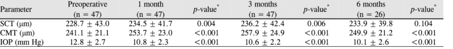
Table 1. Preoperative and postoperative measurements
Parameter Preoperative(n = 47)
1 month
(n = 47) p-value* 3 months
(n = 47) p-value* 6 months
(n = 26) p-value*
SCT (μm) 228.7 ± 43.0 234.5 ± 41.7 0.004 236.2 ± 42.4 0.006 233.9 ± 39.8 0.104
CMT (μm) 241.1 ± 21.1 253.7 ± 23.0 <0.001 257.9 ± 24.9 <0.001 249.9 ± 21.2 <0.001
IOP (mm Hg) 12.8 ± 2.7 10.8 ± 2.3 <0.001 10.6 ± 2.2 <0.001 10.1 ± 2.6 <0.001
Values are presented as mean ± SD unless otherwise indicated.
SCT = subfoveal choroidal thickness; CMT = central macular thickness; IOP = intraocular pressure.
*Compared with preoperative value using paired t-test.
화는 술 후 굴절력에도 영향을 미칠 수 있을 것이다. 따라 서 본 연구에서는 스펙트럼영역 빛간섭단층촬영을 이용하 여 백내장 수술이 중심와아래 맥락막 두께에 미치는 영향 을 알아보고, 중심와아래 맥락막 두께의 변화와 술 후 굴절 력 오차와의 관련성에 대해 조사하고자 하였다.
대상과 방법
2012년 3월부터 2014년 2월까지 본원에서 수정체유화술 및 인공수정체삽입술을 받은 환자들을 후향적으로 조사하 였다. 양안을 수술 받은 경우에 먼저 수술 받은 눈을 대상 으로 하였고, 나이관련황반변성, 중심장액맥락망막병증, 당 뇨망막병증 등의 망막질환을 가진 눈 또는 술 중 후낭파열 같은 합병증이 발생한 환자들은 대상에서 제외하였다. 스 펙트럼영역 빛간섭단층촬영 영상은 신호강도(signal strength) 가 6 이상인 경우만 분석에 포함하였다.
백내장 수술은 모두 한 명의 술자에 의해 시행되었다. 점 안마취 후 2.8 mm 각막절개도를 이용하여 투명각막절개를 시행한 뒤, 전방 유지를 위해 점탄물질(Viscoat, Alcon Laboratories, Fort Worth, TX, USA)을 주입하였다. 이후 원형전낭절개를 시행하고 평형염액을 이용하여 수력분리 술과 수력분층술을 시행하였다. 초음파유화기(Stellaris, Bausch and Lomb, Rochester, NY, USA)로 수정체유화술 을 시행하고 관류흡인기로 피질을 제거한 뒤, 수정체낭 내 에 비구면 일체형 인공수정체 TECNIS ZCB00 (AMO Inc., Santa Ana, CA, USA)를 삽입하였다. 수술 후 levofloxacin 0.5% 점안액과 1% prednisolone 점안액을 한 달간 사용하 였다.
스펙트럼영역 빛간섭단층촬영은 Cirrus HD-OCT (Carl Zeiss Meditec, Dublin, CA, USA)를 사용하여, 술 전, 술 후 1개월, 3개월, 6개월에 검사하였다. 맥락막 두께는 EDI 촬영법으로 획득한 영상을 이용하여, 중심와아래 망막색 소상피의 바깥쪽 경계에서 공막의 안쪽 경계까지 거리를 측정하였다.7 2명의 검사자가 독립적으로 각각 2회씩 측정 하였다.
수술 전 IOLMaster (Carl Zeiss Meditec, Jena, Germany)
로 안축길이, 각막굴절력을 측정하고 Sanders-Retzlaff-Kraff (SRK/T) 공식으로 계산하여 인공수정체 도수를 결정하였 고, 사용했던 인공수정체에 해당하는 목표굴절력을 조사하 였다. 또한 술 후 1개월, 3개월, 6개월에 자동굴절검사 (RK-F1, Canon, Tokyo, Japan)로 측정한 구면렌즈대응치를 조사하고, 목표굴절력과의 차이를 계산하였다. 안압은 비접 촉안압계(TX-F, Canon, Tokyo, Japan)를 이용하여 측정하 였고 중심황반두께는 스펙트럼영역 빛간섭단층촬영의 mac- ular cube 512 × 128 combination scan으로 측정하였다.
통계적 분석은 SPSS version 20.0 (SPSS Inc., Chicago, IL, USA)을 이용하였다. 맥락막 두께 측정에 대한 검사자 간 또는 검사자 내 일치도는 유목내상관계수(intraclass cor- relation coefficient)를 이용하여 평가하였으며, 모든 측정치 의 평균값을 이후 분석에 활용하였다. 술 전과 술 후의 측 정치를 비교하기 위하여 paired t-test 또는 Wilcoxon signed rank test를 시행하였고, 상관관계분석은 Pearson correla- tion을 이용하였다. p-value가 0.05 미만인 경우 통계적 유 의성이 있다고 해석하였다.
결 과
총 47명이 연구되었으며, 이 중 남자는 15명 15안, 여자 는 32명 32안이었다. 평균 연령은 69.4 ± 8.59세였고, 우안 20안, 좌안 27안이 포함되었다. 맥락막 두께 측정에 대한 검 사자 내 유목내상관계수는 0.993 (95% 신뢰구간 0.933-0.938), 0.942 (95% 신뢰구간 0.898-0.967)였으며, 검사자 간의 유목 내상관계수는 0.938 (95% 신뢰구간 0.892-0.965)이었다.
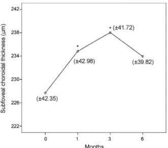
술 전 중심와아래 맥락막 두께는 228.7 ± 43.0 μm였으 며, 술 전과 비교하여 술 후 1개월에 평균 5.9 ± 13.3 μm, 술 후 3개월에 평균 7.6 ± 18.1 μm만큼 통계적으로 유의한 증가를 보였으나(p=0.004 and p=0.006, respectively), 술 후 6개월에는 유의한 차이를 보이지 않았다(p=0.104, Table 1, Fig. 1).
술 전 목표굴절력은 평균 -0.34 ± 0.43 diopters (D)였고, 구면렌즈대응치는 술 후 1개월에 평균 -0.34 ± 1.02D, 술 후 3개월에 평균 -0.27 ± 0.83D, 술 후 6개월에 평균 -0.12 ±
Figure 1. Changes in subfoveal choroidal thickness after cata-
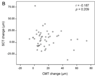
ract surgery. The asterisks (*) indicate significant changes compared with preoperative values (paired t-test).Figure 2. Scatterplots showing the correlation between differ-
ence in the postoperative spherical equivalent (SE) and target refraction (TR) and the change in subfoveal choroidal thick- ness (SCT). (A) At 1 month after surgery. (B) At 3 months af- ter surgery. (C) At 6 months after surgery.0.43D 로 술 전 목표굴절력과 유의한 차이는 없었다 (p=0.087, p=0.154 and p=0.389, respectively). 중심와아래
맥락막 두께의 변화량과 술 후 구면렌즈대응치와 술 전 목 표굴절력과의 차이는 술 후 1개월, 6개월에는 유의한 상관 관계를 보이지 않았으나(r=0.042, p=0.781 and r=-0.376, p=0.058), 술 후 3개월에는 유의한 양의 상관관계를 보였다 (r=0.310, p=0.034, Fig. 2). 술 후 중심와아래 맥락막 두께 가 30 μm 이상 증가한 환자 3명의 경우에, 술 후 3개월에 구면렌즈대응치와 술 전 목표굴절력과의 차이가 평균 0.428 ± 0.430D로 원시편위를 보였으나 통계적으로 유의하 지는 않았다(p=0.285).
안압은 술 전 12.8 ± 2.7 mmHg였으며, 술 후 1개월에 평 균 2.0 ± 2.6 mmHg, 술 후 3개월에 평균 2.2 ± 2.5 mmHg, 술 후 6개월에 평균 2.7 ± 3.3 mmHg만큼 유의하게 감소하 였다(all p values <0.001). 중심황반두께는 술 전 241.1 ± 21.1 μm였으며, 술 전과 비교하여 술 후 1개월에 평균 12.6
± 12.5 μm, 술 후 3개월에 평균 16.8 ± 17.4 μm, 술 후 6개 월에 평균 12.9 ± 15.1 μm만큼 유의하게 증가하였다(all p values <0.001). 중심와아래 맥락막 두께의 변화량은 술 후 1개월, 3개월, 6개월에 각각의 안압 변화량, 중심황반두께 변화량과는 유의한 상관관계를 보이지 않았다(all p values
A B
C
Figure 3. Scatterplots showing the correlation between post-
operative changes in intraocular pressure (IOP) and subfoveal choroidal thickness (SCT). (A) At 1 month after surgery. (B) At 3 months after surgery. (C) At 6 months after surgery.A B
C
>0.05, Fig. 3, 4).
고 찰
본 연구에서는 중심와아래 맥락막 두께가 백내장 수술 후 1개월, 3개월에 유의하게 증가하였으며, 술 후 6개월에 는 유의한 차이를 보이지 않았다. 이전 연구들 중에 백내장 수술 후 맥락막 두께가 증가하지 않았다는 연구결과도 있 지만,9 몇몇 다른 연구들에서는 맥락막 두께가 증가하였다 고 보고하였다.10-12 Pierru et al10은 95명, 115안을 대상으로 백내장 수술 후 3개월까지 중심와아래 맥락막 두께가 평균 12.0 ± 20.6 μm 증가하였다고 보고하였으며, Ohsugi et al11과 Noda et al12 또한 술 후 6개월까지도 중심와아래 맥락막 두 께가 유의하게 증가하였다고 보고하였다. 본 연구에서는 술 후 6개월에는 유의한 차이를 보이지 않았는데, 이러한 결과의 차이는 인종적 차이, 대상자의 수, 수술방법의 차이 등 때문인 것으로 생각된다.
백내장 수술 후에 맥락막 두께가 증가하는 기전을 정확 하게 설명할 수는 없다. 백내장 수술 후 방수 내에 증가된 프로스타글란딘 등의 염증인자가 유리체강을 통과하여 혈
액망막장벽(blood-retina-barrier)을 파괴한다고 알려져 있는 데, 이러한 염증 반응이 맥락막 두께의 증가에 영향을 미칠 것이라고 생각되고 있다.15,16 또한 수술 중 현미경조명에 의 한 빛 노출도 관련이 있을 것으로 생각되고 있는데,12 현미 경하 수술시간과 맥락막 두께 변화와의 연관성에 대한 추 가 연구가 이를 밝히는 데 도움이 될 것이다.
본 연구에서는 술 후 3개월에 중심와아래 맥락막 두께의 변화가 실제 굴절력과 술 전 예측하였던 목표굴절력과의 차이와 양의 상관관계를 보였다(r=0.335, p=0.028). 게다가 비록 대상수가 작아서 통계적으로 유의하지는 않았지만, 중심와아래 맥락막 두께가 30 μm 이상 증가한 3명의 환자 들은 술 후 3개월에 평균 0.428D의 원시 편위를 보였다. 이 는 술 후 중심와아래 맥락막 두께의 증가가 환자의 안축길 이를 줄여서 원시 편위가 일어난 것으로 추측된다. Bilak et al17은 백내장 수술 1개월 후에 안축길이가 평균 0.14 mm 감소하였다고 하였다. 백내장 수술 후 안축길이 변화와 맥 락막 두께 증가에 대한 추가 연구가 필요할 것으로 생각되 며, 이러한 변화가 수술 후 언제까지 지속되는지에 대한 장 기간의 관찰연구도 필요하다.
백내장 수술 후 중심황반두께가 증가하고, 안압이 감소
Figure 4. Scatterplots showing the correlation between post-
operative changes in central macular thickness (CMT) and sub- foveal choroidal thickness (SCT). (A) At 1 month after surgery.(B) At 3 months after surgery. (C) At 6 months after surgery.
A B
C
한다고 알려져 있다.18-20 본 연구에서도 백내장 수술 후 중 심황반두께가 증가하였고 안압이 감소하였다. 술 후 안압 의 감소는 맥락막 두께 증가의 원인일 수도 있다.21 술 후 안압의 감소가 눈관류압(ocular perfusion pressure) 감소와 관련이 있고, 이는 맥락막 두께 증가에도 영향을 미칠 수 있기 때문이다.22 Ohsugi et al11의 연구에서는 술 후 맥락막 두께 증가와 술 후 안압의 감소가 유의한 상관관계를 보였 지만, 본 연구에서는 그렇지 않았다.
본 연구는 후향적으로 의무기록을 조사했다는 점과 이로 인해 경과관찰 기간 동안 환자 수가 일정하지 않은 점 등의 몇 가지 제한점을 가진다. 맥락막 두께를 측정할 수 있는 자동화된 소프트웨어가 개발되지 않아 수동으로 측정하였 다는 제한점이 있으나, 검사자 간 및 검사자 내 유목내상관 계수가 0.9 이상으로 높은 일치도를 보였다. 또한 맥락막 두께의 일중변화를 고려하지 않았다는 것도 제한점이다.23 백내장은 술 전 빛간섭단층촬영 검사에 영향을 미칠 수 있 다고 알려져 있다.24,25 이전 연구에 따르면 빛간섭단층촬영 영상의 신호강도가 적어도 6 이상이어야 결과를 신뢰할 수 있다고 하였고,26 본 연구에서도 신호강도가 6 이상인 경우 만 연구 대상에 포함하였다. 목표굴절력은 SRK/T 공식을
이용하였는데, 다른 공식들과 비교하지 않은 점 또한 본 연 구의 제한점으로 생각된다. 결론적으로, 백내장 수술은 술 후 3개월까지 중심와아래 맥락막 두께를 증가시켰다. 이러 한 맥락막 두께의 변화는 술 후 굴절력에 영향을 미칠 수 있을 것이다. 이를 확인하기 위해서는 추가 연구가 필요할 것으로 생각된다.
REFERENCES
1) Grossniklaus HE, Green WR. Choroidal neovascularization. Am J Ophthalmol 2004;137:496-503.
2) Iida T, Kishi S, Hagimura N, Shimizu K. Persistent and bilateral choroidal vascular abnormalities in central serous chorioretinopathy.
Retina 1999;19:508-12.
3) Torres VL, Brugnoni N, Kaiser PK, Singh AD. Optical coherence tomography enhanced depth imaging of choroidal tumors. Am J Ophthalmol 2011;151:586-93.e2.
4) Ho M, Liu DT, Chan VC, Lam DS. Choroidal thickness measure- ment in myopic eyes by enhanced depth optical coherence tomography. Ophthalmology 2013;120:1909-14.
5) Ciardella AP, Donsoff IM, Huang SJ, et al. Polypoidal choroidal vasculopathy. Surv Ophthalmol 2004;49:25-37.
6) Spaide RF, Koizumi H, Pozzoni MC. Enhanced depth imaging
= 국문초록 =
백내장 수술 후 맥락막 두께 변화와 굴절력 오차
목적: 백내장 수술이 맥락막 두께에 미치는 영향을 알아보고 맥락막 두께 변화와 술 후 굴절력 오차와의 관련성에 대해 조사하고자 하였다.
대상과 방법: 2012년 3월부터 2014년 2월까지 수정체유화술 및 인공수정체삽입술을 받은 47명 47안을 후향적으로 조사하였다. 술 전 또는 술 후 1개월, 3개월, 6개월에 시행한 스펙트럼영역 빛간섭단층촬영 사진을 이용하여 중심와아래 맥락막 두께를 측정하였다.
또한 목표굴절력과 술 후 1개월, 3개월, 6개월에 측정한 구면렌즈대응치와의 차이, 매 방문 시에 안압, 중심황반두께 등도 조사하였다.
결과: 중심와아래 맥락막 두께는 술 전보다 술 후 1개월에 평균 5.9 ± 13.3 μm, 술 후 3개월에 평균 7.6 ± 18.1 μm만큼 유의하게 증가하였으나(p=0.004 and p=0.006, respectively), 술 후 6개월에는 유의한 차이를 보이지 않았다(p=0.104). 중심와아래 맥락막 두께의 변화량은 술 후 구면렌즈대응치와 술 전 목표굴절력과의 차이값과 술 후 1개월, 술 후 6개월에는 유의한 상관관계를 보이지 않았으나, 술 후 3개월에는 유의한 양의 상관관계를 보였다(r=0.310, p=0.034). 중심와아래 맥락막 두께의 변화량은 안압 변화량, 중심황반두께 변화량과는 유의한 상관관계를 보이지 않았다.
결론: 백내장 수술은 술 후 3개월까지 중심와아래 맥락막 두께를 증가시켰다. 중심와아래 맥락막 두께의 변화는 술 후 굴절력에 영향 을 미칠 수 있을 것이다.
<대한안과학회지 2016;57(6):924-929>
spectral-domain optical coherence tomography. Am J Ophthalmol 2008;146:496-500.
7) Margolis R, Spaide RF. A pilot study of enhanced depth imaging optical coherence tomography of the choroid in normal eyes. Am J Ophthalmol 2009;147:811-5.
8) Olsen T. Calculation of intraocular lens power: a review. Acta Ophthalmol Scand 2007;85:472-85.
9) Falcão MS, Gonçalves NM, Freitas-Costa P, et al. Choroidal and macular thickness changes induced by cataract surgery. Clin Ophthalmol 2014;8:55-60.
10) Pierru A, Carles M, Gastaud P, Baillif S. Measurement of sub- foveal choroidal thickness after cataract surgery in enhanced depth imaging optical coherence tomography. Invest Ophthalmol Vis Sci 2014;55:4967-74.
11) Ohsugi H, Ikuno Y, Ohara Z, et al. Changes in choroidal thickness after cataract surgery. J Cataract Refract Surg 2014;40:184-91.
12) Noda Y, Ogawa A, Toyama T, Ueta T. Long-term increase in sub- foveal choroidal thickness after surgery for senile cataracts. Am J Ophthalmol 2014;158:455-9.e1.
13) Chakraborty R, Read SA, Collins MJ. Diurnal variations in axial length, choroidal thickness, intraocular pressure, and ocular biometrics. Invest Ophthalmol Vis Sci 2011;52:5121-9.
14) Oh JH, Oh J, Togloom A, et al. Biometric characteristics of eyes with central serous chorioretinopathy. Invest Ophthalmol Vis Sci 2014;55:1502-8.
15) Miyake K, Ibaraki N. Prostaglandins and cystoid macular edema.
Surv Ophthalmol 2002;47 Suppl 1:S203-18.
16) Xu H, Chen M, Forrester JV, Lois N. Cataract surgery induces reti- nal pro-inflammatory gene expression and protein secretion. Invest
Ophthalmol Vis Sci 2011;52:249-55.
17) Bilak S, Simsek A, Capkin M, et al. Biometric and intraocular pres- sure change after cataract surgery. Optom Vis Sci 2015;92:464-70.
18) von Jagow B, Ohrloff C, Kohnen T. Macular thickness after un- eventful cataract surgery determined by optical coherence tomography. Graefes Arch Clin Exp Ophthalmol 2007;245:1765-71.
19) Sourdille P, Santiago PY. Optical coherence tomography of mac- ular thickness after cataract surgery. J Cataract Refract Surg 1999;25:256-61.
20) Jahn CE. Reduced intraocular pressure after phacoemulsification and posterior chamber intraocular lens implantation. J Cataract Refract Surg 1997;23:1260-4.
21) Saeedi O, Pillar A, Jefferys J, et al. Change in choroidal thickness and axial length with change in intraocular pressure after trabeculectomy. Br J Ophthalmol 2014;98:976-9.
22) Bayhan HA, Bayhan SA, Gürdal C. Long-term increase in sub- foveal choroidal thickness after surgery for senile cataracts. Am J Ophthalmol 2015;159:406-7.
23) Usui S, Ikuno Y, Akiba M, et al. Circadian changes in subfoveal choroidal thickness and the relationship with circulatory factors in healthy subjects. Invest Ophthalmol Vis Sci 2012;53:2300-7.
24) Falavarjani KG, Modarres M, Nikeghbali A. OCT and cataract.
Ophthalmology 2010;117:849; author reply 849-50.
25) van Velthoven ME, van der Linden MH, de Smet MD, et al.
Influence of cataract on optical coherence tomography image qual- ity and retinal thickness. Br J Ophthalmol 2006;90:1259-62.
26) Na JH, Sung KR, Lee Y. Factors associated with the signal strengths obtained by spectral domain optical coherence tomography.
Korean J Ophthalmol 2012;26:169-73.