ISSN 0378-6471 (Print)⋅ISSN 2092-9374 (Online)
https://doi.org/10.3341/jkos.2017.58.4.437
Original Article
시신경유두 좌우 뒤바뀜이 있는 눈의 망막신경섬유층 및 황반부 신경절세포 두께
Retinal Nerve Fiber Layer and Macular Ganglion Cell Thicknesses in Situs Inversus of Optic Discs
김민경⋅김재민⋅황영훈
Min Kyung Kim, MD, Jae Min Kim, MD, Young Hoon Hwang, MD
건양대학교 의과대학 김안과병원 안과학교실 명곡안연구소
Myung-Gok Eye Research Institute, Department of Ophthalmology, Kim’s Eye Hospital, Konyang University College of Medicine, Seoul, Korea
Purpose: To investigate circumpapillary retinal nerve fiber layer (RNFL) and macular ganglion cell-inner plexiform layer (GCIPL) thicknesses as measured by optical coherence tomography in eyes with situs inversus of optic discs.
Methods: RNFL and macular GCIPL thicknesses were measured in eyes with situs inversus of optic discs without other ocular abnormalities (situs inversus group) and in age- and refractive error-matched healthy eyes (control group). RNFL thickness (global area, superior, nasal, inferior, and temporal quadrants) and GCIPL thickness (global area, superior-temporal, superior, superior-nasal, inferior-nasal, inferior, and inferior-temporal sectors and minimum) were compared between the groups.
Results: Nine eyes of 5 subjects with situs inversus of optic discs and 20 healthy eyes of 20 subjects (10 eyes for control groups A and B, respectively) were enrolled. No significant difference was found in superior or inferior quadrant RNFL thickness (p >
0.05); however, the situs inversus group showed a thicker RNFL in the nasal quadrant and a thinner RNFL in the temporal quad- rant (p < 0.01). In macular GCIPL thickness, no significant difference was found in the superotemporal or inferiotemporal sector or for minimum thickness (p < 0.05); however, the situs inversus group showed thicker GCIPL in the global area, superior, super- onasal, inferonasal, and inferior sectors than the control groups (p < 0.01).
Conclusions: In eyes with situs inversus of optic discs, distribution of circumpapillary RNFL and macular GCIPL thickness were different from eyes without this condition. When assessing RNFL and macular GCIPL thicknesses in eyes with situs inversus of optic discs, caution is needed.
J Korean Ophthalmol Soc 2017;58(4):437-442
Keywords: Optic nerve head, Retinal ganglion cell, Retinal nerve fiber layer, Situs inversus
■Received: 2016. 12. 1. ■ Revised: 2017. 1. 10.
■Accepted: 2017. 3. 16.
■Address reprint requests to Young Hoon Hwang, MD Department of Ophthalmology, Kim’s Eye Hospital, #136 Yeongsin-ro, Yeongdeungpo-gu, Seoul 07301, Korea Tel: 82-2-2639-7777, Fax: 82-2-2633-3976 E-mail: [email protected]
ⓒ2017 The Korean Ophthalmological Society
This is an Open Access article distributed under the terms of the Creative Commons Attribution Non-Commercial License (http://creativecommons.org/licenses/by-nc/3.0/) which permits unrestricted non-commercial use, distribution, and reproduction in any medium, provided the original work is properly cited.
시신경유두 좌우 뒤바뀜은 안경(optic stalk)이 안소포 (optic vesicle) 내에 비정상적으로 부착되면서 생기는 드문
선천이상으로, 정상과 비교했을 때 코쪽-귀쪽의 모양이 반 전된 시신경유두의 형태를 가지게 된다.1-3 즉, 정상안에서 시신경유두 주위 혈관의 주행방향이 시신경유두 중심으로 부터 귀쪽으로 포물선을 그리면서 나타나는 반면에, 좌우 뒤바뀜이 있는 경우, 혈관이 시신경유두 중심으로부터 코 쪽으로 주행하다가 급격하게 귀쪽으로 꺾이는 특징적인 양 상을 보인다.1-5 시신경유두 주위의 망막신경섬유층이 망막 혈관 주행을 따라 분포하는 것을 고려해보았을 때, 시신경 유두 좌우 뒤바뀜 환자에서 망막신경섬유층의 분포가 정상
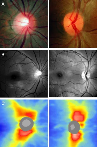
A
B
C
Figure 1. An example of optic disc in a healthy eye (left col-
umn) and an eye with situs inversus of optic disc (right col- umn). Disc photograph (A), red-fee fundus photograph (B), and circumpapillary retinal nerve fiber layer thickness dis- tribution as measured by Cirrus high–definition optical coher- ence tomography(C).인과 다를 것을 유추해볼 수 있다. 이에 대해 Kang et al4은 빛간섭단층촬영을 통해서 시신경유두 좌우 뒤바뀜이 있는 눈에서 정상안에 비해서 시신경유두 주변의 코쪽 망막신경 섬유층이 두껍고, 상이측, 이측, 하이측 망막신경섬유층 두 께가 얇다고 보고한 바 있고, Han and Hwang5은 시신경유 두 좌우 뒤바뀜과 녹내장이 병합된 증례를 통해 녹내장이 진행하면서 시신경유두 주변의 상비측과 하비측 망막신경 섬유층 손상이 생기는 양상을 보고한 바 있다.
녹내장은 시신경유두주변의 망막신경섬유층 두께의 감 소 및 그에 대응하는 시야결손이 특징적인 질병으로 최근 영상장비의 발달로 황반부의 망막신경절세포 두께의 감소 또한 녹내장의 평가에 중요한 역할을 하는 것으로 알려져 있다.6-8 하지만 시신경유두 좌우 뒤바뀜이 있는 눈에서의 황반부 망막신경절세포의 두께에 대한 보고는 아직 없는 상황이다. 따라서 본 연구에서는 빛간섭단층촬영을 이용하 여 시신경유두 좌우 뒤바뀜이 있는 눈의 시신경유두주위 망막신경섬유층 및 황반부 신경절세포-내망상층의 두께에 대해서 알아보고자 한다.
대상과 방법
본 연구는 본원 임상시험심사위원회의 승인을 받은 것으 로 2014년 1월부터 2016년 1월까지 본원 외래를 방문한 환 자들 중, 시신경유두 좌우 뒤바뀜이 있는 눈(뒤바뀜군)과 정상안(대조군)을 대상으로 빛간섭단층촬영을 이용하여 시 신경유두주위 망막신경섬유층 및 황반부 신경절세포-내망 상층의 두께를 측정하였다. 시신경유두 좌우 뒤바뀜은 시 신경유두의 중심에서 시작된 위, 아래 망막 혈관이 시신경 의 코쪽으로 나와 귀쪽으로 급격히 꺾여 주행하는 것으로 정의하였다(Fig. 1). 대조군은 건강검진 목적으로 본원에 내 원한 환자 중 뒤바뀜군과 나이 차이가 5세 이내, 성별 분포 가 같고, 구면렌즈대응치(spherical equivalent)의 차이가 2 디옵터 이내이면서, 시신경유두 좌우 뒤바뀜 및 다른 이상 이 없는 정상안으로 선정하였다.
모든 대상자에서 병력조사, 시력측정, 굴절검사, 골드만 압평안압계를 이용한 안압 측정, 세극등현미경을 이용한 전안부 검사, 90디옵터 렌즈를 이용한 시신경유두 및 안저 검사, 험프리 시야 검사계(model 750; Carl Zeiss Meditec, Dublin, CA, USA)의 24-2 Swedish Interactive Threshold Algorithm을 이용한 시야검사, 빛간섭단층촬영(Cirrus high- definition optical coherence tomography; Carl Zeiss Meditec, Dublin, CA, USA)을 이용한 망막신경섬유층 및 황반부 신 경절세포-내망상층 두께 측정을 시행하였다.
이상의 검사에서 (1) 최대교정시력이 20/30 이상이고, (2)
안압이 정상범위이면서(<21 mmHg), (3) 녹내장성 시신경 변화(시신경유두함몰비 증가, 시신경유두 출혈, 국소적이거 나 전반적인 시신경 테의 얇음 혹은 패임, 등)를 보이지 않 고, (4) 시야검사에서 이상이 없는 환자를 대상으로 하였다.
시야나 시신경에 영향을 줄 수 있는 녹내장을 비롯한 다른 망막 질환이 있는 경우는 분석에서 제외하였다.
빛간섭단층촬영의 Optic Disc Cube 200 × 200 스캔 및 Macular Cube 512 × 128 스캔을 이용해 시신경유두 주위 망막신경섬유층 및 황반부의 신경절세포-내망상층의 두께 를 측정하였다. Optic Disc Cube 200 × 200 스캔은 시신경 유두 중심을 기준으로 하여 6 × 6 mm2 영역의 망막신경섬 유층 두께 및 3.46 mm의 직경을 가지는 원 부위에 해당되 는 망막신경섬유층 두께를 제공하는데, 이를 통해 얻은 평 균 및 각 사분면, 12시간 영역의 망막신경섬유층 두께를 분 석에 이용하였다. Macular Cube 512 × 128 스캔은 황반부
Table 1. Comparison of clinical data between control and situs inversus groups
Control group A(n = 10)
Control group B (n = 10)
Situs inversus group
(n = 9) p-value* p-value†
Age (years) 43.0 (39.0, 46.3) 44.5 (29.0, 52.0) 48.0 (33.0, 49.0) 0.905 0.842
Sex (female:male) 5:5 5:5 4:6 1.000 1.000
Refractive error (diopter) -0.63 (-1.00, -0.38) -1.00 (-1.57, -0.69) -1.00 (-2.00, -0.25) 0.315 0.720 Visual field indices
Mean deviation (dB) -0.79 (-1.89, 0.30) -1.19 (-1.68, -0.08) -0.85 (-1.28, 0.72) 0.497 0.182 Pattern standard deviation (dB) 1.55 (1.48, 1.94) 1.44 (1.14, 1.89) 1.67 (1.30, 2.21) 0.661 0.243 Visual field index (%) 99.0 (99.0, 99.25) 99.5 (99.0, 100.0) 100.0 (98.5, 100.0) 0.400 0.968 Data are presented as median and interquartile range or number of eyes.
* Mann-Whitney U-test: comparison between the control group A and situs inversus group; †Mann-Whitney U-test: comparison between the control group B and situs inversus group.
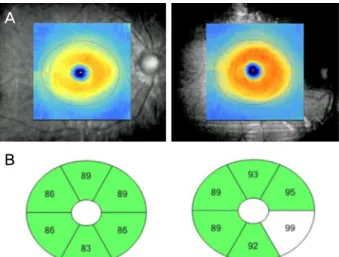
A
B
Figure 2. An example of macular ganglion cell-inner plexi-
form layer thickness distribution. Presented by thickness map (A) and sector map (B) of Cirrus high–definition optical co- herence tomography in a healthy eye (left column) and an eye with situs inversus of optic disc (right column) presented in Figure 1.6 × 6 mm2에 해당하는 부위를 512 A-scan을 구성하는 128 개의 horizontal B-scan으로 측정하는 방식이며, 이를 통해 얻어진 데이터를 ganglion cell analysis (GCA) 알고리즘을 통해 분석하였다. GCA 알고리즘은 내망상층의 바깥쪽 경 계선과 망막신경섬유층의 바깥쪽 경계선을 인식하고 이를 통해 신경절세포층과 내망상층의 두께를 분석하는 방식으 로, 이를 통해 얻은 황반부 14.13 mm2 영역의 평균, 최소값 및 6개 구역(상측, 상이측, 하이측, 하측, 하비측, 상비측)의 두께를 분석에 활용하였다(Fig. 2). 검사 결과에서 신호강도 가 6 미만이거나 중심이 이탈되어 있는 경우, 그 외에 다른 오류가 있는 경우는 분석에서 제외하였다.
뒤바뀜군과 대조군의 나이, 성별 분포, 굴절값, 시야검사 결과 지표 및 빛간섭단층촬영을 통해 측정한 시신경유두주 위 망막신경섬유층 두께 및 황반부 신경절세포-내망상층의 두께를 비교하였다. 변수들의 분포는 정규분포를 보이지 않
았기 때문에 중위값(median) 및 사분위수 범위(interquartile range)를 이용하여 기술하고, 두 군 사이의 변수 비교는 chi squared test 및 Mann-Whitney U-test를 이용하였다. 통계분 석은 SPSS Windows version 12.0 (SPSS Inc., Chicago, IL, USA)를 이용하였으며, p값이 0.05 미만인 경우를 통계적으 로 유의한 것으로 판정하였다.
결 과
총 5명 9안(여성 4안, 남성 5안)이 시신경유두 좌우 뒤바 뀜군에 포함되었고, 20명의 20안이 대조군(대조군 A 10안, 대조군 B 10안)에 포함되었다. 뒤바뀜군 5명 중 1명의 1안 은 황반부에 망막전막이 동반되어 있어서 분석에서 제외하 였다. 대상안 모두 안과 수술을 받은 과거력이 없었다. 뒤 바뀜군의 나이는 48.0세(40.0, 49.0세) (범위, 26–52세)였고, 굴절값은 -1.00디옵터(-2.00, -0.50디옵터) (범위, -2.00~+0.50 디옵터)였다. 뒤바뀜군과 대조군 A, B 사이에 나이, 성별, 굴절값, 시야검사 결과에 유의한 차이를 보이지 않았다 (p>0.05, Table 1).
시신경유두주위 망막신경섬유층 두께의 경우, 평균 및 위아래 사분면의 두께는 유의한 차이를 보이지 않았지만 (p>0.05) 뒤바뀜군이 대조군에 비해서 코쪽 사분면의 망막 신경섬유층이 더 두껍고, 귀쪽 사분면이 더 얇은 양상을 보 였다(p<0.05, Table 2). 12시간 영역의 망막신경섬유층 두 께의 경우, 1시 방향에서 6시 방향까지는 뒤바뀜군에서 대 조군에 비해 두껍게 측정되었고, 7시 방향에서 11시 방향 의 망막신경섬유층은 대조군에 비해 얇게 측정되었다 (p<0.05, Table 1). 신경절세포-내망상층의 경우, 상이측, 하 이측, 최소값은 유의한 차이를 보이지 않았지만(p>0.05) 전 체 평균, 상측, 상비측, 하비측, 하측 영역에서 뒤바뀜군이 대조군보다 두꺼운 양상을 보였다(p<0.05, Table 3).
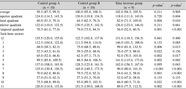
Table 2. Comparison of circumpapillary retinal nerve fiber layer thickness (μm) between control and situs inversus groups
Control group A(n = 10)
Control group B (n = 10)
Situs inversus group
(n = 9) p-value* p-value†
Average 95.5 (87.5, 99.5) 100.0 (95.8, 108.3) 102.0 (94.5, 105.5) 0.211 0.905
Superior quadrant 124.0 (114.5, 143.3) 130.0 (119.8, 134.5) 118.0 (111.0, 143.0) 0.720 0.604
Nasal quadrant 60.0 (51.5, 70.3) 64.5 (62.5, 76.3) 82.0 (71.5, 105.0) 0.004 0.010
Inferior quadrant 122.0 (110.0, 132.5) 130.5 (125.5, 142.5) 126.0 (123.0, 146.0) 0.211 0.661
Temporal quadrant 70.5 (61.0, 77.0) 79.0 (73.0, 84.5) 56.0 (52.0, 60.5) 0.001 <0.001
Clock-hour sectors
12 135.5 (120.0, 155.0) 122.5 (102.8, 137.8) 131.0 (118.5, 156.5) 0.661 0.400
1 112.5 (104.8, 122.0) 111.5 (94.8, 114.0) 146.0 (101.5, 188.0) 0.133 0.065
2 68.0 (58.3, 82.3) 75.0 (68.5, 88.0) 99.0 (81.0, 132.5) 0.006 0.013
3 52.5 (43.5, 61.0) 59.5 (55.0, 68.0) 76.0 (57.5, 88.0) 0.022 0.156
4 65.0 (52.0, 66.8) 62.5 (57.3, 73.3) 92.0 (70.5, 103.0) 0.002 0.017
5 89.5 (85.8, 109.5) 88.5 (84.8, 106.5) 141.0 (115.0, 173.0) 0.002 0.002
6 137.0 (108.0, 163.8) 128.5 (123.8, 161.5) 162.0 (156.5, 167.0) 0.095 0.043
7 135.0 (130.8, 156.5) 165.0 (149.5, 173.5) 90.0 (80.0, 101.5) <0.001 <0.001
8 70.0 (62.8, 80.8) 79.5 (72.0, 92.3) 54.0 (41.5, 58.0) 0.001 <0.001
9 57.0 (51.0, 62.3) 57.5 (51.3, 70.0) 52.0 (47.0, 58.0) 0.133 0.133
10 75.5 (68.8, 89.0) 93.0 (89.5, 103.5) 66.0 (60.5, 71.0) 0.022 <0.001
11 120.0 (114.8, 133.0) 151.5 (139.0, 168.0) 89.0 (77.5, 112.5) 0.002 <0.001
Data are presented as median and interquartile range.
*Mann-Whitney U-test: comparison between the control group A and situs inversus group; †Mann-Whitney U-test: comparison between the con- trol group B and situs inversus group.
Table 3. Comparison of macular ganglion cell-inner plexiform layer thickness (μm) between control and situs inversus groups
Control group A(n = 10)
Control group B (n = 10)
Situs inversus group
(n = 9) p-value* p-value†
Average 82.0 (81.0, 87.0) 84.5 (81.8, 86.0) 90.0 (83.5, 91.5) 0.035 0.028
Minimum 81.5 (79.8, 83.8) 82.0 (80.0, 85.0) 84.0 (80.5, 87.0) 0.315 0.447
Superotemporal 82.5 (79.8, 85.3) 84.0 (79.8, 86.0) 83.0 (81.5, 88.0) 0.356 0.549
Superior 82.5 (81.5, 85.8) 84.0 (82.3, 85.3) 89.0 (86.0, 93.0) 0.010 0.008
Superonasal 85.0 (83.8, 88.5) 87.0 (84.75, 88.0) 94.0 (88.5, 95.5) 0.004 0.013
Inferonasal 83.5 (81.8, 87.3) 85.5 (82.3, 86.0) 93.0 (85.0, 96.5) 0.028 0.017
Inferior 80.0 (78.8, 85.3) 82.0 (79.5, 85.3) 89.0 (81.0, 91.5) 0.033 0.059
Inferotemporal 81.5 (79.0, 86.5) 83.5 (81.5, 86.3) 86.0 (80.0, 88.5) 0.315 0.661
Data are presented as median and interquartile range.
*Mann-Whitney U-test: comparison between the control group A and situs inversus group; †Mann-Whitney U-test: comparison between the con- trol group B and situs inversus group.
고 찰
본 연구 결과에 의하면 시신경유두 주위 망막신경섬유층 두께의 경우, 뒤바뀜군이 대조군에 비해서 코쪽 사분면의 망막신경섬유층이 더 두껍고, 귀쪽 사분면이 더 얇은 양상 을 보였다. Kang et al4은 시신경유두 좌우 뒤바뀜이 있는 12안의 망막신경섬유층 분포를 대조군과 비교하여 발표한 바 있는데, 본 연구의 결과와 마찬가지로 시신경유두 좌우 뒤바뀜이 있는 군에서 코쪽 망막신경섬유층이 더 두껍고, 귀쪽 망막신경층이 더 얇은 경향을 보였으며, 위, 아래 망 막신경섬유층의 가장 두꺼운 지점 또한 더 코쪽으로 이동 한 양상을 보고한 바 있다. 이는 혈관주행과 마찬가지로, 시신경유두 좌우 뒤바뀜군에서 망막신경섬유의 주행 또한
시신경유두의 코쪽으로 시작하여 귀쪽으로 주행이 급격히 바뀌기 때문에 시신경 유두주변 망막신경섬유층이 코쪽에 서 상대적으로 더 밀집되어 있고, 귀쪽이 덜 밀집되어서 나 타나는 현상으로 보인다.
황반부 신경절세포-내망상층의 경우, 상이측, 하이측, 최 소값은 유의한 차이를 보이지 않았지만, 상측, 상비측, 하비 측, 하측 영역에서는 뒤바뀜군이 대조군보다 두꺼운 양상 을 보였다. 이는 시신경유두 좌우 뒤바뀜이 있는 눈에서 시 신경유두 주변의 망막신경섬유층뿐만 아니라 황반부의 신 경절세포-내망상층의 분포도 정상안과 다르다는 점을 시사 한다. 즉 망막신경섬유층에서와 마찬가지로 황반부의 신경 절세포-내망상층도 정상안에 비해서 코쪽으로 더 밀집하게 분포하고 있을 것으로 예측할 수 있다. 반면, 상이측과 하
이측 및 최소 신경절세포-내망상층 두께는 정상안과 유의 하게 다르지 않았다. 녹내장에서 관찰되는 황반부 신경절 세포-내망상층의 변화가 상이측과 하이측에서 먼저 나타나 는 경우가 많고, 최소 신경절세포 두께가 높은 녹내장 진단 력을 가진다는 점을 고려한다면6-8 시신경유두 좌우뒤바뀜 이 있는 눈에서 초기 녹내장 진단 시에는 시신경유두 주변 의 망막신경섬유층보다 황반부 신경절세포-내망상층 두께 분석이 더 유리할 수 있을 것으로 생각한다. 하지만 상이측 과 하이측에서 시작된 신경절세포-내망상층의 변화가 상하 측, 상비측, 하비측으로 진행하는지를 판단할 때는 시신경 유두 좌우뒤바뀜이 있는 눈의 상대적으로 두꺼운 신경절세 포-내망상층이 진행의 저평가를 유발할 수 있기 때문에 주 의가 필요할 것으로 생각한다.
본 연구 결과에 의하면 시신경유두 좌우뒤바뀜이 있는 눈에서 코쪽 황반부 신경절두께가 정상안보다 두꺼웠지만 이 부위 신경절세포의 신경섬유에 해당하는 귀쪽 망막신경 섬유층 두께는 정상안보다 얇았다. 이는 시신경유두 좌우 뒤바뀜이 있는 눈에서 귀쪽 망막신경섬유가 정상안보다 더 위아래로 퍼져서 분포하기 때문일 것으로 유추해 볼 수 있 다. 이에 대해서는 더 많은 눈을 대상으로 하는 추가 연구 가 필요할 것으로 생각된다.
본 연구는 적은 수의 눈을 대상으로 했으며, 망막신경섬 유층 및 황반부 신경절세포의 두께 측정에 영향을 줄 수 있 는 안축장, 시신경유두 크기, 망막 기울기9 등에 대해서 추 가로 분석하지 못한 한계점을 가지고 있다. 따라서 결과 해 석 시 이러한 한계점을 고려해야 할 것이다.
요약하면, 시신경유두 좌우뒤바뀜이 있는 눈에서는 시신 경유두주위 망막신경섬유층과 황반부 신경절세포-내망상 층의 분포가 정상안과 달랐다. 특히 코쪽 망막신경섬유층 이 더 두껍고, 귀쪽이 더 얇은 분포를 보였고, 황반부 신경
절세포-내망상층의 경우, 상이측과 하이측, 최소값을 제외 한 나머지 부분이 더 두꺼운 분포를 보였다. 따라서 시신경 유두 좌우뒤바뀜이 있는 눈의 망막신경섬유층과 황반부 신 경절세포-내망상층 분석 시에 이러한 분포 차이를 고려해 야 할 것이다.
REFERENCES
1) Brodsky MC. Congenital optic disk anomalies. Surv Ophthalmol 1994;39:89-112.
2) Witmer MT, Margo CE, Drucker M. Tilted optic disks. Surv Ophthalmol 2010;55:403-28.
3) Kothari M, Chatterjee DN. Unilateral situs inversus of optic disc as- sociated with reduced binocularity and stereoacuity resembling monofixation syndrome. Indian J Ophthalmol 2010;58:241-2.
4) Kang S, Jin S, Roh KH, Hwang YH. Peripapillary retinal nerve fi- ber layer and optic nerve head characteristics in eyes with situs in- versus of the optic disc. J Glaucoma 2015;24:306-10.
5) Han SY, Hwang YH. Glaucoma in an eye with situs inversus of the optic disc. Semin Ophthalmol 2014;29:172-4.
6) Mwanza JC, Durbin MK, Budenz DL, et al. Glaucoma diagnostic accuracy of ganglion cell-inner plexiform layer thickness: compar- ison with nerve fiber layer and optic nerve head. Ophthalmology 2012;119:1151-8.
7) Jeoung JW, Choi YJ, Park KH, Kim DM. Macular ganglion cell imaging study: glaucoma diagnostic accuracy of spectral-domain optical coherence tomography. Invest Ophthalmol Vis Sci 2013;
54:4422-9.
8) Hwang YH, Jeong YC, Kim HK, Sohn YH. Macular ganglion cell analysis for early detection of glaucoma. Ophthalmology 2014;
121:1508-15.
9) Yamashita T, Sakamoto T, Yoshihara N, et al. Correlations between retinal nerve fiber layer thickness and axial length, peripapillary retinal tilt, optic disc size, and retinal artery position in healthy eyes. J Glaucoma 2017;26:34-40.
= 국문초록 =
시신경유두 좌우 뒤바뀜이 있는 눈의 망막신경섬유층 및 황반부 신경절세포 두께
목적: 시신경유두 좌우 뒤바뀜(situs inversus)이 있는 눈의 시신경유두주위 망막신경섬유층 및 황반부 신경절세포-내망상층의 두께 에 대해서 알아보고자 한다.
대상과 방법: 시신경유두 좌우 뒤바뀜이 있으면서 다른 이상이 없는 눈(뒤바뀜군)과 나이와 굴절값이 같은 정상안(대조군)을 대상으로 빛간섭단층촬영을 이용하여 시신경유두주위 망막신경섬유층 및 황반부 신경절세포-내망상층의 두께를 측정하였다. 뒤바뀜군과 대조 군 사이에 망막신경섬유층(전체 평균 및 상측, 비측, 하측, 이측 사분면) 및 신경절세포-내망상층 두께(전체 평균 및 상이측, 상측, 상비측, 하비측, 하측, 하이측, 최소값)의 차이를 비교하였다.
결과: 총 5명의 9안이 뒤바뀜군에 포함되었고, 20명의 20안이 대조군(대조군 A, B 각각 10안)에 포함되었다. 망막신경섬유층 두께의 경우, 전체 평균 및 위아래 사분면의 두께는 유의한 차이를 보이지 않았지만(p>0.05) 뒤바뀜군이 대조군에 비해서 코쪽 사분면의 망막 신경섬유층이 더 두껍고, 귀쪽 사분면이 더 얇은 양상을 보였다(p<0.01). 신경절세포-내망상층의 경우, 상이측, 하이측, 최소값은 유의 한 차이를 보이지 않았지만(p>0.05) 전체 평균, 상측, 상비측, 하비측, 하측 영역에서 뒤바뀜군이 대조군보다 두꺼운 양상을 보였다 (p<0.01).
결론: 시신경유두 좌우 뒤바뀜이 있는 눈의 경우, 대조군과 다른 시신경유두주위 망막신경섬유 및 황반부 신경절세포-내망상층의 분 포를 보였다. 따라서 시신경유두 좌우 뒤바뀜이 있는 눈에서 빛간섭단층촬영으로 측정한 망막신경섬유층 및 황반 신경절세포-내망상 층 두께 분석 시 주의가 필요하다.
<대한안과학회지 2017;58(4):437-442>