ABSTRACT
Several studies have reported a good correlation between levels of serum hepatitis B virus surface antigen (HBsAg) and covalently closed circular DNA (cccDNA) before and after antiviral therapy. As a result, the quantification of HBsAg levels has attracted much attention in recent years as an important approach to evaluate viral activity. In this study, mAbs against HBsAg were generated and 9 mAbs (H17, H30, H31, H67, H73, H97, H101, H118, and H128) were investigated for optimization of HBsAg quantitation ELISA. Determination of the best combinations of mAbs for sandwich ELISA identified H17 and H31 mAbs as the ideal capture and horseradish peroxidase (HRP) conjugate mAbs, respectively. A standard curve for the current assay system exhibited linearity up to 40 ng/ml of HBsAg while a detection limit of approximately 1 ng/ml of HBsAg was also estimated, which was comparable to that of the other commercial ELISA kits. The ELISA system established in this study is particularly differentiated from other commercial kits in using mAbs for both capture and HRP conjugate, which provides a solution to inconsistency of quality and ethical issues in polyclonal antibodies production using laboratory animals.
Keywords: ELISA; Hepatitis B virus surface antigen; Monoclonal antibody; Quantification
INTRODUCTION
Chronic hepatitis B (CHB) is an important health problem that affects over 350 million people worldwide (1). The hepatitis B virus surface antigen (HBsAg) has been shown to be an early marker of infection by the hepatitis B virus (HBV) and therefore, qualitative HBsAg assays like radioimmunoassays (RIAs) and enzyme immunoassays (EIAs) have been used as screening tools for several decades (1).
But in recent years, quantification of HBsAg is in the spotlight as a new potential marker for monitoring on-treatment response as several studies have reported a good correlation between levels of serum HBsAg and covalently closed circular DNA (cccDNA) before and after antiviral therapy (2-7). Serum HBsAg levels reflect the concentration of intrahepatic HBV cccDNA and may ultimately prove valuable in the management of patients with CHB (8).
Brief Communication
Received: Jul 31, 2017 Revised: Sep 28, 2017 Accepted: Oct 5, 2017
*Correspondence to Se-Ho Kim
Division of Biomedical Convergence, College of Biomedical Science, Kangwon National University, 1 Kangwondaehak-gil, Chuncheon 24341, Korea.
E-mail: kimsho@kangwon.ac.kr
Copyright © 2017. The Korean Association of Immunologists
This is an Open Access article distributed under the terms of the Creative Commons Attribution Non-Commercial License (https://
creativecommons.org/licenses/by-nc/4.0/) which permits unrestricted non-commercial use, distribution, and reproduction in any medium, provided the original work is properly cited.
Conflict of Interest
The author declares no potential conflicts of interest.
Abbreviations
BSA, bovine serum albumin; cccDNA, covalently closed circular DNA; CHB, chronic hepatitis B; HBsAg, hepatitis B virus surface antigen; HBV, hepatitis B virus; HRP, horseradish peroxidase; PBS-T, phosphate-buffered saline, pH 7.4, containing 0.05% Tween 20; PEG-IFN, pegylated interferon; RT, room temperature; TMB, 3,3′,5,5′-Tetramethylbenzidine
Se-Ho Kim1,2,*
1 Division of Biomedical Convergence, College of Biomedical Science, Kangwon National University, Chuncheon 24341, Korea
2Institute of Bioscience and Biotechnology, Kangwon National University, Chuncheon 24341, Korea
ELISA for Quantitative Determination
of Hepatitis B Virus Surface Antigen
There are 2 main techniques to quantitatively measure HBsAg: Architect HBsAg QT (Abbott Diagnostics, Weibaden, Germany) and Elecsys HBsAg II (Roche Diagnostics, Indianapolis, IN, USA) (9). These techniques use microparticles to immobilize antigen-antibody complexes and a chemiluminescence reaction to detect the emitted signals. Additionally, ELISA kits that quantitatively determine HBsAg are also available — HBsAg (native or recombinant) ELISA Kit (Alpha Diagnostics, San Antonio, TX, USA); HBs S Antigen Quantitative ELISA Kit, Rapid-II (Beacle, Inc., Kyoto, Japan); QuickTiter™ HBsAg ELISA Kit (Cell Biolabs, Inc., San Diego, CA, USA). These kits use microtiter plates to immobilize the antigen-antibody complexes and detect their activity by measuring the signal emitted by the complexes when reacted with horseradish peroxidase (HRP) and a 3,3′,5,5′-Tetramethylbenzidine (TMB) substrate. Some kits are only comprised of mAbs while others are comprised of both mAbs and polyclonal antibodies.
In this study, HBsAg-specific mAbs were generated for the HBsAg quantification immunoassay, following which the best combination of mAbs for sandwich ELISA was determined and the assay conditions for the quantification of HBsAg were investigated.
MATERIALS AND METHODS
Generation of monoclonal antibodies
HBsAg-specific mAbs were generated by fusing recombinant HBsAg (ViroStat Inc., Portland, ME, USA)-immunized BALB/c mice spleen cells with mouse myeloma SP2/0 cells, as detailed in a previous study (10).
Preparation of the mAb-HRP conjugate
Each mAb was purified from ascites by DEAE ion exchange chromatography and coupled with HRP (Sigma-Aldrich Co., St. Louis, MO, USA) by applying the sodium periodate method (11), where the carbohydrate moiety of fluorodinitrobenzene-blocked peroxidase was oxidized with sodium periodate to form aldehyde groups. The peroxidase-aldehyde was then unidirectionally bound to free amino groups of mAbs at high efficiencies. Peroxidase-labeled immunoglobulins retained both their immunological and enzymatic activities (11).
Screening of mAb pairs for sandwich ELISA
For primary screening, 9 mAbs (H17, H30, H31, H67, H73, H97, H101, H118, and H128) were diluted to 10 µg/ml in 0.05 M carbonate buffer, pH 9.6. A 100-µl solution of each mAb was added to 96-well plates (Nunc Immuno Module, Maxisorp, Roskilde, Denmark) and incubated overnight at 4°C. The plate was washed once with phosphate-buffered saline, pH 7.4, containing 0.05% Tween 20 (PBS-T), then 200 µl of 1% bovine serum albumin (BSA) in PBS were added and incubated for 1 h at room temperature (RT). The plates were again washed with PBS-T, after which 100 µl of 10 ng/ml HBsAg were added to the plates, which were then incubated for 1 h at RT. After the plate was washed with PBS-T again, 100 µl of each of the 7 anti-HBs mAb-HRP conjugates (H17, H31, H67, H73, H97, H101, and H128) in 1% BSA-PBS were added and incubated for 1 h at RT. After a final wash with PBS-T, 100 µl of TMB solution (KPL, Gaithersburg, MD, USA) was added and the plate incubated for 30 min at RT. Finally, the reactions were stopped by the addition of 100 µl of 1 N sulfuric acid and the OD was measured at 450 nm.
For confirmatory screening with the mAbs selected from the primary screening, ELISA assays were performed as above with 5 immobilized mAbs (H17, H67, H101, H118, and H128) against the 3 HRP-conjugated mAbs (H31, H67, and H101) for 10 ng/ml HBsAg and 1% BSA in PBS.
Feasibility test of the ELISA system with an optimized mAb pair
mAb H17 was selected as the immobilizing antibody while mAb H31 as the HRP conjugate.
HBsAg standard solutions were prepared at concentrations of 0.125, 0.25, 0.5, 1, 5, 10, 20, and 40 ng/ml in 1% BSA-PBS. The rest of the assay procedure was the same as detailed previously. A standard curve of the assay system was examined and compared with those of other ELISA kits. The detection limit of the assay system was determined by comparing the ELISA signal against the background signal.
RESULTS
Determination of optimal mAb pair for ELISA
The purpose of primary screening was to screen out the candidates for further evaluation.
For this, 9 HBsAg-specific mAbs (H17, H30, H31, H67, H73, H97, H101, H118, and H128) were generated and each of the 9 mAbs was immobilized to a 96-well plate. ELISA signals were measured using 10 ng/ml of HBsAg solution against each of the 7 mAb-HRP conjugates (H17, H31, H67, H73, H97, H101, and H128). HRP conjugates of H30 and H118 mAbs were not included because of low binding signals (data not shown). The background signals were not considered in this primary screening assay.
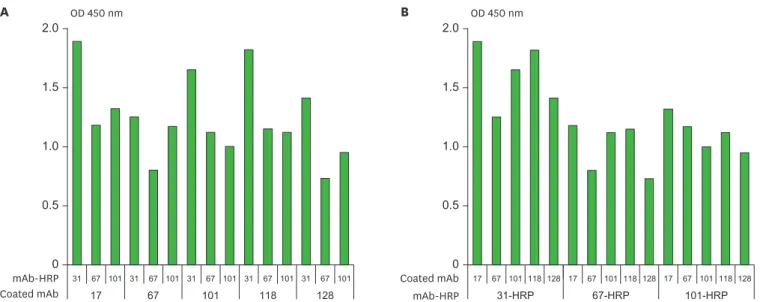
The ELISA signals given by the 9 immobilized mAbs vs. the 7 HRP-conjugated mAbs are shown in Fig. 1. Immobilized mAbs H17, H31, H67, H101, and H128 exhibited OD over 1.3 against H31-HRP and H101-HRP conjugates except H101 & H101-HRP conjugate pair (Fig. 1A). H31-HRP and H101-HRP conjugates exhibited OD over 1.0 against 8 and 6 out of 9 immobilized mAbs respectively (Fig. 1B). As a result, mAbs H17, H31, H67, H101, and H128 exhibited the best traits for immobilization, while mAbs H31 and H101 exhibited the best traits for HRP conjugation. mAbs H17 and H97 proved to be particularly inappropriate for HRP conjugation due to their lower signals (OD below 0.5) against all the immobilized mAbs studied (Fig. 1B).
The purpose of confirmatory screening was to decide the candidates for development of immunoassay kit from the results of primary screening. For this, the immobilized H17, H67, H101, H118, and H128 mAbs were examined against each of the HRP-conjugated mAbs H31, H67, and H101 (Fig. 2). mAb H31 was not included for immobilization due to its best traits for HRP conjugation although it was in the candidates for immobilization in the primary screening. mAbs H67 and H118 were included for HRP conjugation and for immobilization respectively just for comparison although these were not included as candidates for further evaluation in the primary screening. Fig. 2A presents the data with respect to the immobilized mAbs. When the signals of immobilized H67, H101, H118, and H128 against 3 conjugates (H31, H67, and H101) were compared to those of mAb H17, mAbsH101 and H118 exhibited over 85% signals of H17; in contrast H67 and H128 exhibited 65%–75% of signals of H17 (Fig. 2A). As a result, mAbs H17, H101, and H118 exhibit similar efficiencies for immobilization following the accuracy of bioanalytical methods is acceptable within 15%
variation of nominal value (12). Fig. 2B presents the data with respect to the HRP conjugated mAbs. When the signals of H67 and H101 conjugates against 5 immobilized mAbs (H17, H67, H101, H118, and H128) were compared to those of H31 conjugate, both H67 and H101 conjugates exhibited 50%–70% of H31 conjugate except H67 & H101-HRP conjugate pair which exhibited 94% of that of H67 & H31-HRP conjugate pair (Fig. 2B). As a result, the mAb H31-HRP conjugate exhibits the highest signal in all 3 conjugates.
173031677397101118128
1731677397101128
1.0 0.5 0
2.0 1.5 17316773971011281731677397101128173167739710112817316773971011281731677397101128173167739710112817316773971011281731677397101128
OD 450 nmA mAb-HRP Coated mAb 1.0 0.5 0
2.0 1.5
OD 450 nmB 17-HRP31-HRP67-HRP73-HRP97-HRP101-HRP128-HRPmAb-HRP
303167739710111817128303167739710111817128303167739710111817128303167739710111817128303167739710111817128303167739710111817128303167739710111817128Coated mAb Figure 1. The result of primary screening of mAb pairs to screen out the candidates for further evaluation for quantitative HBsAg ELISA. ELISA signals (A) with respect to the immobilized mAbs and (B) with respect to the HRP-conjugated mAbs. ELISA signals were measured for each combination of the 9 immobilized mAbs (H17, H30, H31, H67, H73, H97, H101, H118, and H128) vs. the 7 mAb-HRP conjugates (H17, H31, H67, H73, H97, H101, and H128). Ten ng/ml of HBsAg in 1% BSA-PBS was used as a positive control. Each assay was performed in duplicate. Mean values are shown in the graph.
Therefore, in the following experiment, it was decided that mAb H17 and mAb H31 would be used for immobilization and HRP conjugate preparation, respectively.
In these screening analyses, the ELISA signals were compared against a concentration of 10 ng/ml HBsAg. The background signals given by 1% BSA in PBS were subtracted from the ODs given by 10 ng/ml HBsAg. All the assays were performed in duplicates.
ELISA performance with an optimized mAb pair
mAb H17 was diluted to 10 µg/ml in 0.05 M carbonate buffer, pH 9.6 and immobilized in the plate. HBsAg standards were set in the range of 0.125–40 ng/ml in 1% BSA-PBS. The 1st and 2nd incubations were for 1 h at RT, followed by the substrate reaction for 30 min at RT, which was finally stopped by the addition of 1 N sulfuric acid. The OD was measured at 450 nm.
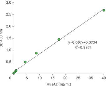
A standard curve was plotted, which exhibited linearity in the range of the standard used (Fig. 3). Back-fitting of the standard curve readout values fell within 20% of the nominal readout value (data not shown) and this satisfied the acceptance criteria that the bias and precision of back-calculated value for at least 75% of the calibration standards should lie within 20% (13).
The detection limit of this system was estimated as 1 ng/ml of HBsAg, which was revealed by a dose response to the ELISA signal and could be differentiated from the background signal (Table 1). The detection limit of HBsAg ELISA Kit (Alpha Diagnostics) and QuickTiter HBsAg ELISA Kit (Cell Biolabs, Inc.) was also 1 ng/ml of HBsAg, as per the product manuals of Alpha Diagnostics & Cell Biolabs, Inc.
DISCUSSION
HBV DNA quantity is the most commonly used marker for therapeutic efficacy during follow-ups in patients chronically infected with HBV when they are usually treated with either
17 67 101 118 128
31 67 101 31 67 101 31
1.0
0.5
0 2.0
1.5
67 101 31 67 101 31 67 101
OD 450 nm
A
mAb-HRP Coated mAb
1.0
0.5
0 2.0
1.5
OD 450 nm
B
31-HRP 67-HRP 101-HRP
mAb-HRP
17 67 101 118 128 17 67 101 118 128 17 67 101 118 128
Coated mAb
Figure 2. The result of confirmatory screening of mAb pairs to decide the candidates for development of quantitative HBsAg ELISA kit. ELISA signals (A) with respect to the immobilized mAbs and (B) with respect to the HRP-conjugated mAbs. ELISA signals were measured for each combination of the 5 immobilized mAbs (H17, H67, H101, H118, and H128) vs. the 3 mAb-HRP conjugates (H31, H67, and H101). Ten ng/ml of HBsAg in 1% BSA-PBS was used as a positive control and 1% BSA-PBS was used as a background control. Each assay was performed in duplicate. Mean values are shown in the graph.
pegylated interferons (PEG-IFNs) or HBV reverse transcriptase nucleos(t)ide analogues (14).
The ultimate goal of anti-HBV treatment is HBsAg clearance, as it leads to the improvement of long-term clinical outcomes, including longer survival (14). However, Anti-HBV treatment cannot be adequately predicted by obtaining an undetectable HBV DNA viral load (14).
Monitoring serum HBsAg levels in patients with CHB may substantially contribute to monitoring HBV DNA (1,14,15). The template for HBV transcription, i.e., the cccDNA, plays a key role in the life cycle of the virus and permits the persistence of infection. HBsAg is translated from mRNA with the transcriptional template-active cccDNA, which has been shown to reflect the number of infected hepatocytes. It has been suggested that HBsAg quantification reflects the concentration of cccDNA in the liver (8). Through this association, HBsAg is hypothesized to be a marker for an immunological response to hepatitis B therapy, independent of the virological response as measured using HBV DNA levels in serum (1).
Quantitative HBsAg was suggested to be helpful in the management of HBV. The recent availability of commercial quantitative assays has reignited an interest in quantitative serum HBsAg as a biomarker for the prognosis and treatment of CHB (16) and stratified the risk of disease progression by predicting treatment responses mainly in patients receiving PEG- IFN therapy (8). HBsAg decline during PEG-IFN therapy of CHB is a strong predictor of a sustained on-treatment response, and patients who fail to achieve an HBsAg decline have a reduced probability of exhibiting a sustained response (1,14,15).
Serum HBV markers are usually detected by EIA, RIA, microparticle enzyme immunoassay (MEIA), or chemiluminescence. The development of automated immunoassay systems has greatly improved the sensitivity, specificity, and accuracy of serum HBV marker detection
Table 1. Varying concentrations of HBsAg exhibiting a dose response during quantitation ELISA after adjusting for background signals
HBsAg (ng/ml) 40 20 10 5 1 0.5 0.25 0.1
ELISA OD* 2.70 1.44 0.86 0.49 0.13 0.07 0.07 0.05
Mean values are shown in the table.
*Each assay was performed in duplicate.
3.0
1.0
0
OD 450 nm
2.5
2.0
1.5
0.5
HBsAg (ng/ml)
5 10 15 20 25 35 40
y=0.067x+0.0704 R2=0.9951
Figure 3. Standard curve for the current HBsAg ELISA system exhibiting linearity within the range of the standards (0–40 ng/ml in 1% BSA-PBS). Each assay was performed in duplicate. Mean values are shown in the graph.
(17). Two fully automated assays are currently commercially available, namely, the Architect HBsAg QT (Abbott Diagnostics) and the Elecsys HBsAg II Quant (Roche Diagnostics) (18).
These assays amplify signals by increasing the surface of immobilization via microparticles and measure these signals by a chemiluminescence reaction using acridinium- or ruthenium- labeled anti-HBs antibodies (monoclonal and polyclonal mixture). As a result, the total assay is very rapid (18 to 35 min) and the sensitivity of the assay is very high, like 0.05 IU/ml, which is equivalent to 0.2 ng/ml of HBsAg (18).
Three ELISA kits for HBsAg quantitation are also commercially available — HBsAg ELISA Kit (Alpha Diagnostics); HBs S Antigen Quantitative ELISA Kit, Rapid-II (Beacle, Inc.);
and QuickTiter HBsAg ELISA Kit (Cell Biolabs, Inc.). HBsAg ELISA Kit and QuickTiter HBsAg ELISA Kit use mAbs for immobilization of HBsAg. Contrastingly, HBs S Antigen Quantitative ELISA Kit, Rapid-II uses polyclonal antibodies. In the secondary reaction of the immunoassay that binds to the captured HBsAg, HBsAg ELISA Kit and HBs S Antigen Quantitative ELISA Kit, Rapid-II, use a polyclonal anti-HBs antibody as an HRP conjugate.
In contrast, QuickTiter HBsAg ELISA Kit uses different detection systems as compared to the other ELISA kits. It includes additional reactions compared to other ELISA kits, by employing FITC-conjugated monoclonal anti-HBs antibody and HRP-conjugated monoclonal anti-FITC antibody.
The ELISA system in this investigation is unique in that it uses mAbs both for capture and HRP-conjugate.
Polyclonal antibodies are employed to increase the sensitivity supposedly in the commercial kits as exampled above. However, using polyclonal antibodies causes problems of
inconsistency of production and quality. Laboratory animals can vary considerably in their ability to respond to different antigens, resulting in significant variation among lots of antibodies (19). Also the ethical aspect of antibodies production using laboratory animals should be considered (20). In contrast, mAbs from hybridoma cells can be produced by bioreactor culture with chemically defined protein-free media without using animals (21,22).
Another commercial kit uses FITC-labeled mAb as a secondary antibody instead of HRP conjugated mAb and this requires one more reaction step and reagent.
The ELISA system in this investigation provides the consistency of quality and simpler reaction step by employing mAbs both for capture and HRP-conjugate. Further mAbs production by bioreactor culture can circumvent the ethical problem of using animals, which could not be achieved in the production of polyclonal antibodies.
The standard ranges of the 3 ELISA kits are as follows: HBsAg ELISA Kit, 0–20 ng/ml; HBs S Antigen Quantitative ELISA Kit, Rapid-II, 0–10 nUnit/ml (where 1 nUnit is defined as the activity expressed by 1 ng of the standard antigen); and QuickTiter HBsAg ELISA Kit, 0–100 ng/ml. In this study, the standard was 0–40 ng/ml.
The detection limit of the HBsAg ELISA Kit and the QuickTiter HBsAg ELISA Kit is 1 ng/ml HBsAg. The detection limit of HBs S Antigen Quantitative ELISA Kit, Rapid-II is 0.05 nUnit/
ml, which is equivalent to 0.05 ng/ml and is far more sensitive than the other ELISA kits and comparable to the chemiluminescence reaction systems such as Architect HBsAg QT and the Elecsys HBsAg II. This kit includes special amplification system to increase the sensitivity.
The detection limit of the current ELISA system in this study was estimated as 1 ng/ml based on the ELISA signals according to the concentration of HBsAg, which is the same as that of HBsAg ELISA Kit and QuickTiter HBsAg ELISA Kit. When the detection limit was estimated by the average baseline value plus 3×standard deviation (23), the detection limit of the current ELISA system was estimated as 0.4 ng/ml. However, this ELISA system did not exhibit dose responses below 0.5 ng/ml of HBsAg (Table 1), as a result the detection limit was estimated as 1 ng/ml.
In conclusion, mAbs against HBsAg were generated and the best pair of mAbs for HBsAg quantitative ELISA was established. The linearity was maintained in 0–40 ng/ml of HBsAg and detection limit was estimated as 1 ng/ml. The ELISA system established in this study is unique in using mAbs both as capture and HRP-conjugate, which can provide the consistency of quality and simpler reaction step compared to other commercial kits.
ACKNOWLEDGEMENTS
This work was supported by the Green Cross Corp., Yongin, Korea.
REFERENCES
1. Sonneveld MJ, Zoutendijk R, Janssen HL. Hepatitis B surface antigen monitoring and management of chronic hepatitis B. J Viral Hepat 2011;18:449-457.
PUBMED | CROSSREF
2. Park H, Lee JM, Seo JH, Kim HS, Ahn SH, Kim DY, Han KH, Chon CY, Park JY. Predictive value of HBsAg quantification for determining the clinical course of genotype C HBeAg-negative carriers. Liver Int 2012;32:796-802.
PUBMED | CROSSREF
3. Werle-Lapostolle B, Bowden S, Locarnini S, Wursthorn K, Petersen J, Lau G, Trepo C, Marcellin P, Goodman Z, Delaney WE 4th. Persistence of cccDNA during the natural history of chronic hepatitis B and decline during adefovir dipivoxil therapy. Gastroenterology 2004;126:1750-1758.
PUBMED | CROSSREF
4. Zoulim F. New insight on hepatitis B virus persistence from the study of intrahepatic viral cccDNA. J Hepatol 2005;42:302-308.
PUBMED | CROSSREF
5. Chan HL, Wong VW, Tse AM, Tse CH, Chim AM, Chan HY, Wong GL, Sung JJ. Serum hepatitis B surface antigen quantitation can reflect hepatitis B virus in the liver and predict treatment response. Clin Gastroenterol Hepatol 2007;5:1462-1468.
PUBMED | CROSSREF
6. Chuaypen N, Sriprapun M, Praianantathavorn K, Payungporn S, Wisedopas N, Poovorawan Y,
Tangkijvanich P. Kinetics of serum HBsAg and intrahepatic cccDNA during pegylated interferon therapy in patients with HBeAg-positive and HBeAg-negative chronic hepatitis B. J Med Virol 2017;89:130-138.
PUBMED | CROSSREF
7. Li J, Sun X, Fang J, Wang C, Han G, Ren W. Analysis of intrahepatic total HBV DNA, cccDNA and serum HBsAg level in chronic hepatitis B patients with undetectable serum HBV DNA during oral antiviral therapy. Clin Res Hepatol Gastroenterol. 2017. doi: 10.1016/j.clinre.2017.03.004.
PUBMED
8. Martinot-Peignoux M, Lapalus M, Asselah T, Marcellin P. HBsAg quantification: useful for monitoring natural history and treatment outcome. Liver Int 2014;34 Suppl 1:97-107.
PUBMED | CROSSREF
9. Karagoz E, Tanoglu A. Clinical usefulness of HBsAg quantification in patients with chronic hepatitis B infection. Hepat Mon 2017;17:e12293.
CROSSREF
10. Hong KW, Kim CG, Lee SH, Chang KH, Shin YW, Ryoo KH, Kim SH, Kim YS. A novel anti-EGFR monoclonal antibody inhibiting tumor cell growth by recognizing different epitopes from cetuximab. J Biotechnol 2010;145:84-91.
PUBMED | CROSSREF
11. Nakane PK, Kawaoi A. Peroxidase-labeled antibody. A new method of conjugation. J Histochem Cytochem 1974;22:1084-1091.
PUBMED | CROSSREF
12. European Medicines Agency. Guideline on Bioanalytical Method Validation. London: European Medicines Agency; 2011.
13. Valentin MA, Ma S, Zhao A, Legay F, Avrameas A. Validation of immunoassay for protein biomarkers:
bioanalytical study plan implementation to support pre-clinical and clinical studies. J Pharm Biomed Anal 2011;55:869-877.
PUBMED | CROSSREF
14. Maylin S, Boyd A, Delaugerre C, Zoulim F, Lavocat F, Simon F, Girard PM, Lacombe K. Comparison between Elecsys HBsAg II and architect HBsAg QT assays for quantification of hepatitis B surface antigen among patients coinfected with HIV and hepatitis B virus. Clin Vaccine Immunol 2012;19:242-248.
PUBMED | CROSSREF
15. Orito E, Fujiwara K, Kanie H, Ban T, Yamada T, Hayashi K. Quantitation of HBsAg predicts response to entecavir therapy in HBV genotype C patients. World J Gastroenterol 2012;18:5570-5575.
PUBMED | CROSSREF
16. Chan HL, Thompson A, Martinot-Peignoux M, Piratvisuth T, Cornberg M, Brunetto MR, Tillmann HL, Kao JH, Jia JD, Wedemeyer H, et al. Hepatitis B surface antigen quantification: why and how to use it in 2011 - a core group report. J Hepatol 2011;55:1121-1131.
PUBMED | CROSSREF
17. İnan N, Demirel A, Ünsur EK, Görmüs U, Sönmez E, Tabak F, Arisoy A. Comparison of
chemiluminescence microparticle immunoassay and electrochemiluminescence immunoassay for detection of HBsAg. Viral Hepat J 2014;20:101-105.
CROSSREF
18. Hadziyannis E. Quantification of HBsAg in serum: characteristics of the assays. OA Hepatol 2013;1:1-6.
19. Andreotti PE, Ludwig GV, Peruski AH, Tuite JJ, Morse SS, Peruski LF Jr. Immunoassay of infectious agents.
Biotechniques 2003;35:850-859.
PUBMED
20. Pohanka M. Monoclonal and polyclonal antibodies production – preparation of potent biorecognition element. J Appl Biomed 2009;7:115-121.
21. Cherlet M, Marc A. Stimulation of monoclonal antibody production of hybridoma cells by butyrate:
evaluation of a feeding strategy and characterization of cell behaviour. Cytotechnology 2000;32:17-29.
PUBMED | CROSSREF
22. Gong X, Li D, Li X, Fang Q, Han X, Wu Y, Yang S, Shen BQ. Fed-batch culture optimization of a growth- associated hybridoma cell line in chemically defined protein-free media. Cytotechnology 2006;52:25-38.
PUBMED | CROSSREF
23. Peng J, Song S, Xu L, Ma W, Liu L, Kuang H, Xu C. Development of a monoclonal antibody-based sandwich ELISA for peanut allergen Ara h 1 in food. Int J Environ Res Public Health 2013;10:2897-2905.
PUBMED | CROSSREF