Diagnostic Whole-Body Scan May Not Be Necessary for Intermediate-Risk Patients with Differentiated Thyroid Cancer after Low-Dose (30 mCi) Radioactive Iodide Ablation
Eon Ju Jeon, Eui Dal Jung
Department of Internal Medicine, Catholic University of Daegu School of Medicine, Daegu, Korea
Background: A diagnostic whole-body scan (WBS) is recommended 6 to 12 months after total thyroidectomy and radioactive iodide ablation in intermediate- or high-risk patients with differentiated thyroid cancer (DTC). The aim of this study was to evalu- ate the necessity of a diagnostic WBS after radioactive iodide ablation in intermediate-risk patients with DTC.
Methods: A total of 438 subjects were included in the study: 183 low-risk subjects and 255 intermediate-risk subjects according to the American Thyroid Association guideline. All subjects diagnosed with DTC received 1,100 MBq (30 mCi) activity of radio- iodine (I-131) following total thyroidectomy. On follow-up, all subjects underwent a diagnostic I-131 WBS after thyroid hor- mone withdrawal.
Results: After initial radioactive iodide ablation, 95.1% of low-risk patients and 91.4% of intermediate-risk patients showed no uptake on diagnostic WBS (P=0.135). Intermediate-risk patients with stimulated thyroglobulin (Tg) levels higher than 2.0 ng/mL showed a greater rate of radioactive iodine uptake on diagnostic WBS. Four intermediate-risk patients showed recurrence during the 16 to 80 months follow-up period. Three of the four patients with recurrence showed no uptake on diagnostic WBS and had a stimulated Tg level less than 2.0 ng/mL.
Conclusion: A diagnostic I-131 WBS after radioactive iodide ablation in intermediate-risk patients with DTC may not be neces- sary. A large prospective study is necessary to determine the necessity of diagnostic WBS in intermediate-risk patients with DTC.
Keywords: Ablation; Iodides; Radioactivity; Thyroid neoplasms
INTRODUCTION
Differentiated thyroid carcinoma accounts for 90% of thyroid cancers including papillary carcinoma and follicular carcino- ma. Major treatment includes surgical treatment and radioio- dine ablation, and can bring positive consequences with a
greater than 90% survival rate [1,2]. Complete ablation of re- sidual thyroid tissue by radioiodine infusion after total thyroid- ectomy can decrease the risk of local recurrence and increase the long-term survival rate [3-8]. Recently, the frequency of thyroid nodules and differentiated thyroid carcinomas has in- creased, and a number of clinical studies have been conducted.
Received: 5 March 2013, Accepted: 20 August 2013 Corresponding author: Eui Dal Jung
Department of Internal Medicine, Catholic University of Daegu School of Medicine, 33 Duryugongwon-ro 17-gil, Nam-gu, Daegu 705-718, Korea Tel: +82-53-650-4098, Fax: +82-53-651-4009, E-mail: [email protected]
Copyright © 2014 Korean Endocrine Society
This is an Open Access article distributed under the terms of the Creative Com- mons Attribution Non-Commercial License (http://creativecommons.org/
licenses/by-nc/3.0/) which permits unrestricted non-commercial use, distribu- tion, and reproduction in any medium, provided the original work is properly cited.
Consequently, the American Thyroid Association (ATA) pub- lished revised guidelines on practice recommendations [4,9, 10]. In these guidelines, the staging system for thyroid cancer uses the American Joint Committee on Cancer/International Union against Cancer-Tumor, Lymph node, Metastasis (AJCC/
UICC-TNM) as a predictor of mortality. Therefore, it is not suitable for determining the intensity of follow-ups and has limited impact on risk of recurrence. For these reasons, patients are classified as low-risk, intermediate-risk, and high-risk groups according to the risk of recurrence [4,11-14]. Diagnostic whole-body scans (WBSs) performed on patients with radioio- dine thyroid ablation for 6 to 12 months have low sensitivity, so although it is not necessary for low-risk patients [15-22], it can be useful for intermediate-risk and high-risk patients at 6 to 12 months [4,23-26]. However, there is no clear evidence suggest- ing the usefulness of diagnostic WBS on intermediate-risk pa- tients. Therefore, this study aims to evaluate whether diagnostic WBS at 6 to 12 months after remnant ablation is necessary for intermediate-risk patients who have differentiated thyroid car- cinoma after total thyroidectomy and have been treated with 1,100 MBq (30 mCi) radioiodine thyroid ablation.
METHODS
This study included 438 patients with differentiated thyroid carcinoma who had undergone 30 mCi radioiodine thyroid ab- lation between January 2003 and December 2011. According to the ATA’s 2009 revised guideline for thyroid nodule and can- cer treatment, low-risk and intermediate-risk groups have been established to reflect the presence of residual lesions and risk of relapse. A low-risk patient is defined by: 1) no local and dis- tant metastasis; 2) all macroscopic tumor has been resected; 3) there is no tumor invasion of locoregional tissues or structures;
4) the tumor does not have aggressive histology (e.g., tall cell, insular, columnar cell carcinoma) or vascular invasion; and 5) if I-131 is given, there is no I-131 uptake outside the thyroid bed on the first posttreatment whole-body radioactive iodide scan. Intermediate-risk patients have any of the following: 1) microscopic invasion of tumor into the perithyroidal soft tissue at initial surgery; 2) cervical lymph node metastases or I-131 uptake outside the thyroid bed on the whole-body radioactive iodide scan done after thyroid remnant ablation; and 3) tumor with aggressive histology or vascular invasion [4].
The post-therapeutic WBS was performed on the third day after administrating 30 mCi radioiodine (I-131) in the sixth week after total thyroidectomy. Pretreatment was as follows:
the patients underwent thyroid hormone withdrawal to increase the radioiodine uptake in residual thyroid tissue. Thyroxine was discontinued 4 weeks before ablation. Instead of thyrox- ine, triiodothyroxine (20 µg) was given orally twice a day for 2 weeks.Triiodothyroxine was withdrawn, and the patients were instructed to follow a low-iodine diet for 2 weeks before abla- tion. Before I-131 administration, serum thyroid stimulating hormone (TSH), free T4, T3, TSH-stimulating thyroglobulin (Tg; reference, 1.4 to 78.0 ng/mL), and antithyroglobulin anti- body (anti-Tg Ab, negative <100 IU/mL) were measured [27].
For all patients, TSH increased to above 30 mU/L after thyroid hormone withdrawal [27]. Diagnostic WBS was performed at 6 to 12 months after radioiodine thyroid ablation. The 3 mCi dose was administered 3 days prior to scanning, and then a γ camera was used to shoot for 13 minutes (Millennium GE, Is- rael). Prior to diagnostic WBS, TSH, free T4, T3, TSH-stimu- lating Tg, and Tg Ab were measured as a follow-up test. The presence of a relapse was confirmed by the results of ultra- sound-induced fine needle aspiration cytology and surgical his- topathology from the suspicious region on neck ultrasonogra- phy. Electric chemiluminescence immunoassays (Roche Diag- nostics, Mannheim, Germany) were used to measure TSH, free T4, and T3, and enzyme immunosorbent assay (Alisei, SEAC, Pomezia, Italy) was used to measure Tg and Tg Ab. The mean comparison of the two groups was performed with t test or Mann-Whitney U test. The categorical variables are presented as numbers and percentages, and they were compared using the chi-square test or Fisher exact test. P<0.05 were statistically considered significant. All the statistical analyses were per- formed using SPSS version 12.0 for Windows (SPSS Inc., Chi- cago, IL, USA).
RESULTS
Clinicopathologic characteristics of low-risk and intermediate-risk patients
Total of 438 patients was observed. Most were diagnosed with papillary carcinoma with no histology of poor prognosis. There were 183 patients in the low-risk group, and 255 patients in the intermediate-risk group. There were no significant differences in age or sex between the two groups (P=0.828 and P=0.106, respectively). The AJCC TNM clinical stage was significantly lower for low-risk patients than for intermediate-risk patients (P<0.001). The size of the thyroid tumor was similar between the two groups: 1.22±0.92 cm in intermediate-risk patients and 1.11±0.78 cm in low-risk patients (P=0.170). The period
during follow-up was 4.42 years (range, 1.33 to 8.75) for low- risk patients and 3.25 years (range, 1.17 to 8.17) for intermedi- ate-risk patients (P<0.001) (Table 1).
Comparison of diagnostic whole-body scan uptake after radioactive iodine ablation according to risk group
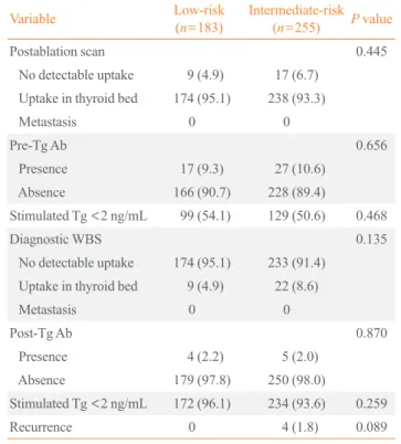
Patients received diagnostic I-131 WBS at 6 to 12 months with radioiodine ablation after total thyroidectomy. Blood tests per- formed at thyroid hormone withdrawal before diagnostic WBS. The 96.1% of low-risk patients and 93.6% of intermedi- ate-risk patients had Tg levels below 2 ng/mL in the absence of Tg Ab, representing no significant difference (P=0.259). At diagnostic WBS, 95.1% of low-risk patients and 91.4% of in- termediate-risk patients had no I-131 uptake outside the thy- roid bed. There was no significant difference between the two groups (P=0.135). The scan result also suggested no local and distant metastasis except the thyroid bed (Table 2). Confirming the presence of a recurrence, 1.8% of intermediate-risk patients and 0.0% of low-risk patients had recurrence of thyroid cancer, showing no statistically significant differences (P=0.089).
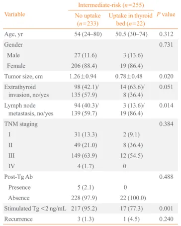
Clinicopathologic characteristics according to diagnostic whole-body scan results in intermediate-risk patients The clinicopathologic characteristics according to the results of diagnostic WBS in intermediate-risk patients were observed
(Table 3). Among 255 intermediate-risk patients, 233 had no uptake in the thyroid bed, and 22 had uptake on the thyroid bed. On diagnostic WBS, the group showing uptake in the thy- roid bed had significantly smaller tumors than the group with no uptake (0.78±0.48 cm vs. 1.26±0.94 cm, respectively; P=
0.020), higher lymph node metastasis (86.4% and 59.7%, re- spectively; P=0.014), Tg levels below 2 ng/mL in the absence of Tg Ab (77.3% and 95.2%, respectively; P=0.001). There was no statistically significant between thyroid intake and age, sex, TNM stage, or Tg Ab.
The presence of uptake in thyroid bed on diagnostic whole-body scan and the recurrence according to thyroglobulin levels in intermediate-risk patients
We performed a blood test before diagnostic WBS after radio- iodine ablation on intermediate-risk patients, and the scan re- sults were compared between patients with negative Tg Ab, di- viding the results of TSH-stimulating Tg levels into two groups based on levels below 2 ng/mL and above 2 ng/mL (Table 4).
Table 1. Clinicopathologic Parameters of the Low- and Inter- mediate-Risk Groups
Variable Low-risk
(n=183) Intermediate-
risk (n=255) P value
Age, yr 52 (22–84) 53 (24–80) 0.828
Gender 0.106
Male 13 (7.1) 30 (11.8)
Female 170 (92.9) 225 (88.2)
Tumor size, cm 1.11±0.78 1.22±0.92 0.170
TNM staging <0.001
I 159 (86.9) 33 (12.9)
II 15 (8.2) 57 (22.4)
III 9 (4.9) 161 (63.1)
IV 0 4 (1.6)
Histology 0.152
Papillary 180 (98.4) 251 (98.4)
Follicular 3 (1.6) 4 (1.8)
Follow-up duration, yr 4.42 (1.33–8.75) 3.25 (1.17–8.17) <0.001 Values are expressed as median (range), number (%), or mean±SD.
TNM, tumor node metastasis.
Table 2. Comparison of Diagnostic Whole-Body Scan Find- ings after Radioactive Iodide Ablation according to Risk Group
Variable Low-risk
(n=183) Intermediate-risk (n=255) P value
Postablation scan 0.445
No detectable uptake 9 (4.9) 17 (6.7) Uptake in thyroid bed 174 (95.1) 238 (93.3)
Metastasis 0 0
Pre-Tg Ab 0.656
Presence 17 (9.3) 27 (10.6)
Absence 166 (90.7) 228 (89.4)
Stimulated Tg <2 ng/mL 99 (54.1) 129 (50.6) 0.468
Diagnostic WBS 0.135
No detectable uptake 174 (95.1) 233 (91.4) Uptake in thyroid bed 9 (4.9) 22 (8.6)
Metastasis 0 0
Post-Tg Ab 0.870
Presence 4 (2.2) 5 (2.0)
Absence 179 (97.8) 250 (98.0)
Stimulated Tg <2 ng/mL 172 (96.1) 234 (93.6) 0.259
Recurrence 0 4 (1.8) 0.089
Values are expressed as number (%). P<0.05.
Pre, before radioactive iodide ablation; Tg, thyroglobulin; Ab, anti- body; WBS, whole-body scan; Post, just before diagnostic WBS.
On diagnostic WBS, 16 out of 250 patients (6.4%) had Tg lev- els higher than 2 ng/mL, and these patients also had a signifi- cantly higher uptake in thyroid bed compared to the below 2 ng/
mL group (31.2% and 7.3%, respectively; P=0.008). However, the recurrence of thyroid cancer was not statistically significant (n=1 [6.2%] and n=3 [1.3%], respectively; P=0.234). The clinical pathology of relapsed patients is described in Table 5.
DISCUSSION
In this study, based on a diagnostic I-131 WBS performed at 6 to 12 months, 255 intermediate-risk patients and 183 low-risk patients diagnosed with differentiated thyroid carcinoma who were initially treated total thyroidectomy and 30 mCi radioio- dine ablation had no statistically significant difference in resid- ual tissue removal rate (95.1% and 91.4%, respectively; P=
0.135). With TSH-stimulating Tg level below 2 ng/mL in the absence Tg Ab, no significant difference was found between intermediate- (93.6%) and low-risk (96.1%) groups (P=0.259).
Intermediate-risk patients were divided into two groups of TSH-stimulating Tg, below 2 ng/mL and above 2 ng/mL in the absence Tg Ab during follow-up before diagnostic WBS. The results of diagnostic WBS were statistically significant differ- ences; 92.7% of the below 2 ng/mL group and 68.8% of the above 2 ng/mL group had no uptake in the thyroid bed (P=0.008). These results suggest that TSH-stimulating Tg im- pacts radioiodine uptake in remnant tissue. During follow-up, four recurrent patients were observed with local recurrence of lymph node metastasis, and most were not observed for thyroid
Table 5. Summary of Clinicopathologic Data of the Four Patients with Recurrence Paitent Age,
yr/sex Tumor
size, cm Extrathyroid invasion LN
Mets TNM
staging
Initial Follow-up
1st WBS Tg, ng/mL Tg Ab Dx WBS Tg, ng/mL Tg Ab
1 75/F 1.5 Y N 3 Remnant <0.5 (–) N <0.5 (–)
2 70/F 2.8 Y Y 3 Remnant 9.1 (–) N 0.803 (–)
3 47/M 0.5 N Y 3 Remnant 8.6 (–) Remnant 2.6 (–)
4 61/F 1.5 N Y 3 Remnant <0.5 (–) N <0.5 (–)
LN, lymph node; Mets, metastasis; TNM, tumor node metastasis; WBS, whole-body scan; Tg, thyroglobulin; Ab, antibody; Dx, diagnostic; F, fe- male; Y, yes; N, no; Remnant, uptake in thyroid bed; M, male.
Table 3. Clinicopathologic Parameters according to Diagnos- tic Whole-Body Scan Results in Intermediate-Risk Patients
Variable
Intermediate-risk (n=255)
P value No uptake
(n=233) Uptake in thyroid bed (n=22)
Age, yr 54 (24–80) 50.5 (30–74) 0.312
Gender 0.731
Male 27 (11.6) 3 (13.6)
Female 206 (88.4) 19 (86.4)
Tumor size, cm 1.26±0.94 0.78±0.48 0.020
Extrathyroid
invasion, no/yes 98 (42.1)/
135 (57.9) 14 (63.6)/
8 (36.4) 0.051 Lymph node
metastasis, no/yes 94 (40.3)/
139 (59.7) 3 (13.6)/
19 (86.4) 0.014
TNM staging 0.384
I 31 (13.3) 2 (9.1)
II 49 (21.0) 8 (36.4)
III 149 (63.9) 12 (54.5)
IV 4 (1.7) 0
Post-Tg Ab 0.488
Presence 5 (2.1) 0
Absence 228 (97.9) 22 (100.0)
Stimulated Tg <2 ng/mL 217 (95.2) 17 (77.3) 0.001
Recurrence 3 (1.3) 1 (4.5) 0.240
Values are expressed as median (range), number (%), or mean±SD.
TNM, tumor node metastases; Post, before diagnostic whole-body scan; Tg, thyroglobulin; Ab, antibody.
Table 4. Diagnostic Whole-Body Scan Results and Recurrence of Intermediate-Risk Patients, according to Thyroid Stimulat- ing Hormone-Stimulated Thyroglobulin Level in the Absence of Antithyroglobulin Antibody (Total, n=250)
Variable Tg <2, ng/mL
(n=234) Tg ≥2, ng/mL (n=16) P value
Diagnostic WBS 0.008
No detectable uptake 217 (92.7) 11 (68.8) Uptake in thyroid bed 17 (7.3) 5 (31.2)
Metastasis 0 0
Recurrence, no/yes 231 (98.7)/3 (1.3) 15 (93.8)/1 (6.2) 0.234 Values are expressed as number (%).
Tg, thyroglobulin; WBS, whole-body scan.
bed uptake from the diagnostic WBS.
Cailleux et al. [28] conducted diagnostic WBS at 6 to 12 months after 3.7 GBq (100 mCi) of radioiodine ablation on dif- ferentiated thyroid carcinoma patients treated with total thy- roidectomy. Ninety-two percent of 256 patients had no uptake in the thyroid bed, while 8% of the patients had uptake. There- fore, measuring Tg after thyroid hormone withdrawal for de- tecting recurrence and metastasis of thyroid cancer has high sensitivity; however, diagnostic WBS performed during fol- low-up had no additional usefulness. Pacini et al. [17] reported similar results from 662 patients with 1,100 MBq to 3.7 GBq (30 to 100 mCi) of radioiodine remnant ablation. In our study, although low-risk (95.1%/4.9%) and intermediate-risk patients (91.4%/8.6%) had no statistically significant differences for uptake in the thyroid bed on diagnostic WBS, the results were similar to that of Cailleux et al. [28]. Therefore, this suggests that a low dose (30 mCi) of radioiodine remnant ablation is not inferior on residual tissue removal compared to a high dose (100 mCi). In addition, the presence of uptake on diagnostic whole-body was similar to the low in intermediate-risk patients as well as low-risk patients, which suggests that diagnostic WBS may not be useful for predicting the recurrence.
In this study, four intermediate-risk patients had recurrence of thyroid cancer during follow-up, and all were suspected to have local lymph node metastasis, based on neck ultrasonogra- phy and confirmed with fine needle aspiration cystology and surgical histology. Neck ultrasonography is a very sensitive ex- amination that can detect cervical metastasis or local recur- rence on differentiated thyroid carcinoma, and it also can find metastatic lesions on a patient who is negative for TSH-stimu- lating Tg [29,30]. Like this, neck ultrasonography is sensitive for confirming recurrence of cancer in intermediate-risk pa- tients. Three out of four patients had no detectable uptake of radioiodine on diagnostic WBS with TSH-stimulating Tg level below 2 ng/mL in the absence Tg Ab. Only one patient had up- take with TSH-stimulating Tg level above 2 ng/mL in the ab- sence Tg Ab. Serum Tg measurement after thyroid hormone withdrawal is known to be highly sensitive test in identifying patients with persistent tumor [31]. However, since the three patients within the group who had recurrence had no uptake on diagnostic WBS, and a TSH-stimulating Tg level below 2 ng/
mL, it turned out to be less useful. Instead, neck ultrasonogra- phy to detect lymph node recurrence seems to be useful even though TSH-stimulating Tg is below 2 ng/mL. Therefore, the result of the diagnostic WBS may not be helpful for surveilling the recurrence risk for intermediate-risk patients. For this rea-
son, diagnostic WBS may not be necessary 6 to 12 months af- ter radioiodine ablation. However, there must be additional fol- low-up studies for patients with a high level of TSH-stimulat- ing Tg measured at first radioiodine ablation.
This study has the following limitations. First, there was a possible selection bias since this study was a retrospective study conducted in single hospital and with a small scale group. Sec- ond, it was not easy to determine the correlation between the poor prognosis and papillary microcarcinoma accompanied by minimally extrathyroid extension. In our study, about 36% of patients showed papillary microcarcinoma with minimal extra- thyroid extension. In these patients, it is controversial whe ther the initially radioiodine ablation is useful, and therefore this challenges the value of the WBS [32,33]. Third, considering the long-term survival rate of thyroid cancer patients, the mean period of follow-up was as short as 3 to 4 years, rendering few patients with recurrence.
In summary, TSH-stimulating Tg level, the most sensitive surveillance of residual thyroid cancer, reflected the radioio- dine uptake on diagnostic WBS. In addition, considering the difficulties of pretreatment including exposure to I-131, ex- pense, and low-iodine diet, diagnostic WBS after radioiodine ablation seems to be of little value for intermediate-risk pa- tients, as it is for low-risk patients. However, a well-designed long-term and large-scale prospective multicenter study is needed to determine the main issues in the future.
CONFLICTS OF INTEREST
No potential conflict of interest relevant to this article was re- ported.
REFERENCES
1. Mazzaferri EL, Jhiang SM. Long-term impact of initial surgical and medical therapy on papillary and follicular thyroid cancer. Am J Med 1994;97:418-28.
2. Sherman SI. Thyroid carcinoma. Lancet 2003;361:501-11.
3. Shah MD, Hall FT, Eski SJ, Witterick IJ, Walfish PG, Freeman JL. Clinical course of thyroid carcinoma after neck dissection. Laryngoscope 2003;113:2102-7.
4. American Thyroid Association (ATA) Guidelines Task- force on Thyroid Nodules and Differentiated Thyroid Can- cer, Cooper DS, Doherty GM, Haugen BR, Kloos RT, Lee SL, Mandel SJ, Mazzaferri EL, McIver B, Pacini F, Sch- lumberger M, Sherman SI, Steward DL, Tuttle RM. Re-
vised American Thyroid Association management guide- lines for patients with thyroid nodules and differentiated thyroid cancer. Thyroid 2009;19:1167-214.
5. Schlumberger MJ. Papillary and follicular thyroid carcino- ma. N Engl J Med 1998;338:297-306.
6. Samaan NA, Schultz PN, Hickey RC, Goepfert H, Haynie TP, Johnston DA, Ordonez NG. The results of various mo- dalities of treatment of well differentiated thyroid carcino- mas: a retrospective review of 1599 patients. J Clin Endo- crinol Metab 1992;75:714-20.
7. Vini L, Harmer C. Radioiodine treatment for differentiated thyroid cancer. Clin Oncol (R Coll Radiol) 2000;12:365- 72.
8. Kim TY, Kim WB, Kim ES, Ryu JS, Yeo JS, Kim SC, Hong SJ, Shong YK. Serum thyroglobulin levels at the time of 131I remnant ablation just after thyroidectomy are useful for early prediction of clinical recurrence in low- risk patients with differentiated thyroid carcinoma. J Clin Endocrinol Metab 2005;90:1440-5.
9. Davies L, Welch HG. Increasing incidence of thyroid can- cer in the United States, 1973-2002. JAMA 2006;295:
2164-7.
10. Cooper DS, Doherty GM, Haugen BR, Kloos RT, Lee SL, Mandel SJ, Mazzaferri EL, McIver B, Sherman SI, Tuttle RM; American Thyroid Association Guidelines Taskforce.
Management guidelines for patients with thyroid nodules and differentiated thyroid cancer. Thyroid 2006;16:109-42.
11. Brierley JD, Panzarella T, Tsang RW, Gospodarowicz MK, O’Sullivan B. A comparison of different staging systems predictability of patient outcome. Thyroid carcinoma as an example. Cancer 1997;79:2414-23.
12. Loh KC, Greenspan FS, Gee L, Miller TR, Yeo PP. Patho- logical tumor-node-metastasis (pTNM) staging for papil- lary and follicular thyroid carcinomas: a retrospective analysis of 700 patients. J Clin Endocrinol Metab 1997;82:
3553-62.
13. Wittekind C, Compton CC, Greene FL, Sobin LH. TNM residual tumor classification revisited. Cancer 2002;94:
2511-6.
14. Sherman SI, Brierley JD, Sperling M, Ain KB, Bigos ST, Cooper DS, Haugen BR, Ho M, Klein I, Ladenson PW, Robbins J, Ross DS, Specker B, Taylor T, Maxon HR 3rd.
Prospective multicenter study of thyroiscarcinoma treat- ment: initial analysis of staging and outcome. National Thyroid Cancer Treatment Cooperative Study Registry Group. Cancer 1998;83:1012-21.
15. Torlontano M, Crocetti U, D’Aloiso L, Bonfitto N, Di Giorgio A, Modoni S, Valle G, Frusciante V, Bisceglia M, Filetti S, Schlumberger M, Trischitta V. Serum thyroglobu- lin and 131I whole body scan after recombinant human TSH stimulation in the follow-up of low-risk patients with differentiated thyroid cancer. Eur J Endocrinol 2003;148:
19-24.
16. Schlumberger M, Berg G, Cohen O, Duntas L, Jamar F, Jarzab B, Limbert E, Lind P, Pacini F, Reiners C, Sanchez Franco F, Toft A, Wiersinga WM. Follow-up of low-risk patients with differentiated thyroid carcinoma: a European perspective. Eur J Endocrinol 2004;150:105-12.
17. Pacini F, Capezzone M, Elisei R, Ceccarelli C, Taddei D, Pinchera A. Diagnostic 131-iodine whole-body scan may be avoided in thyroid cancer patients who have undetect- able stimulated serum Tg levels after initial treatment. J Clin Endocrinol Metab 2002;87:1499-501.
18. Torlontano M, Attard M, Crocetti U, Tumino S, Bruno R, Costante G, D’Azzo G, Meringolo D, Ferretti E, Sacco R, Arturi F, Filetti S. Follow-up of low risk patients with pap- illary thyroid cancer: role of neck ultrasonography in de- tecting lymph node metastases. J Clin Endocrinol Metab 2004;89:3402-7.
19. Frasoldati A, Pesenti M, Gallo M, Caroggio A, Salvo D, Valcavi R. Diagnosis of neck recurrences in patients with differentiated thyroid carcinoma. Cancer 2003;97:90-6.
20. Frilling A, Gorges R, Tecklenborg K, Gassmann P, Bock- horn M, Clausen M, Broelsch CE. Value of preoperative diagnostic modalities in patients with recurrent thyroid carcinoma. Surgery 2000;128:1067-74.
21. Ronga G, Filesi M, Ventroni G, Vestri AR, Signore A. Val- ue of the first serum thyroglobulin level after total thyroid- ectomy for the diagnosis of metastases from differentiated thyroid carcinoma. Eur J Nucl Med 1999;26:1448-52.
22. Toubeau M, Touzery C, Arveux P, Chaplain G, Vaillant G, Berriolo A, Riedinger JM, Boichot C, Cochet A, Brunotte F. Predictive value for disease progression of serum thyro- globulin levels measured in the postoperative period and after (131)I ablation therapy in patients with differentiated thyroid cancer. J Nucl Med 2004;45:988-94.
23. Cobin RH, Gharib H, Bergman DA, Clark OH, Cooper DS, Daniels GH, Dickey RA, Duick DS, Garber JR, Hay ID, Kukora JS, Lando HM, Schorr AB, Zeiger MA; Thyroid Carcinoma Task Force. AACE/AAES medical/surgical guidelines for clinical practice: management of thyroid car- cinoma. American Association of Clinical Endocrinolo-
gists. American College of Endocrinology. Endocr Pract 2001;7:202-20.
24. Taylor T, Specker B, Robbins J, Sperling M, Ho M, Ain K, Bigos ST, Brierley J, Cooper D, Haugen B, Hay I, Hertz- berg V, Klein I, Klein H, Ladenson P, Nishiyama R, Ross D, Sherman S, Maxon HR. Outcome after treatment of high-risk papillary and non-Hurthle-cell follicular thyroid carcinoma. Ann Intern Med 1998;129:622-7.
25. Sawka AM, Thephamongkhol K, Brouwers M, Thabane L, Browman G, Gerstein HC. Clinical review 170: a system- atic review and metaanalysis of the effectiveness of radio- active iodine remnant ablation for well-differentiated thy- roid cancer. J Clin Endocrinol Metab 2004;89:3668-76.
26. Durante C, Haddy N, Baudin E, Leboulleux S, Hartl D, Travagli JP, Caillou B, Ricard M, Lumbroso JD, De Vathaire F, Schlumberger M. Long-term outcome of 444 patients with distant metastases from papillary and follicular thy- roid carcinoma: benefits and limits of radioiodine therapy.
J Clin Endocrinol Metab 2006;91:2892-9.
27. Edmonds CJ, Hayes S, Kermode JC, Thompson BD. Mea- surement of serum TSH and thyroid hormones in the man- agement of treatment of thyroid carcinoma with radioio- dine. Br J Radiol 1977;50:799-807.
28. Cailleux AF, Baudin E, Travagli JP, Ricard M, Schlum- berger M. Is diagnostic iodine-131 scanning useful after total thyroid ablation for differentiated thyroid cancer? J Clin Endocrinol Metab 2000;85:175-8.
29. David A, Blotta A, Rossi R, Zatelli MC, Bondanelli M, Roti E, Braverman LE, Busutti L, degli Uberti EC. Clini- cal value of different responses of serum thyroglobulin to recombinant human thyrotropin in the follow-up of pa- tients with differentiated thyroid carcinoma. Thyroid 2005;
15:267-73.
30. Pacini F, Molinaro E, Castagna MG, Agate L, Elisei R, Ce- ccarelli C, Lippi F, Taddei D, Grasso L, Pinchera A. Re- combinant human thyrotropin-stimulated serum thyro- globulin combined with neck ultrasonography has the highest sensitivity in monitoring differentiated thyroid car- cinoma. J Clin Endocrinol Metab 2003;88:3668-73.
31. Eustatia-Rutten CF, Smit JW, Romijn JA, van der Kleij- Corssmit EP, Pereira AM, Stokkel MP, Kievit J. Diagnostic value of serum thyroglobulin measurements in the follow- up of differentiated thyroid carcinoma, a structured meta- analysis. Clin Endocrinol (Oxf) 2004;61:61-74.
32. Kim HJ, Kim NK, Choi JH, Kim SW, Jin SM, Suh S, Bae JC, Min YK, Chung JH, Kim SW. Radioactive iodine abla- tion does not prevent recurrences in patients with papillary thyroid microcarcinoma. Clin Endocrinol (Oxf) 2013;78:
614-20.
33. Lim DJ, Baek KH, Lee YS, Park WC, Kim MK, Kang MI, Jeon HM, Lee JM, Yun-Cha B, Lee KW, Son HY, Kang SK. Clinical, histopathological, and molecular characteris- tics of papillary thyroid microcarcinoma. Thyroid 2007;
17:883-8.