• 검색 결과가 없습니다.

Intracardiac Foreign Body (Bone Cement) after Percutaneous Vertebroplasty

N/A
N/A
Protected

Academic year: 2021

Share "Intracardiac Foreign Body (Bone Cement) after Percutaneous Vertebroplasty"

Copied!
4
0
0

로드 중.... (전체 텍스트 보기)

전체 글

(1)

Korean J Thorac Cardiovasc Surg 2013;46:72-75 □ Case Report □ http://dx.doi.org/10.5090/kjtcs.2013.46.1.72 ISSN: 2233-601X (Print) ISSN: 2093-6516 (Online)

− 72 −

1

Department of Thoracic and Cardiovascular Surgery, Gyeongsang National University School of Medicine,

2

Institute of Health Science, Gyeongsang National University

Received: August 14, 2012, Revised: September 11, 2012, Accepted: September 13, 2012

Corresponding author: Jong Woo Kim, Department of Thoracic and Cardiovascular Surgery, Gyeongsang National University Hospital, Gyeongsang National University School of Medicine, 79 Gangnam-ro, Jinju 660-702, Korea

(Tel) 82-55-750-8124 (Fax) 82-55-750-8138 (E-mail) cs99kjw@hanmail.net

C

The Korean Society for Thoracic and Cardiovascular Surgery. 2013. All right reserved.

CC
This is an open access article distributed under the terms of the Creative Commons Attribution Non-Commercial License (http://creative- commons.org/licenses/by-nc/3.0) which permits unrestricted non-commercial use, distribution, and reproduction in any medium, provided the original work is properly cited.

Intracardiac Foreign Body (Bone Cement) after Percutaneous Vertebroplasty

Jun Ho Yang, M.D.

1,2

, Jong Woo Kim, M.D.

1,2

, Hyun Oh Park, M.D.

1,2

, Jun Young Choi, M.D.

1,2

, In Seok Jang, M.D.

1,2

, Chung Eun Lee, M.D.

1,2

Percutaneous vertebroplasty is a relatively easy and minimally invasive procedure used in treating vertebral fractures. However, the procedure has many complications, one of which is bone cement leakage, which happens frequently. Leakage to the paravertebral venous system, in particular, may lead to especially devastating consequences. Here we report a case of a 65-year-old male patient with an intracardiac foreign body (bone ce- ment) that generated a perforation on the right ventricle, and result in hemopericardium after percutaneous vertebroplasty. We performed open heart surgery to remove the foreign body.

Key words: 1. Foreign body 2. Pericardial effusion

3. Percutaneous vertebroplasty 4. Bone cements

CASE REPORT

A 65-year-old male was admitted to the emergency room with the chief complaint of epigastric pain and dyspnea that had started 10 days earlier. The patient had a history of un- dergoing percutaneous vertebroplasty (PV) in the third lumbar vertebra a month earlier, which was treated at a local clinic.

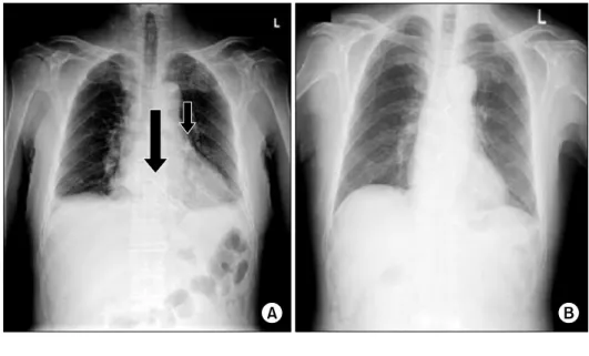
A chest roentgenogram showed a long linear radio-opaque foreign body in the right ventricle (RV) and a shorter one in the pulmonary artery (Fig. 1A). A computed tomography (CT) scan showed the foreign body in the RV, perforating the RV wall near the aortic root and just to the left lateral to the right coronary artery (Fig. 2A), and a moderate quantity of hemopericardium was detected (Fig. 2B). The cardiac echography findings showed hemopericardium and the foreign

body, but no cardiac wall motion dysfunction or valvular dys- function was seen. We planned open heart surgery for the re- moval of the bone cement. Sternotomy was performed in a routine fashion. When the pericardium was opened, a moder- ate amount of hemopericardium was evacuated (200 mL).

The sharp end of the bone cement was perforating the RV wall as we expected (Fig. 3A). By using bi-caval venous can- nulation and single aortic cannulation, a cardiopulmonary by- pass was conducted. The body temperature was lowered to mild hypothermia. The bone cement perforating the RV wall was embedded strongly. Right atriotomy was performed, and the tricuspid valve was checked and shown to be unharmed.

Except for the perforated site, the RV showed no signs of

injury. By retracting the tricuspid valve leaflet, the bone ce-

ment could be seen in the RV chamber. The foreign body

(2)

Intracardiac Foreign Body (Bone Cement) after Percutaneous Vertebroplasty

− 73 −

Fig. 3. (A) Sharp end of a foreign body perforating the right ventricle (arrow). (B) Foreign bodies removed (9 and 2 cm in length, respectively).

Fig. 1. (A) Initial chest roentgeno- gram showing the foreign bodies (ar- rows). (B) Postoperative chest roe- ntgenogram showing no remnants of the foreign bodies.

Fig. 2. (A) Foreign body perforating the right ventricular wall. (B) Hemo- pericardium due to perforation of the right ventricular wall.

was removed by delicately mobilizing the bone cement from inside of the RV chamber toward the outside (Fig. 3B). The perforated site was repaired using a pledgeted Prolene suture.

Total systemic circulatory arrest was performed for the re-

moval of the bone cement in the left pulmonary artery, and

the pulmonary trunk was opened. The bone cement was re-

(3)

Jun Ho Yang, et al

− 74 − moved with biliary forceps in a blind manner (Fig. 3B). After repair of the pulmonary artery, the body temperature was raised back to normal, and the patient was weaned from the cardiopulmonary bypass successfully. Postoperative recovery was uneventful and fast. On postoperative day 7, a follow-up CT scan, a chest roentgenogram, and cardiac echography were performed. The procedures showed no remnants of the foreign bodies (Fig. 1B) or functional disorder. The patient was discharged the next day.

DISCUSSION

PV currently seems to be the standard procedure for verte- bral compression fractures [1,2]. PV is a fairly safe and easy procedure involving an injection of polymethylmethacrylate (PMMA), otherwise known as bone cement, into the col- lapsed vertebrae for pain control and stabilization of the ver- tebral body [1]. During the procedure, bone cement leaks out quite often, but because PV is performed under the guidance of fluoroscopy or CT, the leaks are easily detected during the procedures [1]. Bone cement leakage into the paravertebral venous system can lead to pulmonary embolism and even worse, intracardiac injury, as described in our report. Cement leakages have been detected with CT scans at a frequency of 55.4% and 75.1% [1,2], and among those leakages, the most devastating consequences resulted from leakages into the par- avertebral venous system. Venous leakages have occurred at a frequency of 7.2% [1], and such leakages can lead to pulmo- nary embolisms (0.8%) [1]. Pulmonary embolism caused by bone cement leakage can initially be managed using heparin, followed by warfarin for 6 months in symptomatic patients [3]. In rare cases, the cement embolus can migrate to the car- diac chambers or to the main pulmonary arteries. While still in a semi-fluid state, the bone cement probably migrates to the chambers of the heart through the venous system, and the cement solidifies in the cardiac chambers, resulting in the for- mation of a foreign body. In one case, an endovascular pro- cedure under fluoroscopic observation was performed to re- move a piece of bone cement in the RV [4]. This case was very fortunate because a surgical procedure was avoided.

However, when the cement embolus perforates the cardiac chamber, open heart surgery is inevitable [5,6]. In cases

where the cement embolus had been stuck in the main pul- monary artery, a surgical procedure with cardiopulmonary by- pass were performed [7]. Our experience was a combination of the two situations described above. In a different case, a cardiac tamponade resulted from cement embolism.

Fortunately, in this case the patient was treated only with pericardiostomy because the emboli were small. The result was uneventful [8].

Intracardiac bone cement is not a complication that car- diovascular surgeons encounter every day, but the number of PV cases is growing, and it is possible that intracardiac ce- ment will not be so rare in the future. To prevent complica- tions, PMMA should be injected slowly, and low viscosity PMMA should be avoided [2]. During a PV procedure, close monitoring with a CT scan or fluoroscopic guidance is essential. However, when complications occur, surgical re- moval is recommended, except for a few exceptional cases.

CONFLICT OF INTEREST

No potential conflict of interest relevant to this article was reported.

REFERENCES

1. Pitton MB, Herber S, Koch U, Oberholzer K, Drees P, Duber C. CT-guided vertebroplasty: analysis of technical re- sults, extraosseous cement leakages, and complications in 500 procedures. Eur Radiol 2008;18:2568-78.

2. Nieuwenhuijse MJ, Van Erkel AR, Dijkstra PD. Cement leakage in percutaneous vertebroplasty for osteoporotic ver- tebral compression fractures: identification of risk factors.

Spine J 2011;11:839-48.

3. Krueger A, Bliemel C, Zettl R, Ruchholtz S. Management of pulmonary cement embolism after percutaneous verte- broplasty and kyphoplasty: a systematic review of the literature. Eur Spine J 2009;18:1257-65.

4. Braiteh F, Row M. Right ventricular acrylic cement embo- lism: late complication of percutaneous vertebroplasty. Heart 2009;95:275.

5. Kim MN, Jung JS, Kim SW, Kim YH, Park SM, Shim WJ.

A sword-like foreign body lodged in the ventricular septum:

a rare complication of percutaneous vertebroplasty. Eur Heart J 2010;31:1006.

6. Son KH, Chung JH, Sun K, Son HS. Cardiac perforation

and tricuspid regurgitation as a complication of percuta-

(4)

Intracardiac Foreign Body (Bone Cement) after Percutaneous Vertebroplasty

− 75 − neous vertebroplasty. Eur J Cardiothorac Surg 2008;33:508- 9.

7. Kollmann D, Hoetzenecker K, Prosch H, et al. Removal of a large cement embolus from the right pulmonary artery 4 years after kyphoplasty: consideration of thrombogenicity. J

Thorac Cardiovasc Surg 2012;143:e22-4.

8. Caynak B, Onan B, Sagbas E, Duran C, Akpinar B. Cardiac

tamponade and pulmonary embolism as a complication of

percutaneous vertebroplasty. Ann Thorac Surg 2009;87:299-

301.

수치

Fig. 1. (A) Initial chest roentgeno- roentgeno-gram showing the foreign bodies  (ar-rows)

참조

관련 문서

: Bone response to unloaded titanium implants with a sandblasted and acid-etched surface : A histomorphometric study in the canine mandible.. : Early

• The fundamental relation between the forces acting on a rigid body in plane motion and the acceleration of its mass center and the angular acceleration of the body is

Methods to overcome insufficient bone due to poor bone quality, the pneumatization of a maxillary sinus and other anatomical limitations of implant placement

Alveolar ridge augmentation with titanium mesh and a combination of autogenous bone and anorganic bovine bone: a 2-year prospective study.. Corinaldesi G, Pieri F, Sapigni

success rates of dental implants placed at the time of or after alveolar ridge augmentation with an autogenous mandibular bone graft and titanium mesh: a 3-to

In 4-week group, the group filled with bone graft with decortication revealed larger new bone formation area than shown in the group that had a defect area

• The fundamental relation between the forces acting on a rigid body in plane motion and the acceleration of its mass center and the angular acceleration of the body

The result of using PLGA nanofiber membrane with bone graft material showed that the PLGA nanofiber membrane in the experimental group of 2 weeks were ten times more new