CASE REPORT
고압전기화상 환자에서 손바닥에 비골동맥천공지유리피판수술과 등의 이물질제거수술: 증례 보고
정승원ㆍ이승제 한일병원 성형외과
Peroneal Artery Perforator Free Flap on the Palm and Removal of Back Foreign Body in High Voltage Electrical Burn Patient: A Case Report
Sung Won Jung, M.D., Ph.D. and Seung Je Lee, M.D.
Department of Plastic and Reconstructive Surgery, Hanil General Hospital, Seoul Korea
Wound caused by high-tension electrical burns is difficult to manage because the wound is deep and complex. The wound is progressively necrotic due to microvascular injury resulting in deep tissue exposure. So, coverage of the wound at the entry point and the exit point is cumbersome, often requiring flap coverage. We experienced a case of one patient for peroneal ar- tery perforator free flap coverage on the palm of the right hand of the entry point of electrical burn. The left foot wound of elec- trical exit point was covered by full thickness skin graft. Also a small wound was on the left side of the lower back was the exit point of electrical burn. The lower back wound was healed and recurred repeatedly after burn. On postburn day 6 month, through the radiologic exam, metal shadow was identified in the left gluteus muscle forming chronic sinus. We explored the wound of sinus and a foreign body was identified in the sac as multi braid wires thin as hair. According to the patient’s past his- tory, we suspected that the back wound was caused by electrical burn injury through the wires. (J Korean Burn Soc 2019;22:
58-65)
Key Words: High voltage electrical burn, Peroneal artery perforator free flap, Metal foreign body
Received: 2019. 10. 28, Revised: 2019. 11. 15, Accepted: 2019. 11. 21 Corresponding author: Sung Won Jung, Department of Plastic and Reconstructive Surgery, Hanil General Hospital, 308 Uicheon-ro, Dobong-gu, Seoul 01450, Korea Tel: 82-2-901-3109, Fax: 82-2-901-3114
E-mail: [email protected]
INTRODUCTION
High voltage (high-tension) electrical burn frequently causes wounds at the entry point and exit point of elec- trical currents. The wound often becomes progressively necrotic due to microvascular injury by thermal damage.
The wound will become deep and complex with exposure of the deep structures of the body requiring flap coverage or skin graft. If deep tissue is exposed, flap coverage is recommended for function and esthetics.
Also during electrical burn injury, metal causes more
significant deep injury at or near the contact sites of the human body. At the metal wearing site, more electrical flows causes more thermal damage. We experienced high voltage (22,900 volts) electrical burn injury patient in this case. The patient was transferred from another university hospital on postburn day 7. The entry point of the electric current was the right hand, and the exit point was the left foot and left lower back. Peroneal artery perforator free flap was done on the palm of the right hand, and full-thickness skin graft (FTSG) was done on the left foot sequentially. A small left lower back wound was healed and recurred repeatedly from the time of electrical burn.
On postburn day 6 month, radiologic studies of the lower back wound showed of metal image in the lower back within the gluteus muscle. The wound was explored and the sinus was excised. Within the sinus, multi braid wires thin as hair were found. We studied for correlation of this metals with this time electrical burn injury.
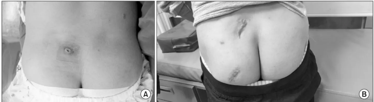
Fig. 3. (A) Another exit point of electrical burn at the left posterior superior iliac spine. The sutured wound was 1.5 cm long. It healed and recurred repeatedly for 6 months after burn injury. (B) 6 months after sinus removal. The scar on posterior superior iliac spine (upper) was caused by sinus. excision 6 month ago, the scar on buttock (lower) was caused by trauma at 9 years old of the patient.
Fig. 1. The right hand (on postburn day 4 week). Tendon and neurovascular structures were exposed after debridement.
Fig. 2. The bone and tendon of 5th toe were exposed.
At the left foot, about 6×6 cm sized wound with eschar was noted on the dorsal side of the 4, 5 metatarsal and toes (Fig. 2). In the left side of the lower back, at the pos- terior superior iliac spine site, about 1.5 cm long wound was noted, which had been closed by a 4-0 nylon at an- other hospital (Fig. 3). The sutured wound showed no
was 1.0 mm. The recipient artery was the third common palmar digital artery, and the recipient veins were two comitant veins. One artery and two veins was anasto- mosed each by a 10-0 nylon by end-to-end anastomosis.
Postoperatively, the flap was well without complications.
At 12 months after operation, range of motion of the right
Fig. 6. After microanastomosis and insetting of the flap.
Fig. 7. Twelve months after the operation, the flap was good for hand function.
Fig. 4. The design for peroneal artery perforator flap on the right lower leg.
Fig. 5. The length of vascular pedicle of harvested flap was about 4.0 cm long.
hand was normal (Fig. 7, 8).
The wound of the left foot at the electrical output site was progressively necrotic with exposure of the bone and tendons. The wound was in the 3rd, 4th, and 5th dorsal metatarsal areas and 3rd, 4th, and 5th toes (Fig. 2). At the 5th toe, the bone and tendon were exposed, and at the 4th toe, the tendon was exposed. Several times of de- bridement and wet dressings were done. On postburn day 6 week, the phalanges of the 5th toe were necrotic.
So, amputation of the little toe, and debridement of the adjacent necrotic tissue was done. Under a microscope, we explored for recipient vessels. The 4th and 5th dorsal metatarsal arteries were too small for microanastomosis.
So, full thickness skin graft was performed on the foot dorsum instead of free flap (Fig. 9). The wound was healed well. During postoperative 12 months, the wound was mildly hypertrophic, and it was controlled by triamcinolone
intralesional injection monthly for 5 times.
The back wound was 1.5 cm long and was sutured be- fore he was transferred to our hospital. It was located around the left posterior superior iliac spine (Fig. 3A). A little amount of serous discharge was noted initially. The wound was healed on postburn day 5 week. But, there- after, it recurred without active inflammation and with small amount of discharges. The wound was healed spontaneously by simple dressing but, it recurred again repeatedly. Around the wound site, there was no scars, no active inflammatory signs, and no palpable masses.
We rechecked the patient’s past history, but no foreign body injury history in the past or this time of electrical burn was noted.
We requested the radiologist for a sinogram, and it showed no specific findings except a 2∼3 cm long sinus.
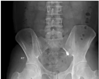
Fig. 10. 3.5 cm×2.5 cm sized radiopaque density at the left side of the pelvic cavity.
Fig. 8. Twelve months after the operation, the range of motion was full.
Fig. 11. Computed tomography showed suspicious foreign body of strong radiopaque density such as metals within the left gluteus maximus muscle.
Fig. 9. Full thickness skin graft was performed instead of a free flap because the diameter of the recipient artery was significantly small.
But, the anteroposterior pelvis radiograph showed a 3 cm×4 cm sized radiopaque density in the left pelvic area (Fig. 10). The raiologists regarded that metallic radio- paque material as an artifact during sinogram. But the pelvic computed tomography (CT) scan revealed strong radiopaque foreign body density within the left gluteus maximus muscle (Fig. 11, 12). Under general anesthesia, the wound was explored after applying gentian violet of the sinus. The sinus was excised, and its size was 4.0 cm
×3.0 cm within the gluteus maximus muscle (Fig. 13).
Within the sinus, multi braid wires thin as hair measuring 2.5×3.0 cm (Fig. 14). Black carbon dusts were scattered around the wires. The wound was closed after irrigation with drain. The wound was healed well during 6 months after the operation, wound problem was not observed (Fig. 3B).
Postoperatively we rechecked the patient's past history and reexamed the patient lower back and hip. So, on the left side of buttock, about 15 cm caudal to the sinus wound of this time electrical burn injury, about 4 cm long oblique posttraumatic scar was found (Fig. 3B). The pa- tient said that scar had been caused by trauma at 9 years old of his age. But he did not remember precisely about the trauma.
DISCUSSION
Electrical burns causes subdermal damage more than flame burn or chemical burn. The severity of electrical
Fig. 12. CT scan showed radiopaque foreign body within the gluteus maximus muscle.
Fig. 13. 3 cm×4 cm sized sinus sac containing wires was removed.
Fig. 14. 3 cm×4 cm multi braid wires thin as hair were removed.
burn is determined by voltage, current, resistance, and frequency. The electrical burn is divided by low tension (below 1,000 volts) injury and high-tension (above 1,000 volts) injury by volts. The hand is the most frequent con- tact point (entry of electrical burn), and the foot is the most common exit of electrical burn in high voltage elec- trical burn1).
The wound caused by high voltage (>1,000 volts) elec- trical burn is progressively necrotic due to intravascular thermal damage and thrombosis. Finally, the wound be- comes deep, exposing important deep structures. So, deep tissue of the palm of the hand beneath the palmar fascia would be exposed. As a result, deep structures of the
palm, the common digital artery, nerve, and tendons are exposed, and then subsequently, flap coverage is required.
For palm resurfacing due to skin and soft tissue defect, a thin flap is desirable. Also the same texture and color matching is required. Considering these aspects, a thin medial plantar flap is good for palm resurfacing2). But, the plantar flap causes some problems such as donor site morbidity, it is a relatively difficult technique, and skin grafting is necessary for the donor site. Another thin flaps such as radial forearm free flap3), superficial circumflex iliac artery perforator flap4) from the groin, and medial sural artery perforator flap5) from the calf may be avail- able for palm resurfacing. But, these flaps also have some disadvantages. The disadvantages of radial forearm free flap are donor site morbidities, skin grafting is usually necessary for the donor site, and scar on the distal fore- arm is usually present. The disadvantages of superficial circumflex iliac artery perforator flap are difficult to har- vest, anatomical variation of the vessels exists, and lymph node injury is observed. The disadvantages of medial su- ral artery perforator flap are donor site scar, and some- times skin grafting is required to donor sites if larger flap is required, and flap thinning may be required.
The peroneal artery perforator free flap has the follow- ing advantages as palm resurfacing. This flap is relatively thin. Also, the anatomical variation is low, primary clo- sure of the donor site is possible, and the length of the
comitant veins of the palm, for the vessel diameters of the harvested perforator peroneal arterial flap were similar. The flap was survived well, and the range of mo- tion of the metacarpophalangeal joints and interphalangeal joints was full, that was checked at postoperative day 12 month. But, disadvantages of this flap are color dismatch- ing (Fig. 7), and the flap was thicker than the palm skin.
Sometimes, perforator vessels are injured, and micro- vascular thrombosis may occur. But, defatting of the flap and careful dissection during flap harvest may overcome these disadvantages. Also full thickness skin graft was re- garded as a available method if deep tissue under the pla- mar aponeurosis was not exposed because of avoiding these disadvantages of peroneal artery perforator free flap.
The exit point of electrical burn was mainly at the left foot dorsum of the 3rd, 4th, and 5th metatarsal areas and dorsal side of the 4th , 5th toes (Fig. 2). The wound was progressively necrotic, and the extensor tendon and digi- tal bones of the 4th, 5th toes were exposed, requiring flap coverage. On postburn day 6 week, under general anes- thesia, debridement of the necrotic tissue and amputation of the phalangeal bones of the 5th toes were done. Under a microscope, the recipient vessels of the 4th, 5th dorsal metatarsal artery and vein was were exposed. But, the 5th dorsal metatarsal artery was necrotic, and the 4th arterial diameter was too small (0.3 mm) to perform micro- anastomosis. So, we performed full thickness skin graft instead of free flap. If free flap would be done, more proximal dissection of the 4th dorsal metatarsal artery would be required and long vascular pedicle for the har- vested flap might be required.
The foot wound was healed well (Fig. 9). After dis- charge postoperative two months, mild hypertrophic
day third week. But, the wound was recurred several days after stitch out. The discharge amount was small, not infected. So, daily dressing was enough. The wound was healed after several days after recurrence. We asked the patient about the injury to the back wound sites. But, there had been no injury to the wound site at this time of electrical burn. The patient had been fallen by the elec- trical shock, but there was had been no foreign material or metals on the ground. He had no remembrance of any- thing regarding the back injury he had sustained during recent 10 years.
After discharge, on postburn day three month, the back wound was recurred. The discharge was scanty, and wound was similar as the previous wound. We evaluated the wound through the radiologic studies. Pelvis AP plain film showed a 2.5 cm×3.5 cm sized metal density in the left side of the pelvic cavity (Fig. 10). Sinogram showed nonspecific findings except the sinus tract. But, pelvic CT showed strong radiopaque density like metals within the left gluteus maximus muscle (Fig. 11, 12).
On postburn day 6 month, under general anesthesia, exploration of the wound was performed. After gentian violet coating on the sinus tract was applied, careful dis- section and removal of the sac was done. The sinus tract and sac was 3.0 cm×4.0 cm sized (Fig. 13) and in there was a lump of multi braid wires measuring of 2.5 cm×3.5 cm were detected (Fig. 14). At the outer surface of the wires, dark carbon dusts were scattered. The wound was closed layerly with a closed suction drain. The wound was healed well without recurrence till now for 6 months postoperatively.
We precisely rechecked patient’s past history of lower back trauma. The patient had a past history of falling down injury on the back (buttock) at his early childhood
Fig. 17. Sural nerve (3.0 cm long) graft was performed on the middle phalanx.
Fig. 18. On postoperative day 4 month.
Fig. 15. A 58 year-old female patient sustained electrical arc burn on her right ring finger on postburn day 4 week. The ring on the proximal phalanx was removed, and the main wound was on the middle phalanx. The flexor digitorum profundus tendon, both the digital arteries and nerves were necrotized.
Fig. 16. The exit point of electrical arc burn on her left foot.
when he had been 9 years old (Fig. 3B). After then , there had been no problems in the back or hip injury site ex- cept the scar on the buttock (Fig 3B). According to the patient’s past history, we suspected that the wires were introduced into the gluteus maximus muscle at the time of falling down injury during his early childhood (about 26 years ago) through the buttock wound, not through the sinus wound of this time burn injury (Fig 3A, 3B).
The electrical current of this time caused the back sinus wound at posterior superior iliac spine. The electrical cur- rent entered through his palm and flew to the metal wires in the gluteus maximus muscle and exploded in his lower back (at posterior superior iliac spine) by heat. Maybe the metal tools of the patient’s waist belt on his clothes did the role of traveling of electrical currents at this time of
electrical burn injury.
There are some reports, not many, about electrical burn injury by metals of by finger ring7) touched by positive or negative terminal of a 12-volt car battery. Ring burn by contact of metal ring on the finger to the car battery (12 volts) or electrodes of low voltage (20∼30 voltage) electrical ring burn may cause deep tissue damage by heat of 1,000 degrees Celsius requiring flap coverage.
Also, there is a report of electrical burn by aluminium- containing transdermal patches by electrical defibrillator or MRI imaging8).
We also had experienced an interesting case of elec- trical arc burn in the hand (Fig. 15). A 58 year-old female patient underwent electrical arc burn on the ring finger of her right hand. Without direct contact of her hand or
tient of sustaining electrical arc burn on the finger, theo- retically, we would suspect that the metal wires within gluteus muscle of the above mentioned male patient caused electrical burn at the back resulting in chronic sinus. The electrical currents pass through low-resistance tissues preferentially. In this patient, more electrical cur- rent flew through the wires within the gluteus muscle.
This current caused high thermal damage to the adjacent tissues due to the high resistance of tissues as like fat, and skin, and bone around the metals, finally forming sinus wound9).
The reports about the electrical burn damage caused by metals within body (as a foreign body) is very rare or absent. Also, further study is required in this chronic back sinus case because we suspected the back wound accord- ing to the patient’s past history. But, it is recommended that the patients wearing metals on their body surface or within the body should be careful about electrical burn.
Many medical devices within the body contain metals, such as metal plates in orthopedics, implants in teeth, car-
3) Michelle G, Sandip H, Marco M, Mohamed S, Ali J A. Flap Decisions and Options in Soft Tissue Coverage of the Upper Limb. Open Orthop J. 2014;8:409-414
4) Berner JE, Nikkhah D, Zhao J, Prousskaia E, Teo TC. The Versatility of the Superficial Circumflex Iliac Artery Perforator Flap: A Single Surgeon’s 16-Year Experience for Limb Reconstruction and a Systemic Review. J Reconstr Microsurg.
2019;Sep 2. doi:10.1055/s-0039-1695051. [Epub ahead of print]
5) Taufique ZM, Daar DA, Cohen LE, Thanik VD, Levine JP, Jacobson AS. The medial sural artery perforator flap: A better option in complex head and neck reconstruction? Laryngoscope.
2019 Jun;129:1330-1336.
6) Shubhra C, Sachin C, Naveen Hedne C, and Naveen B S.
Perforator Peroneal Artery Flap for Tongue Reconstruction.
J Maxiilofac Oral Surg. 2017 Mar; 16:123-126.
7) Sibley PA, Godwin KA. Electrothermal ring burn from a car battery. Orthopedics. 2013 Aug;36:e1096-8.
8) Brown MR, Denman R, Platts D. Analgesic patches and defibrillators: a cautionary tale. Europace. 2009 Nov.(11):
1552-3. doi: 10.1093/europace/eup261. Epub 2009 Oct 3 9) Cheema SA. Pattern and Profile of Electric Burn Injury Cases
at a Burn Centre. J Ayub Med Coll Abbottabad 2016;28.