Cerebral vein thrombosis (CVT) is uncommon disease and known risk factors of CVT are heredi- tary thrombophilia, pregnancy, puerperium, and use of oral contraceptives.1 However, 25% of all cases are considered to be idiopathic.2 Thyro- toxicosis may be a predisposing factor of CVT due to a hypercoagulable state.3,4 Possible associa- tions between thyrotoxicosis and CVT have been described in several case reports.5-8 Here, we re- port a case of CVT in a patient with Graves’
disease.
CASE
A 39-year-old male presented to the emergency room with generalized tonic-clonic seizure and right hemiplegia. He had no history of seizures or cerebrovascular disease, and his medical his- tory was unremarkable except for Graves’ disease, for which he had been treated with methimazole for 1 year prior to admission. Recently, his thyro- toxic state had become aggravated and his methi- mazole dose was increased to 7.5㎎ per day 2 weeks prior (from 2.5㎎ per day). His vital signs were stable except for sinus tachycardia (110 beats/min), and he complained of palpitation. He exhibited mild thyroid enlargement and mild Graves’ ophthalmopathy.
https://doi.org/10.7180/kmj.2016.31.2.179 KMJ
Case Report
A Case of Cerebral Venous Thrombosis in a Patient with Graves’ Disease
Bo Ra Kim1, Jung Hwa Jung1,2, Jong Ryeal Hahm1,2, Jaehoon Jung1,2, Hee Jung Park1, Soo Kyoung Kim1,2
1Department of Internal Medicine, Gyeongsang National University School of Medicine, Jinju, Gyeongsangnam-do, Korea
2Gyeongsang Institute of Health Science, Gyeongsang National University School of Medicine, Jinju, Gyeongsangnam-do, Korea
Superior sagittal sinus thrombosis is an uncommon disease, and 25% of cases are considered to be idiopathic.
Hypercoagulability, local bloodstream stasis, and vessel wall abnormalities may contribute to the development of this condition. The thyrotoxic phase of Graves’ disease is associated with venous thrombosis caused by hypercoagulability, which is in turn induced by increased levels of homocysteine and factor VIII and decreased fibrinolytic activity. Here, we report the case of a 39-year-old male who presented with superior sagittal sinus thrombosis and concomitant hyperthyroidism.
Key Words: Cerebral venous thrombosis, Thyrotoxicosis, Protein C Deficiency
Corresponding Author: Soo Kyoung Kim, Department of Internal Medicine, Institute of Health Sciences, Gyeongsang National University School of Medicine, 79, Gangnam-ro, Jinju-si, Gyeongsangnam-do 52727, Korea TEL: +82-55-750-8874 FAX: +82-55-758-9122 E-mail: [email protected]
Received:
Revised:
Accepted:
Jul. 14, 2015 Sep. 17, 2015 Sep. 21, 2015
Laboratory tests confirmed a thyrotoxic status:
free T4, 4.01 ng/dL (normal 0.93-1.70 ng/dL);
tri-iodothyronine 271 ng/dL (normal 80-200 ng/dL); TSH, below 0.01 mIU/L (normal 0.27-4.2 mIU/L); and thyroid-stimulating antibody, 40 IU/L (normal 0-1.75 IU/L).
Hematological and coagulation tests showed that platelet counts, prothrombin time, partial thromboplastin time, and the levels of antith- rombin III and coagulation factor VIII were within normal ranges. The factor V Leyden mutation was absent, and lupus anticoagulant, anti-cardiolipin antibodies, and anti-phospholipid antibodies were absent. However, the level of protein C (24%, normal 72-160%) and protein S (55%, normal 60-150%) were low. Abnormal serum glucose and
electrolyte value was not detected.
Electrocardiography revealed sinus tachycardia and echocardiography showed normal left ven- tricular size and function with no evidence of an intra-cardiac thrombus. The electroencephalo- gram was normal.
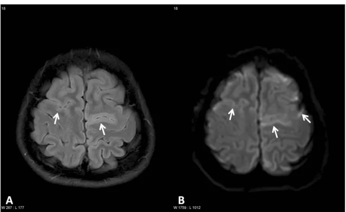
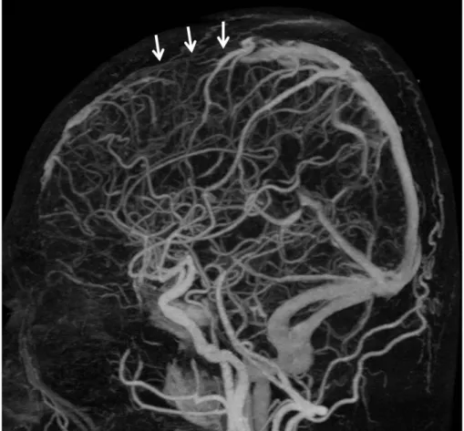
Brain magnetic resonance imaging (MRI) re- vealed hyperintensity associated with thrombosis within the superior sagittal sinus (Fig. 1); this was confirmed by brain computed tomography an- giography (Fig. 2).
The patient was admitted to our intensive care unit, and treatment with intravenous heparin and methimazole 7.5 ㎎/day was commenced. After 3 days, his clinical condition gradually improved.
The focal neurological deficits disappeared after
Fig. 1. Fluid-attenuated inversion recovery (FLAIR, A) and diffusion-2 weighted MR images(DWI, B) demonstrate multiple hyperintense lesions in both high frontal cortices (arrows).
The lesions are more clearly demonstrated on DWI (B) than on FLAIR image (A).
several days of treatment, and his consciousness became normalized. He was treated with methi- mazole 7.5 ㎎/day and an oral anticoagulant to obtain an INR (International Normalized Ratio) between 2 and 3. Six months after discharge, the patient has been doing well. He exhibits complete neurological recovery and has maintained normal thyroid function with methimazole.
DISCUSSION
CVT is very uncommon condition and it’s mor- tality rate is about 5%.9 Instances of CVT in Graves’
disease patients are also rare. Several previous studies suggested that hyperthyroidism is asso- ciated with increased risk of arterial and venous thromboembolism.10 Observation from several case reports have shown the increased risk of CVT5-12 or pulmonary thromboembolism in pa- tients with hyperthyroidism.13,14 Rau et al. re- ported a case of Graves’ disease with CVT pre- sented with headache and general weakness in old man.6 He exhibited a high level of fibrinogen, low protein C activity, and atrial fibrillation.6 Verberne et al. reported the case of Graves’ dis- ease with CVT presenting as a viral encephalitis in young woman through a factor VIII-medicated
Fig. 2. Brain computed tomography (CT) angiography shows segmental 2 occlusion of the superior sagittal sinus due to thrombosis (arrows).
hypercoagulability.7 Grien et al. reported two case of Graves’ disease complicated by pulmonary embolism. Of these patients, young woman showed the increased level of coagulation factor VIII.14
Systemic literature review reported that the most frequently involved sites was cerebral venous veins, and over 60% of these cases reported the additional thrombophilic risk factor such as factor V Leiden mutation or protein C deficiency.10
Although the precise mechanism underlying CVT or other thromboembolic events in Graves’
disease remains unclear, several possibilities have been suggested. These include hypercoagulation, venous stasis, and abnormalities of the venous walls.4,15,16 Many abnormalities of blood coagu- lation during thyrotoxic state have been described. The patient with hyperthyroidism had the shorten activated partial thrombo-plastin time, higher fibrinogen levels, increased levels of factor VIII and homocysteine, and decreased fi- brinolytic activity during thyrotoxicosis in the previous studies.5-7,16 Increased fibrinogen levels and reduced levels of protein C were also asso- ciated with CVT development in Graves’ disease patients.6 The factor V Leiden mutation has been found in another case of thyrotoxic patient with CVT.17 Our case also showed a reduced protein C and protein S level. Protein C inhibits coagu- lation by inactivating factors VIIIa and Va.
Patients with protein C deficiency may be at in- creased risk of thrombo-embolic events.6 Apart from such changes in the coagulation system, sev-
eral inherited or acquired risk factors for throm- bosis are known in thyrotoxic patients.11 Thyrotoxic state could induce vascular endothe- lial dysfunction,11 and venous stasis caused by goi- ter could affect CVT development.5 In our case, except for protein C and protein S deficiency, other thrombophilic features were normal and no other risk factor for venous thrombosis was identified. Furthermore, the goiter was not large that could trigger venous stasis. Overt hyper- thyroidism is associated with thromboembolic events through several mechanisms evidenced by several case series. Therefore, CVT should be sus- pected in thyrotoxic patients with neurological symptoms. In addition, such patients should be screened for any accompanying underlying coagulopathy. Future large observational study is needed to provide the more information about the association between hyperthyroidism and co- agulation-fibrinolytic abnormalities.
REFERENCES
1. Allroggen H, Abbott RJ. Cerebral venous sinus thrombosis. Postgrad Med J 2000;76:12-5.
2. Stam J. Cerebral venous and sinus thrombosis:
incidence and causes. Adv Neurol 2003;92:225–
32.
3. Squizzato A, Gerdes VE, Brandjes DP, Büller HR, Stam J. Thyroid diseases and cerebrovascular disease. Stroke 2005;36:2302-10.
4. Erem C, Ersoz HO, Karti SS, Ukinç K, Hacihasanoglu
A, Değer O, et al. J Blood coagulation and fi- brinolysis in patients with hyperthyroidism.
Endocrinol Invest 2002;25:345-50.
5. Siegert CE, Smelt AH, de Bruin TW. Superior sagittal sinus thrombosis and thyrotoxicosis.
Possible association in two cases. Stroke 1995;26:496-7.
6. Ra CS, Lui CC, Liang CL, Chen HJ, Kuo YL, Chen WF. Superior sagittal sinus thrombosis in- duced by thyrotoxicosis. Case report. J Neu- rosurg 2001;94:130-2.
7. Verberne HJ, Fliers E, Prummel MF, Stam J, Brandjes DP, Wiersinga WM. Thyrotoxicosis as a predisposing factor for cerebral venous thrombosis. Thyroid 2000;10:607–10.
8. Colleran KM, Ratliff DM, Burge MR. Potential association of thyrotoxicosis with vitamin B and folate deficiencies, resulting in risk for hyper- homocysteinemia and subsequent thromboem- bolic events. Endocr Pract 2003;9:290-5.
9. Coutinho JM, Zuurbier SM, Stam J. Declining mortality in cerebral venous thrombosis: a sys- tematic review. Stroke 2014;45:1338-41.
10. Franchini M, Lippi G, Targher G. Hyperthy roidism and venous thrombosis: a casual or caus- al association? A systematic literature review.
Clin Appl Thromb Hemost 2011;17:387-92.
11. Bensalah M, Squizzato A, Ould Kablia S, Menia H, Kemali Z. Cerebral vein and sinus thrombosis and hyperthyrodism: a case report and a system-
atic review of the literature. Thromb Res 2011;
128:98-100.
12. Kim DD, Chunilal S, Young S, Cutfield R. A study of venous thrombosis incidence in patients with acute hyperthyroidism. Intern Med J 2013;
43:361-5.
13. Lin HC, Yang LY, Kang JH. Increased risk of pulmonary embolism among patients with hy- perthyroidism: a 5-year follow-up study. J Th- romb Haemost 2010;8:2176-81.
14. Grine S, Charfi N, Kamoun M, Mnif F, Naceur BB, Rekik N, et al. Hyperthyroidism: A rare cause of pulmonary embolism: Report of two cases.
Indian J Endocrinol Metab 2013;17:1104-7.
15. Squizzato A, Romualdi E, Büller HR, Gerdes VE.
Clinical review: Thyroid dysfunction and effects on coagulation and fibrinolysis: a systematic review. J Clin Endocrinol Metab 2007;92:2415 -20.
16. Lippi G, Franchini M, Targher G, Montagnana M, Salvagno GL, Guidi GC, et al. Hyperthyroidism is associated with shortened APTT and increased fibrinogen values in a general population of un- selected outpatients. J Thromb Thrombolysis 2009;28:362-5.
17. Molloy E, Cahill M, O'Hare JA. Cerebral venous sinus thrombosis precipitated by Graves' disease and Factor V Leiden mutation. Ir Med J 2003;
96:46-7.