J. Exp. Biomed. Sci. 15 (2009) 67–72
Paclitaxel Suppress Dedifferentiation via Mitogen-activated Protein Kinase Pathway in Rabbit Articular Chondrocyte
Jeong Hee Im and Song Ja Kim
†Department of Biological Sciences, College of Natural Sciences, Kongju National University, Korea
Microtubule-interfering agents (MIAs), including paclitaxel, have been attributed in part to interference with microtubule assembly, impairment of mitosis, and changes in cytoskeleton. But the signaling mechanisms that link microtubule disarray to destructive or protective cellular responses are poorly understood. This study investigated the effect of paclitaxel on differentiation such as type II collagen expression and sulfated proteoglycan accumulation in rabbit articular chondrocytes. Paclitaxel caused differentiated chondrocyte phenotype as demonstrated by increment of type II collagen expression and proteoglycan synthesis Paclitaxel treatment stimulated activation of ERK-1/2 and p38 kinase. Inhibition of ERK-1/2 with PD98059 enhanced paclitaxel-induced differentiation, whereas inhibition of p38 kinase with SB203580 suppressed paclitaxel-induced differentiation. Our findings suggest that ERK-1/2 and p38 kinase oppositely regulate paclitaxel-induced differentiation in chondrocytes.
Key Words: Dedifferentiation, Paclitaxel, MAP kinase, Microtubule
서 론
연골 세포외기질 (Extracellular matrix, ECM)은 여러 가 지 세포조절신호가 연골 특정 유전자들을 자극하여 발 현 조절되어진다 (DeLise et al., 2000; Fukui et al., 2001).
관절 연골세포에서 세포외기질의 합성은 연골조직의 발 생, 유지, 재생에 매우 중요한 역할을 한다고 알려져 있 다. 관절 연골세포에 있는 ECM 분자 발현의 항상성은 퇴행성 관절염과 같은 연골 질병이 발생하는 동안 연골 세포의 분화된 표현형의 손실 등을 포함하며 파괴된다 (Samdell and Aigner, 2001). 연골세포의 탈분화는 type II collagen의 발현이 중단되고 섬유아세포성 type I과 type III collagen의 발현이 증가되기 시작한다. 연골세포는 염 증성 사이토카인인 Interleukin - 1 (Martel-pelletier et al., 1999), 산화질소 (Amin and Abramson, 1998), 레티노산 (Cash et al., 1997; Weston et al., 2000), 계대배양 (Lefebvre et al., 1990; Yoon et al., 2002) 등 여러 가지 원인에 의해 외 부 기질 합성의 불균형을 야기시키며, 이는 연골세포의
탈분화 및 세포사멸을 일으킨다. 이러한 세포 수준의 손 상으로 인해 조직이 파괴되면 이는 류마티스 관절염 혹 은 퇴행성 관절염의 발병 원인이 된다.
최근, 세포골격의 구조는 연골세포의 표현형을 결정하 는 중요한 인자로 작용한다고 보고되었다. Retinoic acid 나 세포 계대배양에 의해 탈분화된 연골세포에서는 actin 세포골격의 변화를 관찰할 수 있다. 또한 dihydrocyto- chalasin B에 의한 actin 세포골격의 붕괴는 탈분화된 연 골세포의 재분화를 일으키고 (Brown and Benya, 1988), 이러한 actin 세포골격은 관절 연골세포에서 골형성 단 백질이나 산화질소에 의한 표현형의 변화를 야기시켰다 (Vinall et al., 2002). 이처럼, actin 세포골격 단백질과 함 께 또 다른 세포골격의 주요 구성인자인 미세소관은 α 와 β tubulin으로 구성된 heterodimer subunit으로 조합된 중합체이다. 미세소관은 모든 진핵세포에 존재하며 분화 (Surajit et al., 2007), 염증반응 (Kotha et al., 2000), 유사분열, 세포내 수송, 세포이동, 세포극성유지 등에 중요한 역할 을 한다.
Paclitaxel은 1963년 처음 주목나무 (Yew, Taxus brevifolia) 의 껍질에서 분리되어 1971년에 화학구조가 규명되었 고 탁월한 항암효과를 나타내는 시약 중의 하나로 B16 melanoma, L1210 (Wan et al., 1971)과 p388 leukemia, human MX-1 mammary, CX-1 colon과 LX-1 lung tumor (Wiernik et al., 1987) 세포 등에서 항암효과를 나타내는 것으로 알
*논 문 접 수: 2009년 2월 25일 수정재접수: 2009년 3월 7일
†교신저자: 김송자, (우) 314-701 충남 공주시 신관동 182번지, 국립공주대학교 자연과학대학 생명과학과
Tel: 041-850-8507, Fax: 041-850-0927 e-mail: [email protected]
려져 있다. Paclitaxel은 β-tubulin과 결합하여 미세소관을 중합체 상태로 안정화시킴으로써 세포를 후기 G2/M기 에 머물게 하여 세포분열을 억제한다 (Arnal and Wade, 1995; Nogales et al., 1995). 이 유사분열의 억제 기능을 통 해 항암제로서의 기능을 하는 것으로 생각된다 (Horwitz, 1992). 또 다른 연구에서, paclitaxel은 콜라겐 유도 관절 염을 완전하게 저해하였으며 (Brahn and Banqurigo, 1994;
Houri and O'Sullivan, 1995), 류마티스 관절염이 유도된 쥐 에 paclitaxel을 처리하였을 때, 관절염과 활액성 병리가 거의 일어나지 않는다고 보고되었다. 이러한 결과들은 paclitaxel이 관절염 억제제로서의 기능을 가지고 있다는 것을 보여주는 것이라 하겠다. 그러나, paclitaxel의 이러 한 작용 기작에 대해서는 앞으로 많은 연구가 필요하다.
본 연구에서는 토끼 무릎관절 연골세포에서 미세소관 의 탈중합을 억제하는 paclitaxel을 처리하여 미세소관의 세포골격 구조의 변화가 연골세포의 분화를 어떻게 조 절하는지에 대해 알아보았다.
재료 및 방법
1. Cell culture
토끼 무릎 연골세포의 추출은 2주된 New Zealand White종의 토끼로부터 추출하였다. 추출방법은 치사량 의 Diethyl ether로 토끼를 치사 시킨 후 뒷다리 관절부분 을 포함한 요골과 장골을 적출한다. 적출한 관절로부터 연골을 절편하여 0.2% collagen 분해효소 (381 U/mg 고상, Sigma)가 함유된 PBS 완충용액에 넣고 37℃에서 7시간 동안 반응시켜 세포를 조직에서 분리시킨다. 녹지 않은 조직과 추출된 세포는 원심분리를 이용하여 나눈 후, 상층액을 모은다. 상층액에 분리되어 있는 세포는 원심 분리로 다시 분리 후 상층액을 제거하여 세포만 모은다.
모은 세포는 10%의 FBS (fetal bovaine serum), 50 μg/ml의 Streptomycin, 50 units/ml의 Penicillin이 함유된 Dulbecco's modified Eagle's medium (DMEM, Gibco-BRL, Gaithersburg, MD)에 녹인 후 배양 접시에 5×10⁴의 밀도로 배양을 시 작한다. 배양액은 이틀에 한번씩 교체하여 주었으며, 배 양접시의 세포밀도가 거의 80%가 되었을 때 시약을 처 리하였다.
2. Western blot analysis
단백질의 정량분석을 위하여 세포를 50 mM Tris-HCl, pH 7.4, 150 mM NaCl, 1% Nonidet P-40, 0.1% Sodium
dodecylsulfate가 함유된 세포 용해용액에 단백질 분해효 소 억제제 [10 μg/ml leupeptin, 10 μg/ml pepstatin A, 10 μg/ml aprotinin, 1 mM 4-(2-aminoethyl) benzensulfonyl fluoride]와 인산 분해효소 억제제 (1 mM NaF, 1 mM Na3VO4)를 첨가하여 세포 단백질을 추출하였다. 추출한 단백질은 SDS-polyacrylamide gel에 전기영동하여 단백질 을 크기 별로 분리한 후 Nitrocellulose 막으로 이동시켰 다. Type II collagen, pp38, pERK과 actin 항체는 Santa Cruz Biotechnology사에서 구입하여 사용하였다 (Santa Cruz, CA).
3. Reverse transcriptase polymerase chain reaction (RT-PCR)
배양된 연골세포에 Paclitaxel (1 μM)을 처리한 후 모든 RNA는 Trizol (Invitrogen, Gronongen, Netherland)을 이용하 여 추출하였다. 추출한 RNA는 Maxim RT-PCR premix kit (Intron Biotechnology)을 이용하여 type II collagen cDNA 합성과 증폭을 위하여 RT-PCR을 수행하였으며 primer는 5'-GAC CCC ATG CAG TAC ATG CG-3' (sense)와 5'-AGC CGC CAT TGA TGG TCT CC-3' (antisense)를 사용하여 45℃ annealing 온도에서 증폭하였다.
4. Immunofluorescence
연골세포에서 paclitaxel에 의한 type II collagen의 발현 과 분포를 관찰하기 위하여 Immunofluorescence를 수행하 였다. 배양된 연골세포는 3.5% paraformaldehyde로 10분 동안 상온에서 고정시켰으며, 0.1% Triton X-100을 이용하 여 세포막 투과성을 높여주었다. 고정된 세포는 type II collagen antibody (Santa Cruz, CA)을 사용하여 염색하였으 며, 형광현미경으로 관찰하였다.
5. Immunohistochemistry
토끼 관절연골조직 (~125 mm3)을 4% paraformaldehyde 용액에 24시간 동안 4℃에 담가놓는다. 다양한 농도 (100~30%)의 ethanol로 세척 후 파라핀에 완전히 잠기게 놓고 굳힌다. 4 μm로 얇게 절편하여 0.1% Alcian Blue로 염색한 후, sulfated protoglycan 축적량을 현미경으로 확인 하였다.
6. Determination of chondrocyte phenotype
연골세포의 분화 정도는 0.1% Alcian Blue 용액을 사용 하여 sulfated proteoglycan의 축적량을 측정하였다. 세포를
1 × PBS로 두 번 세척 후 Kahle's solution에서 10분 동 안 고정한 뒤 다시 1 × PBS로 두 번 세척한다. 그 후 Alcian Blue solution에 12시간 이상 염색한다. 0.1 N HCl로 한 번 세척 후 4 M Guanidine HCl을 넣고 12 시간 동안 shaking한다. 96 well plate에 약 180 μl/well 씩 넣어 ELISA reader에서 600 nm로 값을 측정하였다.
7. Statistics
본 실험의 결과는 여러 차례의 실험 중 평균적인 결과 를 사용하였다.
결 과
1. Paclitaxel은 토끼 관절연골 세포의 분화를 유도한다
Tubulin 안정화를 촉진하는 것으로 알려진 paclitaxel을 토끼관절 연골세포에 처리하여 tubulin의 중합 안정화가 연골세포의 분화에 미치는 영향을 알아보았다. 1 μM paclitaxel을 각 시간 별로 처리하거나, 농도 별로 paclitaxel 을 24시간 동안 처리하여 연골세포 분화 표지인자로 알 려진 type II collgaen의 발현 정도를 Western blot과 RT- PCR로 확인하였다. Type II collagen의 발현은 시간과 농 도 의존적으로 증가하는 양상을 보였으며 (Fig. 1A, 1B),
이는 전사 수준에서도 같은 양상을 보여 주었다 (Fig.
1B). 이에, 연골세포의 분화 표지인자로서 많이 사용되 B
C A
Fig. 1. Paclitaxel induced differentiation of articular chondrocyte. Chondrocytes were untreated or treated with 1 μM paclitaxel for specified time periods (A) or indicated concentration of paclitaxel for 24 h (B). The expression of type II collagen was determined by Western blot analysis (A and B, upper panel) and RT-PCR (B, lower panel). Actin and GAPDH were used as loading controls.
Accumulation of sulfated proteoglycan in cells treated with pacltaxel was quantified by Alcian blue staining (C). The data represent the results of a typical experiment conducted at least three times with similar results.
Fig. 2. Paclitaxel promoted differentiation of articular chondro- cyte. Catilage explants were untreated (Control) or treated with 1 μM paclitaxel for 24 h. Localization and expression of type II collagen was determined by fluorescent microscope (left pannel and original magnification ×1000). Sulfated proteoglycan were detected in tissue sections treated with the paclitaxel (1 μM) for 24 h was quantified by Alcian blue staining (right pannel and original magnification ×400). The data represent the results of a typical experiment conducted at least four times with similar results.
어지는 세포외 기질 분자인 sulfated proteoglycan의 축적 을 Alcian Blue staining 후 정량한 결과 paclitaxel 처리 농 도 의존적으로 증가하는 것을 확인할 수 있었다 (Fig. 1C).
위의 결과를 연골세포 및 조직 수준에서 확인하고자 Immunofluorescence와 Immunohistochemistry를 행하였다.
연골세포에 1 μM paclitaxel을 처리한 후 type II collagen 항체를 사용하여 면역형광염색을 하여 형광현미경으로 관찰한 결과 paclitaxel을 처리한 세포군에서 type II colla- gen의 발현이 증가되어짐을 확인할 수 있었다 (Fig. 2, left panel). 또한, 연골조직을 떼어내어 paclitaxel을 24시 간 처리한 후 Alcian blue staining하여 현미경으로 관찰한 결과 paclitaxel 처리한 조직에서 Alcian blue staining이 훨 씬 진하게 염색이 된 것을 확인할 수 있었다 (Fig. 2, right panel). 위의 결과들을 종합해 볼 때, paclitaxel을 처리할 경우, 즉 tubulin 중합 안정화는 연골세포와 조직에 분화 를 유도하는 것을 알 수 있다.
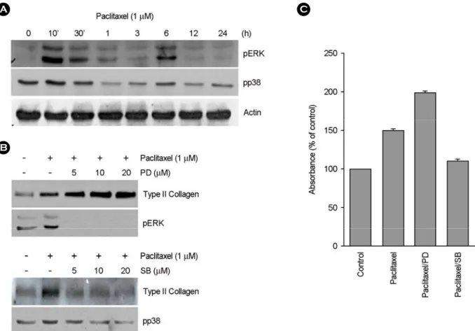
2. Paclitaxel에 의한 연골세포의 분화 유도는 ERK-1/2 와 p38 kinase가 상반되게 조절한다
위의 결과에서 paclitaxel이 연골세포의 분화를 유도한 것을 알 수 있었다. 이에, 연골세포의 분화 유도에 관여 하는 세포신호전달 기전을 알아보고자 세포의 분화에 관 여하는 것으로 알려진 MAP kinase pathway를 조사하였다.
또한, 본 연구자의 이전 연구결과에서 MAP kinase의 두 subtypes인 ERK-1/2와 p38 kinase가 연골세포의 분화를 조절한다는 결과를 기초로 하여 조사하였다 (Kim et al., 2002). 1 μM paclitaxel을 연골세포에 처리한 결과 ERK-1/
2와 p38 kinase의 활성이 10분일 때 최고로 활성화 되었 다가 1시간 뒤 감소하였고, 또한 6시간 뒤에 다시 약한 활성이 나타나다 이후 감소하는 양상을 나타내었다 (Fig.
3A). ERK-1/2와 p38 kinase의 활성과 연골세포의 분화 사 이의 조절 기작을 알아 보기 위하여 ERK-1/2 상위신호 전달의 저해제인 PD98095 (PD)와 p38 kinase의 저해제인 A
B
C
Fig. 3. ERK-1/2 and p38 kinase oppositely regulated for paclitaxel-induced differentiation. Chondrocytes were treated with 1 μM paclitaxel for indicated periods. ERK-1/2 and p38 phosphorylation was detected using Western blot analysis (A). B, Articular chondrocytes were treated with 1 μM paclitaxel in the absence or presence of the indicated concentration of 10 μM PD98059 (PD) or 10 μM SB203580 (SB). Actin was used as loading control. Accumulation of sulfated proteoglycan in cells treated with pacltaxel was quantified by Alcian blue staining (C).
SB203580 (SB)을 paclitaxel과 함께 각각 처리하여 type II collagen의 발현양상을 알아보았다. Paclitaxel에 PD를 함 께 처리하여 ERK-1/2의 활성을 저해하였을 경우, 저해제 의 농도가 증가할수록 type II collagen 발현이 증가하는 것을 확인할 수 있었다 (Fig. 3B, upper panel). 이와 반대 로 p38 kinase 활성 저해제인 SB를 처리하였을 경우 저 해제 농도 의존적으로 type II collagen의 발현이 감소하 는 것을 확인할 수 있었다 (Fig. 3B, lower panel). Sulfated proteoglycan의 축적량도 ERK-1/2와 p38 kinase의 활성 저 해 시 type II collagen의 발현양상과 같은 결과를 보였다 (Fig. 3C). 이는 tubulin 중합 안정화에 의한 연골세포 분 화 유도는 ERK-1/2 kinase에 의해 더 억제되어지며, p38 kinase에 의해 분화가 촉진되는 것으로 보여진다. 즉, paclitaxel에 의한 연골세포의 분화는 두 MAP kinases의 상반된 상호작용에 의해 조절되는 것으로 보여진다.
고 찰
CD에 의한 actin 세포골격의 붕괴는 발생과정 중 날개 부분의 중간엽 줄기세포로부터 연골형성화를 촉진시키며, 분화된 연골세포의 표현형을 유지시킨다고 이전 연구결 과에서 발표하였다. 이는 PI3-kinase, AKT, MAP kinase, PKC-α와 PKC-ξ와 같은 세포신호 전달과정을 조절하여 산화질소에 의한 연골세포의 탈분화와 세포 자가사멸을 억제하며 COX-2의 발현을 저해하는 것으로 알려져 있으 나, JAS에 의한 actin 세포골격의 재구성은 오히려 연골 세포의 탈분화와 COX-2의 발현을 유도하는 것으로 밝 혀졌다 (Kim et al., 2002). 위의 연구결과를 토대로 하여, 세포골격 단백질의 변형에 의한 연골세포의 분화 또는 탈분화에 관여하는 조절 기작에 대해 더 알아보기 위해 미세소관인 tubulin의 중합 상태를 안정화시키는 paclitaxel 을 연골세포에 처리하였다.
Paclitaxel은 세포내에서 tubulin의 소단위에 우선적으로 붙어 tubulin dimer의 작용을 향상시켜 미세소관의 화합을 유도하고 탈중합을 억제함으로써 미세소관을 안정화시킨 다 (Horwitz 1992; Arnal and Wade, 1995; Nogales et al., 1995).
류마티스 관절염에서는 활막세포가 종양처럼 비정상적 으로 증가하면서 만성 염증을 일으켜서 결국은 관절이 파괴되는 것을 알려져 있다. Paclitaxel은 활막세포에 선택 적으로 작용하여 cell cycle의 정지와 apoptosis를 유도한 다는 것이 보고되었다. 이는 paclitaxel이 활막세포의 증 식과 분열을 제어함으로써 관절염을 치료할 수 있는 새
로운 약재 개발의 가능성을 제시하였다 (A.HUI et al., 1997). 또한, Paclitaxel은 bovine 연골세포에서도 activator protein-1의 활성을 억제함으로써 Interleukin-1β가 유도한 collagenase와 stromelysin의 발현을 저해하여 관절염치료 제로서의 가능성을 제시하였다 (A.HUI et al., 1998; A.HUI et al., 1997).
흥미롭게도 본 연구에서, paclitaxel은 토끼관절 연골 세포의 증식을 저해하였으며 (data not shown), type II collagen 단백질의 발현량을 증가시켜 연골세포의 분화 를 유도하였다 (Fig. 1). 더군다나, paclitaxel에 의해 유도 된 연골세포의 분화 유도는 주요 MAP kinases subtypes인 ERK-1/2와 p38 kinase의 상반된 조절에 의해 이루어진다 는 것을 알아냈다 (Fig. 3). 위의 연구결과들을 종합해 볼 때, tubulin의 안정화는 세포골격 단백질에 영향을 주며 결국 연골세포의 형태에 영향을 주어 연골세포의 분화를 조절하는 것으로 생각된다. 이러한 연골세포 형태 변화 는 연골세포의 세포증식 및 염증반응, 세포사멸 등에도 관련되어 있다는 보고가 최근에 많이 이루어지고 있으므 로 이에 대한 지속적인 연구가 수행되어야 할 것이다.
REFERENCES
Amin AR, Abramson SB. The role of nitric oxide in articular cartilage breakdown in osteoarthritis. Curr Opin Rheumatol.
1998. 10: 263-268.
Arnal I, Wade RH. How does taxol stabilize microtubules? Curr Biol. 1995. 5: 900-908.
Brahn E, Tang C, Banquerigo ML. Regression of collagen-induced arthritis with taxol, a microtubule stabilizer. Arthritis Rheum.
1994. 37: 839-845.
Brown PD, Benya PD. Alterations in chondrocyte cytoskeletal architecture during phenotypic modulation by retinoic acid and dihydrocytochalasin B-induced reexpression. J Cell Biol.
1988. 106: 171-179.
Cash DE, Bock CB, Schughart K, Linney E, Underhill TM.
Retinoic acid receptor alpha function in vertebrate limb skeletogenesis: a modulator of chondrogenesis. J Cell Biol.
1997. 136: 445-457.
Cohen E, Ophir I, Shaul YB. Induced differentiation in HT29, a human colon adenocarcinoma cell line. J Cell Sci. 1999. 112:
2657-2666.
DeLise AM, Fischer L, Tuan RS. Cellular interaction and signaling in cartilage development. Osteoarthr Cart. 2000. 8: 309-334.
Fukui N, Purple CR, Sandell LJ. Cell biology of osteoarthritis: the chondrocyte's response to injury. Crru Rheumatol Rep. 2001.
3: 496-505.
Hui A, Min WX, Tang J, Cruz TF. Inhibition of activator protein 1 activity by paclitaxel suppresses interleukin-1-induced colla- genase and stromelysin expression by bovine chondrocytes.
Arthritis Rheum. 1998. 41: 869-876.
Horwitz SB. Mechanism of action of taxol. Trends Pharmacol Sci.
1992. 13: 134-136.
Houri JM, O'Sullivan FX. Animal models in rheumatoid arthritis.
Curr Opin Rheumatol. 1995. 7: 201-205.
Kim SJ, Ju JW, Oh CD, Yoon YM, Song WK, Kim JH, Yoo YJ, Bang OS, Kang SS, Chun JS. ERK-1/2 and p38 kinase oppositely regulate nitric oxide-induced apoptosis of chondro- cytes in association with p53, caspase-3, and differentiation status. J Biol Chem. 2002. 277: 1332-1339.
Lefebvre V, Peeters-Joris C, Vaes G. Production of collagens, collagenase and collagenase inhibitor during the dedifferen- tiation of articular chondrocytes by serial subcultures. Biochim Biophys Acta. 1990. 1051: 266-275.
Martel-pelletier J, Alaaeddine N, Pelletier JP. Cytokines and their role in the pathophysiology of osteoarthritis. Front. Biosci.
1999. 4. 694-703.
Sandell LJ, Aigner T. Articular cartilage and changes in arthritis.
An introduction: cell biology of asteoarthritis. Arthr. Res. 2001.
3: 107-113.
Subbaramaiah K, Hart JC, Norton L, Dannenberg AJ. Microtubule- interfering agents stimulate the transcription of cyclooxygenase- 2. Evidence for involvement of ERK1/2 and p38 mitogen- activated protein kinase pathways. J Biol Chem. 2000. 275:
14838-14845.
Wani MC, Taylor HL, Wall ME, Coggon P, McPhail AT. Plant antitumor agents. V.I. The isolation and structure of taxol, a novel antileukemic and antitumor agent from Taxus brevifolia. J Am Chem Soc. 1971. 93: 2325-2327.
Wiernik PH, Schwartz EL, Strauman JJ, Dutcher JP, Lipton RB, Paietta E. Phase I clinical and pharmacokinetic study of taxol.
Cancer Res. 1987. 47: 2486-2493.
Yoon YM, Kim SJ, Oh CD, Ju JW, Song WK, Yoo YJ, Huh TL, Chun JS. Maintenance of differentiated phenotype of articular chondrocytes by protein kinase C and extracellular signal- regulated protein kinase. J Biol Chem. 2002. 277: 8412-8420.
Vinall RL, Lo SH, Reddi AH. Regulation of articular chondrocyte phenotype by bone morphogenetic protein 7, interleukin 1, and cellular context is dependent on the cytoskeleton. Exp Cell Res. 2002. 272: 32-44.