Biomedical Science Letters 2014, 20(3): 173~179 http://dx.doi.org/10.15616/BSL.2014.20.3.173 eISSN : 2288-7415
Effect of Korean Red Ginseng on Artificial Sand Dust (ASD) Induced Allergic Lung Inflammation
Jung-Ha Kim1, Tae-Jin Lee2, Im Jee-Aee3 and Duk-Chul Lee4,†
1Department of Family Medicine, Chung-Ang University Healthcare System, Seoul 156-756, Korea
2Department of Pathology, Chung-Ang University College of Medicine, Seoul 156-755, Korea
3Sports and Medicine Research Center, INTOTO Inc., Seoul 120-160, Korea
4Department of Family Medicine, Severance Hospital, Yonsei University, College of Medicine, Seoul 120-752, Korea
Asian sand dust is known to promote various respiratory symptoms or disorders. For the prevention of harmful health effects by Asian sand dust, the best strategy is known to avoid or reduce exposure to the Asian sand dust. Several studies have shown that Korean red ginseng (RG) has anti-inflammatory and anti-allergic effects. The study aimed to clarify the effect of Korean red ginseng intake on lung inflammation responses to artificial sand dust (ASD) similar to Asian sand dust. BALB/c mice were divided into five groups (n=12) of control (saline), ovalbumin (OVA), OVA with ASD, OVA plus RG with ASD, and OVA plus dexamethasone (DEXA) with ASD. Histopathologic evaluation of lung was conducted.
Interleukin (IL)-5, IL-12, interferon (IFN)-γ, IL-13, monocyte chemotactic protein (MCP)-1, and eotaxin within bronchoalveolar lavage (BAL) fluid were measured by ELISA. OVA+ASD group significantly increased concentrations of IL-5, IL-13, MCP-1, and eotaxin (P<0.01) compared to the control. OVA+ASD+RG group showed significant decreased levels of IL-2, IL-13, MCP-1 and eotaxin (P<0.01) compared with OVA+ASD. Between RG and DEXA treatment groups, there was no significant difference in all cytokines and chemokines. The inflammatory cells were significantly decreased in treatment groups with RG or DEXA compared to OVA+ASD group. This study suggests a beneficial effect of Korean RG administration in preventing inflammation of lung resulting from Asian sand dust.
Key Words: Asian sand dust, Korean red ginseng, Anti-inflammation, Allergic mouse model
Asian sand dust is a meteorological phenomenon that affects much of Northeast Asia. The dust originats in the deserts of China and Mongolia and is carried to the Korean Peninsula by prevailing westerly winds, resulting in social and economic damage (Kwon and Cho, 2004). It is known that particulate matter (PM) less than 10 μm in diameter (PM10) in sand dust affect human health. Increased PM10 invading the lower trachea particularly promotes different
respiratory diseases (Pope et al., 1995; Hong et al., 1999;
Kim, 2004). Moreover, water soluble ions (Park et al., 2004b), inorganic components (mineral elements) (Ichinose et al., 2008b), or organic substances such as bacteria in the dust (Echigo et al., 2005; Ichinose et al., 2008a) have been suggested to be associated with respiratory inflammation responses. The pulmonary function of asthma patients deteriorates and nocturnal asthma symptoms increase during the dust storm season (Min et al., 2001; Song, 2001; Park et al., 2003b). Also, daily mortality rate and hospital admission rate were reported to increase due to respiratory illnesses during the Asian sand dust season (Kwon et al., 2002; Chen et al., 2004; Hwang et al., 2005). Furthermore, recent studies have reported that the dust may enhance
*Received: July 26, 2014 / Revised: September 21, 2014 Accepted: September 24, 2014
†Corresponding author: Duk-Chul Lee. Department of Family Medicine, Severance Hospital, Yonsei University, College of Medicine, 250 Seongsanno, Seodaemun-gu, Seoul 120-752, Korea.
Tel: +82-2-2228-2331, Fax: +82-2-362-2473 e-mail: [email protected]
○CThe Korean Society for Biomedical Laboratory Sciences. All rights reserved.
Brief Communication
allergic rhinitis and rise in Th2 cytokines in particular seem to be associated in this process (Kim et al., 2009).
The main components of red ginseng (RG, the steamed root of Panax ginseng C.A. Meyer, family Araliaceae) are ginsenosides and polysaccharides that have various medicinal effects including anti-inflammatory (Lee and Lau, 2011;
Paul et al., 2012), anti-allergic (Kim and Yang, 2011), and anti-tumor effects (Ho et al., 2012). Strong anti-allergic and anti-inflammatory effects have been identified particularly in ginsenoside Rh1 (Park et al., 2004a), Rh2 (Park et al., 2003a), and Rb1, and its metabolic substance compound K (Park et al., 2005; Yang et al., 2007). In a recent study of mice model of allergic asthma, histological changes such as increased mast cell numbers and thickened basement membrane, epithelium, and subepithelial smooth muscle, in lungs have been improved in the RG group compared with the placebo group. The histologic improvement of
RG was greater than the effect of dexamethasone (DEXA) administration (Babayigit et al., 2008).
Therefore, this study aimed to clarify the effect of Korean RG intake on lung inflammation responses in an allergic mice model using an artificial sand dust (ASD) similar to Asian sand dust.
The study used a total of 60 five-week-old healthy female BALB/c mice (Central Lab. Animal Inc., Korea) weighing between 20~25 g. All mice were raised in the animal facility at 20~23℃ and the humidity of 45~70%
from one week before the experiment. All experiments were performed in accordance with the National Institutes of Health guidelines for laboratory animals. The study was approved by the Institutional Animal Care and Use Com- mittees of the National Veterinary Research & Quarantine Service.
The study used the 5000 mesh yellow soil powder
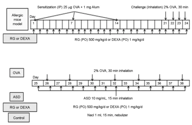
Fig. 1. Study protocol. For allergic mouse model, a mixture of 25 μg OVA and 1 mg Al(OH)3 gel was injected into the peritoneum (systemic immunization) and intranasal sensitization was induced with 2% OVA for 4 days. OVA group inhaled 2% OVA every other day for two weeks. OVA+ASD group inhaled 2% OVA every other day and 1 mL ASD (10 mg/mL) every day for two. OVA+ASD+RG or OVA+ASD+DEXA group were orally administered with a 500 mg/kg/d of RG powder or a 1 mg/kg/d of DEXA, respectively, since the first day of peritoneal injection. OVA and ASD inhalation protocol was the same as the OVA+ASD group. Control group was sprayed with 1 mL of NaCl for two weeks. OVA: ovalbumin, ASD: artificial sand dust, RG: red ginseng, DEXA: dexamethasone
(Yellow soil, Hongikbio, Korea). It was near identical to sand dust collected during the dust storm season of Korea in terms of chemical components, the contents of heavy metals and particle size among commercially manufactured yellow soils (Seo, 2009). ASD is comprised of ions and heavy metals such as Ca2+ 6.1 cmol/kg, K+ 2.5 cmol/kg, Mg2+ 6.2 cmol/kg, Na+ 0.3 cmol/kg, Cd 7.9 mg/kg, Pb 20.4 mg/kg, Zn 90.7 mg/kg, Cu 14.4 mg/kg, Cr 37.2 mg/kg, and Ni 17.6 mg/kg. ASD has pH 6.2 and electrical conductivity of 35.5 dS/m (Seo, 2009).
BALB/c mice were divided into five groups (n=12) each of control (saline), ovalbumin (OVA), OVA+ASD, OVA+
ASD+RG, and OVA+ASD+DEXA. The generation of the OVA-induced allergic animal model was described pre- viously (Kim et al., 2009). For systemic sensitization, 25 μg OVA (Sigma, St. Louis, MO) and 1 mg Al(OH)3 gel (Pierce Chemical Co., Rockford, IL) were mixed with 3 mL of saline and injected with 300 μL into the peritoneal cavity of each mouse on day 1, 7 and 14. Intranasal sensitization was implemented with 2% OVA for 30 minutes using a jet nebulizer (Omron, Japan) from day 21 to day 24 (Fig. 1).
Subsequently, 2% OVA 5 mL inhalation was maintained in allergic mice in the OVA group for 30 minutes per session every other day for two weeks. Along with OVA 5 mL inhalation, the OVA+ASD group inhaled 10 mg/mL ASD for 15 minutes per session. Moreover, RG and DEXA group was orally administered with 500 mg/kg/d of RG powder (KT&G, Korea) and a 1 mg/kg/d of dexamethasone (Dexa-S®, Ilsung, Korea), respectively, since the first day of peritoneal injection for allergic animal model. The control
group was sprayed with 1 mL of NaCl for 15 minutes in each session for two weeks using a nebulizer (Fig. 1).
Among 12 mice in each group, 6 mice were used in histopathological examination of the lung. Lung samples were fixed with 10% paraformaldehyde, and stained with haematoxylin and eosin (H&E) to evaluate the degree of inflammatory cell infiltration. The degrees of observed inflammation were distinguished into three stages in lung tissue specimen. Grade 0 was established as a normal stage with almost no inflammatory cell infiltration while grade 1 was judged to be a mild inflammation stage with slight inflammatory cell infiltration around bronchioles. Moreover, grade 2 was defined to be a stage of inflammatory cell cluster detection with more severe inflammatory cell infiltration. The interpretation of lung tissue samples was carried out by two pathologists who were not informed with the categorized group of specimens.
Among 12 mice in each group, the rest 6 mice were used in obtaining bronchoalveolar lavage (BAL) fluid. The lungs were lavaged with 1 mL of sterile saline at 37℃ by syringe.
The supernatants of BAL fluid were stored at -80℃ until analysis of cytokines and chemokines. Interleukin (IL)-5, IL-12, interferon (IFN)-γ, IL-13, monocyte chemotactic protein (MCP)-1, and eotaxin (R&D systems, Minneapolis, USA) within BAL fluid were assessed using an Enzyme- Linked Immunosorbent Assay (ELISA) kit.
Data are presented as mean ± standard deviation (SD).
Statistical analyses on the pathologic inflammation degrees, the levels of cytokines and chemokines in BAL fluid were conducted using ANOVA followed by Tukey's test. Dif-
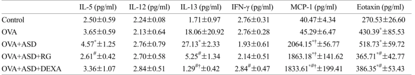
Table 1. Expression of inflammatory cytokines in bronchoalveolar larvage fluid
IL-5 (pg/ml) IL-12 (pg/ml) IL-13 (pg/ml) IFN-γ (pg/ml) MCP-1 (pg/ml) Eotaxin (pg/ml)
Control 2.50±0.59 2.24±0.08 1.71±0.97 2.76±0.31 40.47±4.34 270.53±26.60
OVA 3.65±0.59 2.13±0.64 18.06±20.92 2.76±0.28 45.29±6.47 430.39*±85.53 OVA+ASD 4.57*±1.25 2.76±0.79 27.13*±2.33 1.93±0.61 2064.15*†±56.77 518.73*±59.72 OVA+ASD+RG 2.61#±0.42 2.70±0.58 5.25#±1.34 2.14±0.51 1863.18*†±141.62 365.71*#±42.77 OVA+ASD+DEXA 3.36±1.07 2.84±0.51 1.29#†±0.42 2.84#±0.47 1833.61*#†±199.41 386.35*#±53.43 Each experiment consisted of six observations. All values were expressed as mean ± SE.
*P<0.01 compared to control by ANOVA.
#P<0.01 compared to OVA+ASD by ANOVA.
†P<0.01 compared to OVA by ANOVA.
OVA: ovalbumin, ASD: artificial sand dust, RG: Red ginseng, DEXA: dexamethasone
ferences among groups were determined as statistically significant at a level of P<0.05. All calculations were performed using the SAS 9.1 statistics package (SAS Institute, Inc., Cary, NC, US).
Table 1 shows the levels of cytokines and chemokines in BAL fluid. OVA+ASD group significantly increased con- centrations of IL-5, IL-13, MCP-1, and eotaxin (P<0.01) compared to the control group. OVA+ASD+RG group showed significant decreased levels of IL-5, IL-13, MCP-1 and eotaxin (P<0.01) compared with the OVA+ASD group.
IL-5 and IL-13 levels of OVA+ASD+RG group were not significantly different from the control group. OVA+ASD+
DEXA group also showed decreased concentrations of IL-13, MCP-1, and eotaxin (P<0.01), and increased IFN-γ level (P<0.01) compared to the OVA+ASD group. Between RG and DEXA treatment groups, there were no significant differences in all cytokines and chemokines. Fig. 2 and 3 shows the pathologic change and inflammation degree in lung according to the study groups. The inflammatory cells were significantly decreased in treatment groups with RG or DEXA group compared to the OVA+ASD group (P<0.01).
The purpose of this study was to verify the preventive effect of Korean RG intake on pulmonary diseases generated by sand dust using allergic mice model. Asian sand dust
A B C
D E
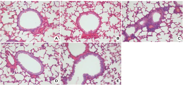
Fig. 2. Histopathologic changes of the lung. A: No pathological changes in the airway treated with saline (control). B: Moderate infiltration of inflammatory cells in the airway treated with OVA alone. C: Marked infiltration of inflammatory cells in the airway treated with OVA+
ASD. D: Significantly decreased infiltration of inflammatory cells in the airway treated with OVA+ASD+RG compared to C. E: Significantly decreased infiltration of inflammatory cells in the airway treated with OVA+ASD+DEXA compared to C. H&E stain (×400). OVA:
ovalbumin, ASD: artificial sand dust, RG: red ginseng, DEXA: dexamethasone
Fig. 3. Inflammation in lung. Each experiment consisted of six observations. Inflammation of OVA+ASD+RG group was signifi- cantly lower than in that of OVA+ASD group.
*P<0.01 compared to control by ANOVA.
#P<0.01 compared to OVA+ASD by ANOVA.
OVA: ovalbumin, ASD: artificial sand dust, RG: red ginseng, DEXA: dexamethasone
induced inflammatory cell infiltration of lung and increased inflammatory cytokines and chemokines in BAL fluid. The administration of RG reduced these lung inflammatory responses, which was effective comparable to steroid, well known as anti-inflammatory drug.
Several active components were reported to be involved in the mechanism of anti-allergic and anti-inflammatory efficacy of RG. Ginsenoside Rh2 was reported to have cell membrane stabilizing effect by inhibiting the secretion of β-hexosaminidase and anti-inflammatory efficacy by in- hibiting the formation of nitrogen oxide (NO) and pro- staglandin E2 (PGE2) (Park et al., 2003a). In particular, it was found to be more powerful in cell membrane stabilizing effect compare with disodium cromolycate (Park et al., 2003a). Furthermore, ginsenoside Rh1 inhibited the release of histamine in peritoneal mast cells of mice and inhibited passive cutaneous anaphylaxis (PCA) by IgE in mice, exhibiting a stronger efficacy than disodium cromolycate (Park et al., 2004a). Moreover, ginsenoside Rb1 and its metabolic substance compound K were also reported to be inhibit the expression of iNOS, COX-2, and NF-κB (Park et al., 2004a; Park et al., 2005). In addition, compound K has been suggested to influence the role of TLR4 and TLR9 in inflammatory responses (Yang et al., 2007).
Previous studies reported that respiratory diseases are generated by eosinophil infiltration, increased eosinophil relevant cytokine and chemokines, and allergic inflam- mations resulting from Th2 predominant response (Chen et al., 2004; Hiyoshi et al., 2005; Ichinose et al., 2008b).
However, the mechanism by which sand dust develops allergic responses is unclear. Although the dust may be one antigen directly involved in inducing allergies, further studies are essential to distinguish antigen substances among various components in sand dust. The sizes of Asian sand dust particles range between 0.1∼20 μm including fine particles under 2.5 μm and ultra-fine particles under 0.1 μm (Choi et al., 2001; Mori et al., 2003). Titanium dioxide, a known fine particle found in sand dust, is assumed to be a direct cause as well as a deteriorating factor in pulmonary diseases. It was suggested that phagocytosis of titanium dioxide by lung macrophages may affect the change and adjustment of respiratory cells (Donaldson et al., 2001).
Some studies have suggested that ions (Park et al., 2004b), such as Na+, Mg2+, Ca2+, NH4+, SO42-, NO3-, toxins including LPS (Ichinose et al., 2008a), and heavy metals (Ichinose et al., 2008b) in the dust act as a new allergic antigen in healthy individuals or an activating factor in allergic patients.
Among the measured cytokines, the levels of MCP-1 and eotaxin of all groups increased in our study compared to those in a previous study (Ichinose et al., 2008a). IL-12 and IL-γ concentrations were generally low in our study (Ichinose et al., 2008a; Ichinose et al., 2008b). These findings may be caused by different sand dusts. Despite the use of ASD, the study obtained similar results consistent with previous studies. The BAL fluids of ASD exposed allergic mice model showed significant increases in not only IL-5, a known key mediator of eosinophilic inflam- mation, but also MCP-1, eotaxin, and the Th2 cytokine IL-13 compared to those of control mice (Ichinose et al., 2008a; Ichinose et al., 2008b). Both cytokines of BAL fluids and inflammatory cell infiltration of lung tissues were significantly decreased in Korean RG-treated (OVA+ASD+
RG) group compared to the ASD-exposed allergic mice (OVA+ASD) group. It is clinically significant outcome that the anti-inflammatory effect of RG is not any different with that of DEXA.
However, RG or DEXA can also influence OVA-induced allergy as well as dust-exposed allergy. In our study, because OVA+RG and OVA+DEXA group were not included, it is difficult to clarify whether the RG or DEXA has a beneficial effect on inflammation by dust-exposed allergy.
In conclusion, the current study shows that sand dust boosts the allergic responses in the allergic mice model.
This study suggests the beneficial effects of Korean RG administration in preventing inflammations of lung resulted from Asian sand dust. Further studies in human subjects are needed to verify the effect of RG intake on preventing respiratory diseases caused by sand dust.
Acknowledgements
This work was supported by the 2010 grant from the Korean Society of Ginseng funded by Korea Ginseng Corporation. The authors would like to thank Seong-Jin Park, Ph.D. at Climate Change & Agroecology Division,
National Academy of Agricultural Science for providing the artificial sand dust.
REFERENCES
Babayigit A, Olmez D, Karaman O, Bagriyanik HA, Yilmaz O, Kivcak B, Erbil G, Uzuner N. Ginseng ameliorates chronic histopathologic changes in a murine model of asthma. Allergy Asthma Proc. 2008. 29: 493-498.
Chen YS, Sheen PC, Chen ER, Liu YK, Wu TN, Yang CY. Effects of Asian dust storm events on daily mortality in Taipei, Taiwan.
Environ Res. 2004. 95: 151-155.
Choi JC, Lee M, Chun Y, Kim J, Oh S. Chemical composition and source signature of spring aerosol in Seoul. Korea J Geophys Res. 2001. 106: 18067-18074.
Donaldson K, Stone V, Clouter A, Ren wick L, MacNee W.
Ultrafine particles. Occup Environ Med. 2001. 58: 211-216.
Echigo A, Hino M, Fukushima T, Mizuki T, Kamekura M, Usami R. Endospores of halophilic bacteria of the family Bacillaceae isolated from non-saline Japanese soil may be transported by Kosa event (Asian dust storm). Saline Systems. 2005. 1: 1-13.
Hiyoshi K, Ichinose T, Sadakane K, Takano H, Nishikawa M, Mori I, Yanagisawa R, Yoshida S, Kumagai Y, Tomura S, Shibamoto T. Asian sand dust enhances ovalbumin-induced eosinophil recruitment in the alveoli and airway of mice.
Environ Res. 2005. 99: 361-368.
Ho YL, Li KC, Chao W, Chang YS, Huang GJ. Korean red ginseng suppresses metastasis of human hepatoma SK-Hep1 cells by inhibiting matrix metalloproteinase-2/-9 and uro- kinase plasminogen activator. Evid Based Complement Alternat Med. 2012. 2012: 965846.
Hong YC, Leem JH, Ha EH, Christiani DC. PM10 exposure, gaseous pollutants, and daily mortality in Inchon, Korea.
Environ Health Perspect. 1999. 107: 873-878.
Hwang SS, Cho SH, Kwon HJ. Effects of the severe Asian dust events of daily mortality during the spring of 2002, in Seoul, Korea. J Prev Med Public Health. 2005. 38: 197-202.
Ichinose T, Yoshida S, Hiyoshi K, Sadakane K, Takano H, Nishikawa M, Mori I, Yanagisawa R, Kawazato H, Yasuda A, Shibamoto T. The effects of microbial materials adhered to Asian sand dust on allergic lung inflammation. Arch Environ Contam Toxicol. 2008a. 55: 348-357.
Ichinose T, Yoshida S, Sadakane K, Takano H, Yanagisawa R, Inoue K, Nishikawa M, Mori I, Kawazato H, Yasuda A, Shibamoto T.
Effects of asian sand dust, Arizona sand dust, amorphous silica and aluminum oxide on allergic inflammation in the murine lung. Inhal Toxicol. 2008b. 20: 685-694.
Kim DY, Yang WM. Panax ginseng ameliorates airway in- flammation in an ovalbumin-sensitized mouse allergic asthma model. J Ethnopharmacol. 2011. 136: 230-235.
Kim MY. Physical and chemical characteristics of Asian dust. J Korean Med Assoc. 2004. 47: 453-464.
Kim ST, Lee EJ, Jung HJ, Gang IG, Cha HE, Kim DY, Do SH.
The effect of Asian sand dust in allergic inflammation of allergic mouse. Korean J Otorhinolaryngol-Head Neck Surg.
2009. 52: 498-505.
Kwon HJ, Cho SH. The health effects of Asian dust event. J Korean Med Assoc. 2004. 47: 465-470.
Kwon HJ, Cho SH, Chun YS, Lagarde F, Pershagen G. Effects of the Asian dust events on daily mortality in Seoul, Korea.
Environ Res. 2002. 90: 1-15.
Lee DC, Lau AS. Effects of Panax ginseng on tumor necrosis factor-α-mediated inflammation: a mini-review. Molecules.
2011. 16: 2802-2816.
Min PK, Kim CW, Yun YJ, Chang JH, Chu JK, Lee KE, Han JY, Park JW, Hong CS. Effect of yellow sand on respiratory symptoms and diurnal variation of peak expiratory flow in patients with bronchial asthma. J Asthma Allergy Clin Immunol.
2001. 21: 1179-1186.
Mori I, Nishikawa M, Tanimura T, Quan H. Change in size distribution and chemical composition of Korea (Asian dust) aerosol during long-range transport. Atmos Environ. 2003.
58: 4523-4563.
Park EK, Choo MK, Han MJ, Kim DH. Ginsenoside Rh1 pos- sesses antiallergic and anti-inflammatory activities. Int Arch Allergy Immunol. 2004a. 133: 113-120.
Park EK, Choo MK, Kim EJ, Han MJ, Kim DH. Antiallergic activity of ginsenoside Rh2. Biol Pharm Bull. 2003a. 26:
1581-1584.
Park EK, Shin YW, Lee HU, Kim SS, Lee YC, Lee BY, Kim DH.
Inhibitory effect of ginsenoside Rb1 and compound K on NO and prostaglandin E2 biosyntheses of RAW264.7 cells induced by lipopolysaccharide. Biol Pharm Bull. 2005. 28:
652-656.
Park JW, Lim YH, Kyung SY, An CH, Lee SP, Jeong SH, Ju YS.
Effects of ambient particulate matter (PM10) on peak expiratory flow and respiratory symptoms in subjects with bronchial asthma during yellow sand period. Tuberc Respir Dis. 2003b. 55: 570-578.
Park SH, Song CB, Kim MC, Kwon SB, Lee KW. Study on size distribution of total aerosol and water-soluble ions during an Asian dust storm event at Jeju Island, Korea. Environ Monit Assess. 2004b. 93: 157-183.
Paul S, Shin HS, Kang SC. Inhibition of inflammations and macro- phage activation by ginsenoside-Re isolated from Korean ginseng (Panax ginseng C.A. Meyer). Food Chem Toxicol.
2012. 50: 1354-1361.
Pope CA 3rd, Bates DV, Raizenne ME. Health effects of particulate air pollution: Time for reassessment? Environ Health Perspect.
1995. 103: 472-480.
Seo KH. Chemical and microbiological characteristics and risk assessment of Asian dust. National Academy of Agricultural Science. 2009.
Song HI. Effect of air pollution on childhood asthma living in Seoul. J Asthma Allergy Clin Immunol. 2001. 21: 28-39.
Yang CS, Ko SR, Cho BG, Lee JY, Kim KH, Shin DM, Yuk JM, Sohn HJ, Kim SY, Wee JJ, Do JH, Jo EK. Compound K (CK) rich fractions from Korean red ginseng Inhibit Toll-like receptor (TLR) 4- or TLR 9-mediated mitogen-activated protein kinases activation and proinflammatory responses in murine macrophages. J Ginseng Res. 2007. 31: 181-190.