http://dx.doi.org/10.3988/jcn.2012.8.3.224 J Clin Neurol 2012;8:224-229
Introduction
The incidence of traumatic brain injury (TBI) is reported to be between 56 and 836 per 100000,1 and the burden of the disease attributable to TBI is high due to it being more com- mon in young adults and causing significant disabilities. One of the distinguishing features of TBI is a wide variation in fun- ctional outcome. In addition, disabilities after TBI are mostly cognitive, behavioral, and subjective symptoms. The assess- ment of the disability associated with TBI and prediction of the outcome are frequently required for claiming damages and
social support. Several prognostic factors of a poor outcome have been proposed, such as old age, low initial Glasgow Coma Scale (GCS) score, anisocoria, hypotension, or shock.2-4 Radio- logical findings, such as obliteration of the third ventricle or basal cistern, midline shift, subarachnoid hemorrhage (SAH),3,4 lesion burden,5 and grade of diffuse axonal injury6 are report- ed as being predictive of a poor outcome.
Computed tomography (CT) has remained the primary imaging tool for TBI in clinical practice because of its sensi- tivity in detecting acute hemorrhage and its rapid scanning ab- ility. However, CT has limited sensitivity for diffuse axonal injury (DAI), nonhemorrhagic contusions, and brainstem le- sions.7 Although many studies have used delayed magnetic resonance imaging (MRI) to assess clinical outcome, few st- udies have used early MRI for the assessment of the severity or probable outcome of trauma.2,6,8,9 Early diffusion-weighted
Prediction of Outcome after Traumatic Brain Injury Using Clinical and Neuroimaging Variables
Seo-Young Lee,a Sam Soo Kim,b Choong-Hyo Kim,c Seung-Woo Park,c Jae Hyo Park,d Minjoo Yeoa
aDepartments of Neurology, bRadiology and cNeurosurgery, Kangwon National University Hospital, Chuncheon, Korea
dDepartment of Neurosurgery, Dongguk University Ilsan Hospital, Goyang, Korea
Received August 17, 2011 Revised January 16, 2012 Accepted January 16, 2012 Correspondence Choong-Hyo Kim, MD Department of Neurosurgery, Kangwon National University Hospital, 156 Baengnyeong-ro, Chuncheon 200-722, Korea Tel +82-33-258-2410 Fax +82-33-258-2221 E-mail [email protected] Minjoo Yeo, MD
Department of Neurology, Kangwon National University Hospital, 156 Baengnyeong-ro, Chuncheon 200-722, Korea Tel +82-33-258-2413 Fax +82-33-258-2103 E-mail [email protected]
Background and PurposezzThe functional outcome of traumatic brain injury (TBI) varies wi- dely. The aim of this study was to identify the factors predicting outcome following TBI.
MethodszzWe prospectively enrolled acute TBI patients, and assessed them clinically and radio- logically using brain magnetic resonance imaging (MRI). Functional outcome was measured using the Glasgow Outcome Scale (GOS) at 3 months after TBI. A GOS score of ≤4 was regard- ed as an unfavorable outcome. We performed multivariate analysis to investigate the association between clinicoradiological variables and outcome.
ResultszzForty-two patients completed the clinical evaluation in the acute phase and outcome measurement at 3 months. Motorcycle accident was associated with unfavorable outcome [odds ratio (OR)=38.3, p=0.022]. If the patients were the victims of the accident, they were more likely to have an unfavorable outcome (OR=21.3, p=0.037). All seven patients with a low Glasgow Coma Scale (GCS) score (i.e., ≤8) at 24 or 48 h after TBI were also found to have an unfavorable outcome. The presence of diffuse axonal injury (DAI) was a significant predicting factor of an un- favorable outcome (OR=8.48, p=0.042).
ConclusionszzMotorcycle accident, being an accident victim, and a lower GCS score at 24 hours or more after the accident were found to be unfavorable prognostic variables. DAI was the only radiologic variable predicting an unfavorable outcome. Thus, it is important to identify DAI by applying MRI in the acute phase. J Clin Neurol 2012;8:224-229 Key Wordszz traumatic brain injury, outcome, magnetic resonance imaging.
Open Access
cc This is an Open Access article distributed under the terms of the Cre- ative Commons Attribution Non-Commercial License (http://creative- commons.org/licenses/by-nc/3.0) which permits unrestricted non-com- mercial use, distribution, and reproduction in any medium, provided the ori- ginal work is properly cited.
image (DWI) is particularly valuable in evaluating DAI.10 We performed a prospective study of TBI, including clini- cal measurements and MRI in the acute phase and at 3 months postinjury. The objectives of the current study were to de- scribe the clinical and radiological characteristics of TBI and to elucidate its early prognostic factors.
Methods
Population and procedures
TBI patients were prospectively evaluated and followed up for more than 3 months. We consecutively enrolled patients who had clinical or radiologic evidence of TBI and where this evidence was obtained within 1 week after the time of injury.
Exclusion criteria were being older than 75 years and having previous central nervous system (CNS) illness or concussion.
All subjects received a brain CT scan immediately after visit- ing the hospital. We performed brain MRI within 2 weeks of the initial visit (mean, 4.1 days) if the patient’s condition allow- ed it. We investigated the cause of trauma, clinical symptoms, neurological signs, presence of seizure during hospitalization, and GCS score at the time of presentation, and at 24 h, 48 h, and 1 week later using a structured case-recording form. The GCS scores of patients who were sedated were excluded from the analysis of prognostic factors.
Functional outcome was evaluated using the Glasgow Out- come Scale (GOS) at 3 months after TBI in an outpatient clinic or by telephone interview. We dichotomized the outcome into favorable (GOS score=5, able to return to work or school) and unfavorable (GOS score ≤4).
Between October 2007 and September 2009, 93 individu- als with TBI were registered. Of these, 51 patients were exclud- ed for the following reasons: visiting the hospital more than 1 week after the injury (3 patients), being older than 75 years (10 patients), CNS illness (16 patients), concussion (10 pati- ents), and refusal to provide consent or a missed follow-up (12 patients). Ultimately, 42 patients completed the clinical evalu- ation during the admission and at the 3-month follow-up.
Our institutional review board approved this study. Inform- ed consent to participate was obtained from the patients and their caregivers.
MRI and CT protocols
All subjects were scanned using a 1.5-T magnetic resonance (MR) scanner (Gyroscan ACS-NT, Philips Medical Systems, Best, The Netherlands). Axial T1, T2, fluid-attenuated inver- sion recovery, gradient-echo, and diffusion, supplemented with coronal T2 and sagittal T1 images were obtained.
Single-shot spin-echo echo-planar DWI sequences were obtained by applying diffusion gradients in three orthogonal
directions for each section, with two diffusion weightings (b=
0 s/mm2 and b=1000 s/mm2). The parameters for DWI were as follows: repetition time/echo time, 3298/61 ms; matrix, 128×
128; field of view, 24 cm; slice thickness, 5 mm; and intersec- tion gap, 1.5 mm.
All CT scans were obtained using a helical CT scanner (Li- ghtSpeed Plus, GE Medical Systems, Milwaukee, WI, USA).
The scanning parameters were 120 kV and 300 mA, with an image matrix of 512×512, a field of view of 22 cm, and a sec- tion thickness of 5 mm.
Imaging analysis
An experienced neuroradiologist (Kim SS) and neurologists (Lee SY and Yeo MJ) interpreted the imaging findings by con- sensus based on visual inspection, in accordance with prede- fined variables. We evaluated the presence of a contusion, sub- dural hemorrhage (SDH), epidural hemorrhage (EDH), SAH, DAI, midline shift, diffuse brain swelling, and transtentorial herniation, and the location and size of lesions using brain CT and MR images. Combined injury was defined when it had two or more component of TBI among contusion, SDH, and EDH.
DAI appears on MR images as multiple, small, deeply sit- uated, and elliptical lesions that spare the overlying cortex.11 DAI was defined as punctuate lesions of profound hypoin- tensity on T2-weighted gradient-echo imaging, and as le- sions of profound hyperintensity on fluid-attenuated inver- sion recovery and DWI.12,13 DAI was classified as stage 1, 2, or 3 using the modified Adams classification,14 according to its location. SAH was graded according to the 4-point Fisher scale (Table 1).15 The presence of diffuse brain swelling was determined based on the observation of diffuse cisternal oblit- eration on the brain CT image obtained during the acute pe- riod. Contusion was defined as parenchymal patchy lesion, which manifests as a low density on CT and a hyperintensity on T2-weighted MRI with or without hemorrhagic foci. We calcu-lated the size of the contusion by measuring the diam- eter of the high signal intensity on T2-weighted MRI, since many of the contusions were not sufficiently visible on CT.
Statistics
We identified the independent predictors of unfavorable out- come by applying multiple logistic regression with stepwise selection. The regression model included the clinicoradiologi- cal variables that appeared to be associated with an unfavor- able outcome in univariate analysis (p<0.1) or predictors sug- gested in previous studies. Multicollinearity was tested using the contingency coefficient, which represents the strength of association between categorical variables. A separate model was fit for each associated variable to avoid multicollinearity.
The magnitude of associations between the potential pre-
dictive variables and outcome was quantified using the odds ratio (OR) and the corresponding 95% confidence interval. A probability value of p<0.05 was considered significant in mul- tivariate analysis. Statistical analysis was performed using the SPSS program (version 19.0, IBM SPSS Statistics, Chicago, IL, USA).
Results
General characteristics
The 3-month outcome after TBI was available for 42 patients.
Their mean age was 44.5 years (range, 8-73 years), and 28 (66.7%) were men and 14 (33.3%) were women. The causes of the TBIs were falling (19 patients, 45.2%), motorcycle acci- dent (1 patients, 26.2%), pedestrian injury (6 patients, 14.3%), bicycle accident (3 patients, 7.14%), and car accident (3 pa- tients, 7.14%). Ten (23.8%) of the patients were victims of the accidents, while others were perpetrators or those whose re- sponsibility of the accident was ambiguous.
Severity according to the GCS at presentation was mild (GCS score, 13-15) in 31 patients (73.8%), moderate (GCS score, 9-12) in 6 patients (14.3%), and severe (GCS score, 3-8) in 5 patients (11.9%). Acute seizure occurred in 6 pa- tients (14.3%), within two weeks of onset of TBI.
We detected a contusion in 34 (81.0%), SAH in 24 (60.0%), SDH in 30 (71.4%), EDH in 7 (16.7%), and DAI in 16 (41.0%) patients. Thirty-three patients (78.6%) had combined injuries with multiple pathologic features. The contusion was located in the frontal, temporal, occipital, and parietal lobes in 28, 25, 5, and 4 patients, respectively. Fourteen, six, two, and two pa- tients had grade 1, 2, 3, and 4 SAH, respectively. Ten, two, and four patients had grade 1, 2, and 3 DAI, respectively. Mi-
dline shift (>5 mm) was detected in 16 (38.1%) patients, tr- anstentorial herniation was observed in 7 (16.7%) patients, and diffuse swelling was observed in 7 (16.7%) patients.
Three months after their accident, 27 patients (64.3%) were able to return to work or school (GOS score=5), 8 pa- tients had moderate disability (GOS score=4), and 2 patients could not live independently (GOS score=3). Five patients died within 3 months (GOS score=1); the time to death ranged from 2 to 37 days. Two patients died as a result of systemic tr- auma, two patients died because of either the TBI itself or sep- sis after a prolonged bedridden state due to the TBI, and one patient died unexpectedly of an unknown cause (Table 2).
Predictive factors Clinical variables
The cause of motorcycle accident was an independent prog- nostic factor of an unfavorable outcome (OR=44.6; p=0.012, multivariate analysis). If the patients were victims of the ac- cidents they were more likely to have an unfavorable out- come (OR=68.7; p=0.013, multivariate analysis). Four out of five patients who had an initial GCS score of ≤8 had an un- favorable outcome, and six patients with a GCS score of ≤8 at 24 h and seven patients with a GCS score of ≤8 at 48 h had all unfavorable outcomes. Age and gender were not as- sociated with outcome. Acute seizure did not predict an un- favorable outcome (Table 3 and 4).
Neuroimaging variables
DAI was a prognostic factor of unfavorable outcome (OR=8.48;
p=0.042, multivariate analysis). Grade 2 or more DAI was par- ticularly strongly associated with an unfavorable outcome (OR=30.5; p=0.018, multivariate analysis). However, DAI did not always predict an unfavorable outcome. Seven (43.8%) DAI patients had a good recovery, including one patient with grade 3 DAI. DAI was significantly associated with motorcycle ac- cidents (contingency coefficient, 0.327; p=0.031) and an initial GCS score of ≤8 (contingency coefficient, 0.418; p=0.004).
Diffuse swelling and grade 2 or more SAH were related to an unfavorable outcome in univariate analysis (OR=6.25; p=0.046 and OR=7.00; p=0.015, respectively), but the association was not statistically significant in multivariate analysis. Diffuse sw- elling covaried with an initial GCS score of ≤8 (contingency coefficient, 0.53; p<0.001). All four patients with grade 3 or more severe SAH had an unfavorable outcome.
The presence and size of the SDH or EDH, the presence, size, and location of the contusion, combined injury, and mid- line shift were not significantly associated with an unfavor- able outcome. Seven of the nine patients with a contusion lar- ger than 60 cm3 regained good function (Table 3 and 4).
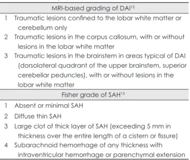
Table 1. Grading system of diffuse axonal injury (DAI) and sub- arachnoid hemorrhage (SAH)
MRI-based grading of DAI12
1 Traumatic lesions confined to the lobar white matter or cerebellum only
2 Traumatic lesions in the corpus callosum, with or without lesions in the lobar white matter
3 Traumatic lesions in the brainstem in areas typical of DAI (dorsolateral quadrant of the upper brainstem, superior cerebellar peduncles), with or without lesions in the lobar white matter
Fisher grade of SAH13 1 Absent or minimal SAH
2 Diffuse thin SAH
3 Large clot of thick layer of SAH (exceeding 5 mm in thickness over the entire length of a cistern or fissure) 4 Subarachnoid hemorrhage of any thickness with
intraventricular hemorrhage or parenchymal extension MRI: magnetic resonance imaging.
Discussion
Relatively few studies have focused on the relationship be- tween the cause of injury and functional outcome. The pres- ent study found that a TBI cause of motorcycle accident pre- dicted an unfavorable outcome. Motorcycle accidents accom- pany marked acceleration-deceleration without protection, and commonly result in severe TBI, including DAI.16 In the IMPACT study,17 falling was associated with a poorer out- come than traffic accidents and assaults. However, this rela- tionship was lost after adjustment for age, and TBI associat- ed with motorcycle accidents was not analyzed separately.
The GCS score was associated with outcome in this study, which is consistent with the results of previous studies. All patients who had low GCS score at 24 or 48 h had an unfa- vorable outcome, which suggests a low probability of favor-
able outcome following coma persisting for 24 h or more after TBI, although the sample was too small to allow generalization.
Being the victim of an accident was shown to be a signifi- cant independent prognostic factor of an unfavorable out- come. Most studies4,18 investigating predictive factors of TBI categorized good and poor outcomes as GOS scores of ≥4 and ≤3, respectively. However, the most frequent and prob- lematic symptoms after TBI are cognitive and behavioral symptoms. Therefore, we classified a GOS score of ≤4 (able to live independently, but unable to return to work or school) as an unfavorable outcome. However, since the level of social functioning is influenced by emotion and can be manipulated by secondary gain, it is important to determine the objective variables that are correlated with outcome in order to establish whether or not social dysfunction is concordant with the TBI.
DAI was the only neuroimaging variable associated with unfavorable outcome. DAI was observed in 41.0% of the sub- jects in our study, and was reported to range between 48% and 72% according to the severity of the subjects’ injury in other studies.14,19 DAI results from the tearing of axons due to shear- ing forces during acceleration, deceleration, and rotation of the brain,13 and appears to be the frequent cause of the poor out- come.6 Brainstem lesions were found to be associated with a poor outcome in previous studies.2,6,8,9 brainstem lesions were detected in only four of the patients in the present study, of Table 2. General characteristics of the patients
Characteristic Statistic
Gender
Male 28 (66.7%)
Female 14 (33.3%)
Age (years) 44.5 (8-73)
60-75 15 (35.7%)
< 60 27 (64.3%)
Cause
Falling 19 (45.2%)
Motorcycle accident 11 (26.2%)
Pedestrian injury 6 (14.3%)
Bicycle accident 3 (7.14%)
Car accident 3 (7.14%)
Severity by the initial level of consciousness
Mild (GCS 13–15) 31 (73.8%)
Moderate (GCS 9–12) 6 (14.3%)
Severe (GCS 3–8) 5 (11.9%)
Type of lesion
Contusion 34 (81.0%)
SAH 24 (60.0%)
SDH 30 (71.4%)
EDH 7 (16.7%)
DAI 16 (41.0%)
Combined 33 (78.6%)
Outcome at 3 months
GOS5 27 (64.3%)
GOS4 8 (19.0%)
GOS3 2 (4.8%)
GOS2 0
GOS1 5 (11.9%)
DAI: diffuse axonal injury, EDH: epidural hemorrhage, GCS: Glas- gow Coma Scale, GOS: Glasgow Outcome Scale, SAH: sub- arachnoid hemorrhage, SDH: subdural hemorrhage.
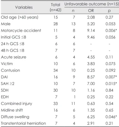
Table 3. Univariate analysis of the clinicoradiological variables that predicted outcomes
Variables Total (n=42)
Unfavorable outcome (n=15)
n OR p
Old age (>60 years) 15 7 2.08 0.27
Male 28 13 5.20 0.053
Motorcycle accident 11 8 9.14 0.006*
Initial GCS ≤8 5 4 9.46 0.056
24 h GCS ≤8 6 6 - -
48 h GCS ≤8 7 7 - -
Acute seizure 6 4 4.55 0.11
Victim 10 6 3.83 0.075
Contusion 34 10 0.25 0.092
DAI 16 9 8.57 0.007*
SAH ≥2 10 7 7.00 0.015*
SDH 30 10 1.16 0.84
EDH 7 1 0.25 0.22
Combined injury 33 11 0.63 0.54
Midline shift 16 6 1.35 0.65
Diffuse swelling 7 5 6.25 0.046*
Transtentorial herniation 7 4 2.91 0.21
*Indicates the results considered statistically significant (p
<0.05).
DAI: diffuse axonal injury, EDH: epidural hemorrhage, GCS:
Glasgow Coma Scale, OR: odds ratio, SAH: subarachnoid hem- orrhage, SDH: subdural hemorrhage.
which three had an unfavorable outcome, although we were unable to estimate the statistical significance of this finding.
Lesion burden was associated with outcome in several stud- ies,6 but it was not applicable to individual patients. In cases of extra-axial hematoma, timely surgery can result in good out- come. Even for parenchymal lesions, seven out of nine patients with a large contusion (>60 cm3) had a good outcome in this study.
Early MRI improves the outcome prediction in TBI be- cause it can detect DAI-which is a frequent form of TBI and a strong prognostic factor-much more sensitively than can CT and delayed MRI. DWI and gradient-echo sequences are the most useful MR sequences for evaluating TBI in the acute stage due to their superior resolution for DAI and the short scan time, which permits the evaluation of clinically unstable patients.
The major limitation of this study was the small number of patients and the absence of a long-term outcome measure- ment. Our population was relatively old and many patients were excluded because of preexisting CNS illness. In addition, loss of follow-up is more frequent in TBI than in chronic ill- ness. Large-scale multicenter studies are therefore needed to confirm our findings. The GOS score is usually determined at 3, 6, and 12 months after TBI;20 we measured it at 3 months as a midterm outcome. Many studies have found that the symp- toms of mild TBI resolve within 3 months.22 The time course of recovery for moderate-to-severe TBI was not closely ex- amined. Three months is sufficient for stabilization of the le- sion and longer times after TBI may permit interventions of confounding factors such as the environment and restoration ability of the individuals, although recovery can occur after more than 3 months following TBI.
In summary, motorcycle accident, being an accident vic-
tim, decreased level of consciousness after 24 h, and DAI ra- ther than the type and size of lesion are unfavorable prognos- tic variables.
Conflicts of Interest
The authors have no financial conflicts of interest.
Acknowledgements
This study was supported by a 2008 Kangwon National University Hos- pital Grant.
REFERENCES
1. Bruns J Jr, Hauser WA. The epidemiology of traumatic brain injury:
a review. Epilepsia 2003;44 Suppl 10:2-10.
2. Lagares A, Ramos A, Pérez-Nuñez A, Ballenilla F, Alday R, Gómez PA, et al. The role of MR imaging in assessing prognosis after severe and moderate head injury. Acta Neurochir (Wien) 2009;151:341-356.
3. MRC CRASH Trial Collaborators, Perel P, Arango M, Clayton T, Edwards P, Komolafe E, et al. Predicting outcome after traumatic brain injury: practical prognostic models based on large cohort of in- ternational patients. BMJ 2008;336:425-429.
4. Murray GD, Butcher I, McHugh GS, Lu J, Mushkudiani NA, Maas AI, et al. Multivariable prognostic analysis in traumatic brain injury:
results from the IMPACT study. J Neurotrauma 2007;24:329-337.
5. Di Stefano G, Bachevalier J, Levin HS, Song JX, Scheibel RS, Fletcher JM. Volume of focal brain lesions and hippocampal forma- tion in relation to memory function after closed head injury in chil- dren. J Neurol Neurosurg Psychiatry 2000;69:210-216.
6. Weiss N, Galanaud D, Carpentier A, Naccache L, Puybasset L. Clini- cal review: Prognostic value of magnetic resonance imaging in acute brain injury and coma. Crit Care 2007;11:230.
7. Gentry LR, Godersky JC, Thompson B, Dunn VD. Prospective com- parative study of intermediate-field MR and CT in the evaluation of closed head trauma. AJR Am J Roentgenol 1988;150:673-682.
8. Weiss N, Galanaud D, Carpentier A, Tezenas de Montcel S, Naccache L, Coriat P, et al. A combined clinical and MRI approach for outcome assessment of traumatic head injured comatose patients. J Neurol 2008;255:217-223.
9. Skandsen T, Kvistad KA, Solheim O, Lydersen S, Strand IH, Vik A.
Prognostic value of magnetic resonance imaging in moderate and se- vere head injury: a prospective study of early MRI findings and one- year outcome. J Neurotrauma 2011;28:691-699.
10. Huisman TA. Diffusion-weighted imaging: basic concepts and appli- cation in cerebral stroke and head trauma. Eur Radiol 2003;13:2283- 2297.
11. Gentry LR. Imaging of closed head injury. Radiology 1994;191:1-17.
12. Ezaki Y, Tsutsumi K, Morikawa M, Nagata I. Role of diffusion- weighted magnetic resonance imaging in diffuse axonal injury. Acta Radiol 2006;47:733-740.
13. Parizel PM, Ozsarlak, Van Goethem JW, van den Hauwe L, Dillen C, Verlooy J, et al. Imaging findings in diffuse axonal injury after closed head trauma. Eur Radiol 1998;8:960-965.
14. Skandsen T, Kvistad KA, Solheim O, Strand IH, Folvik M, Vik A.
Prevalence and impact of diffuse axonal injury in patients with mod- erate and severe head injury: a cohort study of early magnetic reso- nance imaging findings and 1-year outcome. J Neurosurg 2010;113:
556-563.
15. Fisher CM, Kistler JP, Davis JM. Relation of cerebral vasospasm to subarachnoid hemorrhage visualized by computerized tomographic scanning. Neurosurgery 1980;6:1-9.
16. Shuaeib FM, Hamouda AMS, Umar RSR, Hamdan MM, Hashmi MSJ. Motorcycle helmet. Part I. Biomechanics and computational is- Table 4. Multivariate analysis of the clinicoradiological variables
that predicted unfavorable outcome
OR (95% CI) p
Old age (>60 years) 3.10 (0.32-30.2) 0.33
Male 8.66 (0.81-92.2) 0.074
Motorcycle accident 38.3 (1.69-869) 0.022*
Victim 21.3 (1.21-374) 0.037*
Diffuse axonal injury 8.48 (1.08-66.5) 0.042*
Diffuse swelling 16.1 (0.28-943) 0.18
SAH ≥2 3.65 (0.31-43.4) 0.31
Motorcycle accident and diffuse axonal injury were separate- ly analyzed because they are significantly related with each other. Iinitial Glasgow Coma Scale ≤8 was excluded from this analysis, because the number was too small for multivariate analysis and it was significantly colinear with diffuse axonal in- jury and diffuse swelling.
*Indicates the results considered statistically significant (p<
0.05).
SAH: subarachnoid hemorrhage, OR: odds ratio, CI: confidence interval.
sues. J Mater Process Technol 2002;123:406-421.
17. Butcher I, McHugh GS, Lu J, Steyerberg EW, Hernández AV, Mush- kudiani N, et al. Prognostic value of cause of injury in traumatic brain injury: results from the IMPACT study. J Neurotrauma 2007;24:
281-286.
18. Maas AI, Stocchetti N, Bullock R. Moderate and severe traumatic brain injury in adults. Lancet Neurol 2008;7:728-741.
19. Gentry LR, Godersky JC, Thompson B. MR imaging of head trauma:
review of the distribution and radiopathologic features of traumatic lesions. AJR Am J Roentgenol 1988;150:663-672.
20. Zink BJ. Traumatic brain injury outcome: concepts for emergency care. Ann Emerg Med 2001;37:318-332.
21. Steyerberg EW, Mushkudiani N, Perel P, Butcher I, Lu J, McHugh GS, et al. Predicting outcome after traumatic brain injury: develop- ment and international validation of prognostic scores based on ad- mission characteristics. PLoS Med 2008;5:e165; discussion e165.
22. Carroll LJ, Cassidy JD, Peloso PM, Borg J, von Holst H, Holm L, et al. Prognosis for mild traumatic brain injury: results of the WHO Collaborating Centre Task Force on Mild Traumatic Brain Injury. J Rehabil Med 2004;(43 Suppl):84-105.