CASE REPORT
146 Copyright ⓒ 2009 Korean Neurological Association
Print ISSN 1738-6586 / On-line ISSN 2005-5013 10.3988/jcn.2009.5.3.146 J Clin Neurol 2009;5:146-148
Secondary Amyloidosis Associated with Multiple Sclerosis
Seok Jae Kang, MDa; Joo-Hark Yi, MDb; Hyun-Seok Hong, MDb; Si-Hyung Jang, MDc; Moon-Hyang Park, MDc; Ho-Jung Kim, MDb; Kyu-Yong Lee, MDa; Young-Joo Lee, MDa; Sang-Woong Han, MDb; Seong-Ho Koh, MDa
Departments of aNeurology, bInternal Medicine and cPathology, Hanyang University College of Medicine, Guri, Korea
Received March 4, 2009 Revised March 27, 2009 Accepted March 27, 2009 Correspondence Seong-Ho Koh, MD Department of Neurology, Hanyang University College of Medicine, 249-1 Gyomun-dong, Guri 471-020, Korea Tel +82-31-560-2267 Fax +82-31-560-2267 E-mail [email protected]
BackgroundaaMultiple sclerosis (MS) is a demyelinating disease of the central nervous system.
Secondary amyloidosis can occur as a complication of chronic systemic inflammatory and in- fectious diseases. Until now there has been no report of secondary amyloidosis associated with MS. We report herein a case of renal biopsy-proven secondary amyloidosis in a patient with MS.
Case ReportaaA 41-year-old woman with MS was hospitalized due to aggravated quadri- paresis and edema in both lower extremities. Laboratory findings showed nephrotic-range pro- teinuria and hypoalbuminemia. A percutaneous renal biopsy procedure was performed, the re- sults of which revealed secondary amyloid-A-type amyloidosis associated with MS.
ConclusionsaaThis is the first report of secondary amyloidosis associated with MS.
J Clin Neurol 2009;5:146-148 Key Wordsaamultiple sclerosis, secondary amyloidosis, nephrotic syndrome.
Introduction
Multiple sclerosis (MS) is a demyelinating autoimmune dis- ease of the central nervous system. Its pathological triad com- prises central nervous system inflammation, demyelination, and gliosis.1 Secondary amyloidosis, which develops seconda- rily to chronic inflammatory conditions such as rheumatoid arthritis, is now called amyloid-A (AA) amyloidosis because a major factor in the protein deposition process involves a cl- eaved product of the acute-phase protein, serum amyloid A (SAA).2 Cerebrovascular amyloid deposits in the region of demyelinated plaques without systemic amyloidosis have been reported rarely in cases of MS;3 however, there is no report in the literature of MS related to AA amyloidosis. This article presents a case of MS with secondary AA amyloidosis, presenting with nephrotic syndrome. This is the first report of secondary amyloidosis associated with MS.
Case Report
A 41-year-old Korean woman was hospitalized due to aggra- vated quadriparesis. She had been diagnosed with MS at an age of 33 years. At 26 years of age, the patient experienced her first episode of quadriparesis with sensory changes in both
lower extremities. At 33 years, she noticed decreased visual acuity for several days. At 40 years, she was admitted to the ho- spital due to quadriparesis, dysarthria, and confusion, and her brain MRI showed brain, medullary, and spinal cord lesions (Fig. 1A) ; steroid pulse therapy was conducted. The patient’s neurological symptoms improved, and she was discharged.
One year later, she was hospitalized again due to aggravated quadriparesis; she also complained of edema in both lower extremities. Steroid pulse therapy was performed again and the motor weakness in her upper extremities improved, but she continued to complain of dyspnea, orthopnea, and peri- pheral edema. Her chest X-ray showed cardiomegaly with pulmonary edema, but her echocardiogram showed normal findings. Laboratory studies showed the following param- eters: white blood cell count 6,500/mm3, red blood cell count 2.93×10⁶/mm3, hemoglobin 9.6 g/dL, hematocrit 28.1%, plate- lets 248,000/mm3, serum sodium 144 mEq/L, serum potas- sium 4.7 mEq/L, serum chloride 109 mEq/L, serum creatinine 1.2 mg/dL, serum blood urea nitrogen 78 mg/dL, serum al- bumin 2.5 g/dL, serum total protein 5.0 g/dL, serum choles- terol 218 mg/dL, and serum SAA 44.4 μg/mL (reference level <8 μg/mL). In addition, anti-nuclear antibody, anti-neut- rophil cytoplasmic antibody, rheumatoid factor, and cryoglo- bulin were not detected in this patient’s serum, which was
Kang SJ et al.
www.thejcn.com 147 also negative for hepatitis B surface antigen and anti-hepatitis-
C antibody.
The 24-hour urinalysis revealed a protein excretion of 4,831 mg/day. A percutaneous renal biopsy procedure confirmed the presence of AA-type amyloidosis (Fig. 1B-E). The patient had no family history of amyloidosis. We performed immunofixa- tion electrophoresis on her serum and urine to exclude a plas- ma-cell dyscrasia. M-proteins were not detected and hypoal- buminemia was observed using serum immunofixation elect- rophoresis. Urine immunofixation electrophoresis revealed al- buminuria and increased globulin without an M-spike. AA amyloidosis was confirmed in view of the positive immunohis- tochemical staining for amyloid A and negative staining for kappa and lambda; however, with the exception of a past his- tory of non-febrile asymptomatic bacteriuria and a short- lasting minor decubitus sore on the coccyx, there was no evidence of other systemic infection or inflammatory disease.
Consequently, this case was diagnosed as secondary AA-type amyloidosis associated with MS.
Discussion
We present herein the clinicopathological findings of a patient with MS who developed AA amyloidosis approximately 15 years after the onset of MS. Amyloidosis is caused by the extra- cellular deposition of pathologic, insoluble, fibrillar proteins in organs and tissues. Precursor proteins are known to change into fibrils through multiple mechanisms that differ among the various types of amyloid. Secondary amyloidosis is caus- ed by the deposition of amyloid originating from SAA, which is an acute-phase protein produced in response to inflamma- tion4 and occurs most commonly among patients with chronic inflammatory diseases such as rheumatoid arthritis, juvenile rheumatoid arthritis, and inflammatory bowel disease.5 Fa- milial Mediterranean fever (FMF) is also an inflammatory dis- ease characterized by episodic fever and serositis. Livneh et al.6 reported the development of AA amyloidosis in up to 90% of untreated FMF patients. A high prevalence of FMF was recently reported in one MS cohort in Turkey.7 These
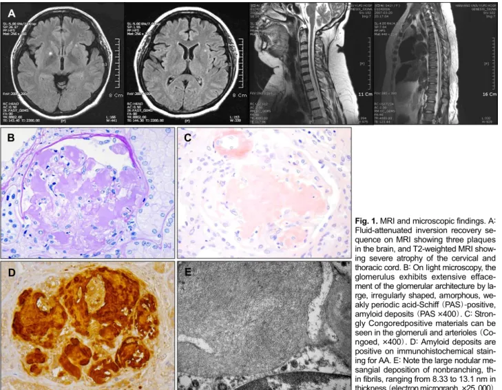
Fig. 1. MRI and microscopic findings. A:
Fluid-attenuated inversion recovery se- quence on MRI showing three plaques in the brain, and T2-weighted MRI show- ing severe atrophy of the cervical and thoracic cord. B: On light microscopy, the glomerulus exhibits extensive efface- ment of the glomerular architecture by la- rge, irregularly shaped, amorphous, we- akly periodic acid-Schiff (PAS)-positive, amyloid deposits (PAS ×400). C: Stron- gly Congoredpositive materials can be seen in the glomeruli and arterioles (Co- ngoed, ×400). D: Amyloid deposits are positive on immunohistochemical stain- ing for AA. E: Note the large nodular me- sangial deposition of nonbranching, th- in fibrils, ranging from 8.33 to 13.1 nm in thickness (electron micrograph, ×25, 000).
A
B C
D E
Amyloidosis in MS
148 J Clin Neurol 2009;5:146-148
data suggest that MS is related to secondary amyloidosis, although until now there have been no reports of secondary amyloidosis in patients with MS.
One previous study found that SAA levels were increased in the peripheral blood of patients with relapsing-remitting type MS. SAA plays an important role in the conversion of innate immunity into the acquired immune response present during periods of acute and chronic inflammation.8 Therefore, in- creases in levels of SAA in MS patients may be considered evidence of the role of inflammation in MS, and may be a precursor of amyloid fibrils. However, there are no reports of increased levels of SAA in asymptomatic bacteriuria or de- cubitus sores.
Amyloid may be deposited either locally or systemically.
The precursor proteins differ from each other in their prima- ry structure and function. The clinical presentations and symp- toms dependupon the distribution pattern and the amount of amyloid deposited. Renal involvement, as in this case, can cause nephrotic syndrome. The main treatment protocol for AA amyloidosis is management of the underlying inflamma- tory disease process, which usually focuses on the surgical debridement of inflammatory tissue, antibiotic treatment of infectious processes, anti-inflammatory medications (colchi- cine and anti-tumor necrosis factor blockade), and immuno- supp ressive (cyclophosphamide) agents.9 However, these treatments, have not yet been established in randomized controlled studies. In our case, disease-modifying agents for MS (e.g., interferon-β, glatiramer acetate) could be consider- ed for the treatment of MS-associated secondary amyloidosis.
However, the effect of these agents has not yet been estab- lished.
There have been two previous reports of localized amyloid deposits in MS;5 however, there have been no reported cases
of associated systemic amyloidosis. This is the first published case of secondary amyloidosis presenting as nephrotic synd- rome associated with MS.
The findings of this case suggest strongly that MS is a ch- ronic inflammatory disease and that secondary amyloidosis can develop in MS.
Acknowledgements
Secondary Amyloidosis Associated with Multiple Sclerosis.
REFERENCES
1. Compston A, Coles A. Multiple sclerosis. Lancet 2002;359:1221-1231.
2. Uda H, Yokota A, Kobayashi K, Miyake T, Fushimi H, Maeda A, et al.
Two distinct clinical courses of renal involvement in rheumatoid pa- tients with AA amyloidosis. J Rheumatol 2006;33:1482-1487.
3. Schroöder R, Nennesmo I, Linke RP. Amyloid in a multiple sclerosis lesion in clearly of Alambda type. Acta Neuropathol 2000;100:709- 711.
4. Falk RH, Comenzo RL, Skinner M. The systemic amyloidoses. N Engl J Med 1997;337:898-909.
5. Lachmann HJ, Goodman HJ, Gilbertson JA, Gallimore JR, Sabin CA, Gillmore JD, et al. Natural history and outcome in systemic AA am- yloidosis. N Engl J Med 2007;356:2361-2371.
6. Livneh A, Zemer D, Langevitz P, Laor A, Sohar E, Pras M. Colchicine treatment of AA amyloidosis of familial Mediterranean fever. An analysis of factor affecting outcome Arthritis Rheum 1994;37:1804- 1811.
7. Tuglular S, Yalcinkaya F, Paydas S, Oner A, Utas C, Bozfakioglu S, et al. A retrospective analysis for aetiology and clinical findings of 287 secondary amyloidosis cases in Turkey. Nephrol Dial Transplant 2002;17:2003-2005.
8. Ristori G, Laurenti F, Stacchini P, Gasperini C, Buttinelli C, Pozzilli C, et al. Serum amyloid A protein is elevated in relapsing-remitting mul- tiple sclerosis. J Neuroimmunol 1998;88:9-12.
9. Schwimmer JA, Joseph RE, Appel GB. Amyloid, fibrillary, and the glomerular deposition disease in therapy, In: Wilcox CS, Brady HR (eds). Nephrology and Hypertension. Philadelphia: Saunders, 2003;
253-261.