서 론
유방의 유두상 병변은 전체 유방 조직검사 검체에서 흔하지 않 은 병변이며 전체 조직검사 중 1-3% 정도이다.(1) 유방의 유두상
병변은 양성종양부터 악성종양까지 다양한 종류의 병변을 포함 하고 있는데 유두상 병변의 종류에는 유두종, 유두종증, 경화성 유두종, 비정형 유두종, 관내 유두암, 침윤성 유두암 등이 있다.(2) 유방병변의 조직검사로는 고식적으로 세침흡인세포검사가 널 리 사용되어 왔으나, 높은 민감도와 특이도의 침생검이 점차적으 로 많이 이용되고 있다. 유방 유두상 병변의 감별에 있어서도 침 생검이 세침흡인세포검사보다 민감도가 더 우수하다고 보고하고 있다.(3) 양성 유두종과 악성 유두종의 감별에 있어 Jacob 등(4) 은 양성 유두종 일부에서 비정형상피증식증이나 관상피내암이 Purpose: It is well recognized that distinguishing benign
papillary lesions from malignant papillary lesions on core needle biopsy may pose difficult diagnostic problems. This study was conducted to define the potential role of ultrasound- guided core biopsy for the diagnosis of papillary lesions of the breast.
Methods: Twelve hundred and seventy nine women con- secutively underwent 14-gauge core biopsy between January 2004 and December 2006. Of the 1,279 patients, 42 patients (3.2%) had papillary lesions of the breast on core needle biopsy. Of these 42 patients, 35 patients underwent surgical excision or sono-guided vacuum assisted excision. We com- pared the pathologic results of the excised specimens with the pathologic results on core needle biopsy.
Results: Of the 35 patients, 23 patients underwent surgical excision and 12 patients underwent sono-guided vacuum assisted excision. Three patients with intraductal papilloma
without atypism on the core needle biopsy were confirmed to have intraductal papilloma accompanied with atypism by the final pathology. All 4 patients with papillomatosis or intra- ductal papilloma with atypism at core needle biopsy were confirmed to have intraductal papilloma with atypism by the final pathology. There were no patients identified to have breast cancer.
Conclusion: Our results revealed the accuracy of core needle biopsy for making the diagnosis of papillary lesions of the breast. Surgical excision may not always be necessary for papillary lesion of the breast that is diagnosed on core needle biopsy. Surgical excision is considered in patients with pap- illomatosis or papillary lesions with atypism seen on core needle biopsy.
Key Words : Papillary neoplasm, Core needle biopsy 중심단어 : 유두상 종양, 침생검
Clinicopathologic Features of the Papillary Breast Lesions Diagnosed on Ultrasonography-guided Core Needle Biopsy
Jung Hyun Park, Ja Seong Bae, Young Jin Suh, Woo Chan Park, Byung Joo Song, Jeong Soo Kim, Sang Seol Jung
Department of Surgery, The Catholic University of Korea, Seoul, Korea
Journal of Breast Cancer
O R I G I N A L A R T I C L E
ISSN 1738-6756 J Breast Cancer 2007; December 10 (4): 269-72
박정현ㆍ배자성ㆍ서영진ㆍ박우찬ㆍ송병주ㆍ김정수ㆍ정상설 가톨릭대학교 의과대학 외과학교실
초음파 유도 하 핵생검으로 진단된 유방 유두상 종양의 임상병리학적 특징
책임저자 : 송병주
139-706 서울시 서초구 반포 4동 505, 가톨릭대학교 강남성모병원 외과 Tel: 02-590-2735, Fax: 02-590-1406
E-mail : bjsong@catholic.ac.kr
접수일 : 2007년 10월 08일 게재승인일 : 2007년 11월 29일
*본 논문의 요지는 2005년 추계외과학회 구연 발표되었음.
269
270 Jung Hyun Park, et al.
존재하는 경우 적은 조직검체에 이러한 병변이 포함되지 않을 수 있기 때문에 세침흡인세포검사나 침생검 모두 양성유두종과 악성 유두종의 감별이 모두 어렵다고 주장하였다. 반면에 다른 저자들 에 의한 보고에서는 침생검으로 양성 유두상 병변을 진단할 수 있 고 침생검 소견이 영상학적 소견과 일치할 경우에 외과적 절제가 필요 없으며 단지 비정형증을 동반하고 있는 유두상 병변일 경우 에 외과적 절제가 필요하다고 하였다(1, 5-7).
이와 같이 유방 유두상 병변은 진단과 치료에서 아직 기준이 정 립이 되어 있지 않다. 이에 저자들은 침생검으로 진단된 유방 유 두상 병변을 조사하여 외과적 절제술 후 진단과 비교하여 정확도 를 예측하고 향후 유방 유두상 병변의 치료방침에 도움을 주고자 한다.
방 법
2004년 1월부터 2006년 12월까지 강남성모병원에서 초음파 유도하 침생검을 시행 받은 1,279명의 환자를 대상으로 후향적으 로 조사하였다. 환자들은 문진과 함께 이학적 검사를 받았으며 초 음파를 시행받았다. 초음파는 5-12 MHz의 탐색자(HDI 5000 Sono CT [Philips medical systems, Bothell, Washington, USA] and HDI 5000 ATL [Advanced Technology Labora- tory, Bothell, Washington, USA])를 사용하였다. 초음파 소 견으로는 병변의 크기, 위치, 경계, 종괴 내부의 에코음영, Breast Imaging and Reporting data system (BIRADS) 분류를 조 사하였다. 침생검은 대부분 BIRADS 분류상 C4a 이상의 소견을 나타내는 병변을 대상으로 실시하였다. 침생검은 14게이지 주사 바늘을 이용하여 적어도 4번 이상 시행하여 조직검체를 얻었다.
침생검상 유두상 병변으로 진단된 환자는 외과적 절제술이나 초 음파 유도 음압 보조 절제술을 시행 받았으며 절제술을 시행하지 않은 환자는 본 연구에서 제외되었다. 침생검상 유두상 병변으로 진단 받은 환자의 임상적 특징을 조사하였고, 초음파 소견과 병리 소견을 조사하였다. 또한 침생검의 병리소견과 절제술 후 병리소 견을 비교 분석하였다.
결 과
1. 임상적특징
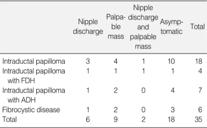
1,279명의 환자 중 42명(3.2%)이 침생검상 유두상 병변으로 진 단되었고 이 중 35명이 외과적 절제술이나 초음파 유도 음압 보조 절제술을 시행 받았으며, 7명은 침생검 실시 후 절제술을 시행하 지 않아 본 연구에서 제외하였다. 환자의 평균 나이는 44.6세(17- 79세)이고 종괴의 평균 크기는 1.0 cm (0.4-3.7 cm)이었다. 종 괴의 치료는 외과적 절제술이 23명에서 시행되었고, 초음파 유도 하 음압 보조 절제술은 12명에서 시행되었다. 환자의 증상에 따 른 분류에서 18명(51%)이 증상이 없었으며 17명은 촉지성 종괴나 유두분비를 동반하고 있었다(Table 1). 각 질환별 분류에서는 관 내유두종에서 유두분비가 3예, 촉지성 종괴가 4예, 유두분비를 동반한 촉지성 종괴 1예 및 무증상이 10예였다. 개화성 관상피 증 식증을 동반한 유두종예에서는 유두분비, 촉지성 종괴, 유두분비 를 동반한 촉지성 종괴와 무증상이 각각 1예였다. 비정형증을 동 반한 유두종예에서는 유두분비가 1예, 촉지성 종괴가 2예, 무증상 이 4예였다(Table 2).
2. 유두병변의초음파소견및최종조직소견과의관계
유두상 병변의 위치는 중앙이 9예, 가장자리가 26예였으며 비 정형증을 동반한 관내 유두종의 경우에 유륜하가 2예, 주변부가 5예였다. 관 확장증은 19예에서 동반되었다. 종괴의 에코 소견은 저에코가 24예, 등에코가 11예이며 이 중 비정형증을 동반한 경우 에는 모두 저에코 소견을 보였다. 병변의 경계는 전체적으로 국한 성 경계가 26예, 비국한성 경계가 9예였으며, 이 가운데 비정형증 을 동반한 유두종예에서는 국한성 경계가 3예, 비국한성 경계가 4예가 있었다. 유방 초음파의 BIRADS 분류에서는 C3가 9예 C4 가 26예였으며 C5는 없었으며 비정형증을 동반한 병변은 모두 C4였다(Table 3).
Table 1.Clinical characteristics of patients with papillary neo- plasm
Age (yr) 44.6±12 (17-79)
Size (cm) 1.0±0.6 (0.4-3.7)
Symptom
Symptomatic 17
Asymptomatic 18
Operation
Surgical excision 23
Vacuum assisted biopsy 12
Table 2.Symptoms associated with papillary neoplasm
Nipple discharge
Palpa- ble mass
Asymp- tomatic Total Nipple
discharge and palpable
mass
Intraductal papilloma 3 4 1 10 18
Intraductal papilloma 1 1 1 1 4
with FDH
Intraductal papilloma 1 2 0 4 7
with ADH
Fibrocystic disease 1 2 0 3 6
Total 6 9 2 18 35
FDH=florid ductal hyperplasia; ADH=atypical ductal hyperplasia.
Characteristics of Papillary Breast Lesions on Core Needle Biopsy 271
3. 침생검의조직소견과외과적절제후조직소견의관계
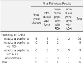
종괴의 수술치료를 시행한 35예에서 최종 조직검사상 관내유 두종은 18예, 개화성 관상피 증식증을 동반한 관내유두종은 4예, 비정형증을 동반한 관내유두종은 7예, 섬유낭종성 질환이 6예였 다. 침생검에서 관내유두종으로 진단 받은 26예 중 2예에서 비정 형을 동반한 관내 유두종으로 판명되었으며, 유두종증으로 진단 받은 1예는 역시 비정형증을 동반한 관내 유두종으로 판명되었다.
침생검에서 비정형증이 없는 것으로 진단받은 32예 중 3예(9.4%) 에서 비정형증을 동반한 유두종으로 판명되었고, 핵생검에서 비 정형증을 동반한 관내 유두종으로 진단받은 3예는 모두 비정형증 으로 동반한 관내 유두종으로 판명되었으며 그 중 1예는 소엽종괴 를 동반한 관내 유두종으로 판명되었다(Table 4).
고 찰
저자들은 초음파 유도 하에 14게이지 침생검을 시행하여 35명 의 유방 유두상 병변을 진단하였으며, 수술적 치료 후 최종 조직 검사 결과 6예(17%)에서 유두상 병변이 아닌 섬유낭종성 질환으 로 판명되었다.
유방 유두상 병변은 침생검을 통한 조직검사에서 드물게 발견 되는 병변이다. 이전의 다른 연구에서는 0.2-5%까지 보고하였 으며(1, 8, 9) 본 연구에서는 전체 침생검을 시행한 조직검사 결과 중 3.2%를 차지하였다. 유방 유두상 병변에 대한 관심이 낮았던 것은 낮은 발생률 때문이지만, 임상 및 영상학적으로 양성 유두병 변과 악성유두암의 감별이 매우 어렵고, 양성 유두병변과 악성유 두암의 구별이 매우 중요하지만 완전 절제를 하지 않는 경우 확진 이 어려우므로 임상 진료에서는 그 관심이 높아지고 있다.
침생검은 세침흡인세포검사와 비교하여 불충분한 검체가 얻어 지는 경우가 적고 위음성률이 낮기 때문에 널리 쓰여지고 있으며 실제로 좋은 성적을 나타내고 있다.(10, 11) 그러나, 침생검으로 양성 유두상 병변이 진단된 후에 치료 방침에 대해서는 아직 일치
된 의견이 없다. Hoda와 Rosen (12)은 침생검으로 진단된 모든 유두상 병변에 대해 반드시 외과적 절제가 필요하다고 주장하였 는데 이는 검체 오류의 위험도가 있으며 지금까지 침생검으로 진 단된 유두상 병변을 오랜기간 동안 추적관찰한 연구가 없다는 사 실을 근거로 주장하였다. 그러나 또 다른 문헌 보고에 의하면 양 성 유두상 병변이 영상학적 소견과 병리학적 소견이 일치할 경우 에는 외과적 절제 필요없이 추적관찰로 충분하며 불일치할 경우 에는 외과적 절제가 필요하다고 보고하였다(6, 8, 13). 본 연구에 서는 유두상 병변의 외과적 절제를 시행하였고 침생검 소견상 비 정형증이 없는 유두상 병변으로 밝혀진 32예 중 28예(87.5%)에 서 외과적 절제를 통한 최종 조직검사 결과상 양성 유두상 병변으 로 판명되었다. 비정형증을 동반한 유두상 병변의 외과적 절제의 중요성은 잘 알려져 있는데 지금까지 문헌 보고에 의하면 침생검 결과에서 비정형을 동반한 유두상 병변이 외과적 절제 후 최종 조 직검사에서암으로판명될위험도가높다고보고하고있다(14, 15).
최근 침생검에 의한 조직 소견의 정확도를 높이기 위한 방법이 연 구되고 있는데, 기존의 H&E 염색보다 p63, cytokeratin 5/6, 혹은 calponin 같은 면역조직화학법을 이용한 조직검사가 더 정 확하다는 보고도 있으나,(16) 본 연구에서는 시행하지 않았다.
유두상 병변을 가진 환자들은 자주 유두 분비를 호소하게 되는 데 병적 유두분비를 호소하는 환자의 40-70%에서 유두종이 있 다고 보고하고 있다.(17, 18) 그러나 유두분비로 양성 병변과 악 성 병변을 감별할 수 없고, 또한 유두분비를 호소하는 환자의 검 사방법으로는 유관조영술이 좋다고 알려져 있으나 유두관 내의 병변을 찾는 데에는 비특이적인 방법으로 보고되고 있다.(19, 20) 본 연구에서는 35예 중 8예(22.9%)에서 유두분비를 호소하였으 Table 4.Relation between results of CNB and final pathologic results
Final Pathologic Results
Fibro- cystic disease
Intra- ductal papil- loma
Intra- ductal papil- loma with FDH
Intra- ductal papil- loma with ADH
Total
Pathology on CNBx
Intraductal papilloma 6 16 2 2 26
Intraductal papilloma 0 2 2 1 5
with FDH
Intraductal papilloma 0 0 0 3 3
with ADH
Papillomatosis 0 0 0 1 1
Total 6 18 4 7 35
CNBx=core needle biopsy; FDH=florid ductal hyperplasia; ADH=
atypical ductal hyperplasia.
Table 3.Ultrasonographic findings of papillary lesion
Ultrasonographic findings
Location Central 9
Periphery 26
Echogenecity Hypoechoic 24
Isoechoic 11
Margin Circumscribed 26
Not circumscribed 9
BIRADS C3 9
C4 26
C5 0
BIRADS=Breast imaging and reporting data system.
272
나, 유두분비물의 세포도말 검사는 시행하지 않았으며 유두분비 의 증상이 다른 문헌보다는 다소 낮은 소견을 보였다.
결 론
저자들은 양성 유두상 병변이 초음파 유도하의 침생검을 통하 여 진단되었을 때 외과적 수술을 통하여 최종 진단된 소견과 얼마 나 일치하는지 알아보고자 하였다. 수술 전 초음파 소견상 양성유 두종과 악성유두종을 감별할 수 있는 특징적인 소견은 없었고, 세 침흡인세포검사나 침생검 모두 단독 검사만으로 정확한 결론을 내리는 데에는 한계가 있으나, 유두상 병변의 진단에 있어 영상학 적 소견과 침생검의 조직 소견이 일치하는 경우에는 침생검 결과 의 신뢰성을 인정할 수 있는 것으로 생각되고, 침생검 결과에서 비정형증을 동반한 유두상 병변은 외과적 절제술을 고려해야 할 것으로 생각된다.
참고문헌
1. Liberman L, Bracero N, Vuolo MA, Dershaw DD, Morris EA, Ab- ramson AF, et al. Percutaneous large-core biopsy of papillary breast lesions. Am J Roentgenol 1999;172:331-7.
2. Tavassoli FA: Papillary lesions; in Tavassoli FA (cd): Pathology of the Breast. Norwalk, Appleton & Lange, 1992;193-227.
3. Lam WW, Chu WC, Tang AP, Tse G, Ma TK. Role of radiologic features in the management of papillary lesions of the breast. Am J Roentgenol 2006;186:1322-7.
4. Jacobs TW, Connolly JL, Schnitt SJ. Nonmalignant lesions in breast core needle biopsies: to excise or not to excise? Am J Surg Pathol 2002;26:1095-110.
5. Ivan D, Selinko V, Sahin AA, Sneige N, Middleton LP. Accuracy of core needle biopsy diagnosis in assessing papillary breast lesions:
histologic predictors of malignancy. Mod Pathol 2004;17:165-71.
6. Carder PJ, Garvican J, Haigh I, Liston JC. Needle core biopsy can reliably distinguish between benign and malignant papillary lesions of the breast. Histopathology 2005;46:320-7.
7. Agoff SN, Lawton TJ. Papillary lesions of the breast with and without atypical ductal hyperplasia: can we accurately predict benign behavior from core needle biopsy? Am J Clin Pathol 2004;122:440-3.
8. Rosen EL, Bentley RC, Baker JA, Soo MS. Imaging-guided core needle biopsy of papillary lesions of the breast. Am J Roentgenol
2002;179:1185-92.
9. Philpotts LE, Shaheen NA, Jain KS, Carter D, Lee CH. Uncommon high-risk lesions of the breast diagnosed at stereotactic core-needle biopsy: clinical importance. Radiology 2000;216:831-7.
10. Jeffrey PB, Ljung BM. Benign and malignant papillary lesions of the breast. A cytomorphologic study. Am J Clin Pathol 1994;101:
500-7.
11. Rubin E, Dempsey PJ, Pile NS, Bernreuter WK, Urist MM, Shumate CR, et al. Needle-localization biopsy of the breast: impact of a selective core needle biopsy program on yield. Radiology 1995;195:627-31.
12. Hoda SA, Rosen PP. Practical considerations in the pathologic diag- nosis of needle core biopsies of breast. Am J Clin Pathol 2002;118:
101-8.
13. Mercado CL, Hamele-Bena D, Singer C, Koenigsberg T, Pile-Spell- man E, Higgins H, et al. Papillary lesions of the breast: evaluation with stereotactic directional vacuum-assisted biopsy. Radiology 2001;
221:650-5.
14. Reynolds HE. Core needle biopsy of challenging benign breast con- ditions: a comprehensive literature review. Am J Roentgenol 2000;
174:1245-50.
15. Philpotts LE, Shaheen NA, Jain KS, Carter D, Lee CH. Uncommon high-risk lesions of the breast diagnosed at stereotactic core-needle biopsy: clinical importance. Radiology 2000;216:831-7.
16. Shah VI, Flowers CI, Douglas-Jones AG, Dallimore NS, Rashid M.
Immunohistochemistry increases the accuracy of diagnosis of benign papillary lesions in breast core needle biopsy specimens. Histopa- thology 2006;48:683-91.
17. Paterok EM, Rosenthal H, Sabel M. Nipple discharge and abnormal galactogram. Results of a long-term study (1964-1990). Eur J Obstet Gynecol Reprod Biol 1993;50:227-34.
18. Dietz JR, Crowe JP, Grundfest S, Arrigain S, Kim JA. Directed duct excision by using mammary ductoscopy in patients with pathologic nipple discharge. Surgery 2002;132:582-7.
19. Woods ER, Helvie MA, Ikeda DM, Mandell SH, Chapel KL, Adler DD. Solitary breast papilloma: comparison of mammographic, galac- tographic, and pathologic findings. Am J Roentgenol 1992;159:487-91.
20. Funovics MA, Philipp MO, Lackner B, Fuchsjaeger M, Funovics PT, Metz V. Galactography: method of choice in pathologic nipple discharge? Eur Radiol 2003;13:94-9.
Jung Hyun Park, et al.