Corresponding author:
Mi-Young ParkDepartment of Clinical Laboratory Science, Suwon Science College, 288 Seja-ro, Jeongnam-myeon, Hwaseong 18516, Korea
E-mail:
[email protected]ORCID:
https://orcid.org/0000-0001-8033-995XORIGINAL ARTICLE
Transplantation of Brain-Derived Neurotrophic
Factor-Expressing Mesenchymal Stem Cells Improves Lower Urinary Tract Symptoms in a Rat Model
Seung Hwan Jeon 1 , Mi-Young Park 2
1
Department of Urology, College of Medicine, The Catholic University of Korea, Seoul, Korea
2
Department of Clinical Laboratory Science, Suwon Science College, Hwaseong, Korea
뇌유래신경영양인자 발현 중간엽 줄기세포의 하부요로증상 개선 효과
전승환 1 , 박미영 2
1
가톨릭대학교 성의교정 비뇨의학과,
2수원과학대학교 임상병리과
ARTICLE INFO ABSTRACT
Received
November 23, 2020Revised
November 30, 2020Accepted
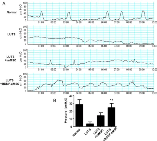
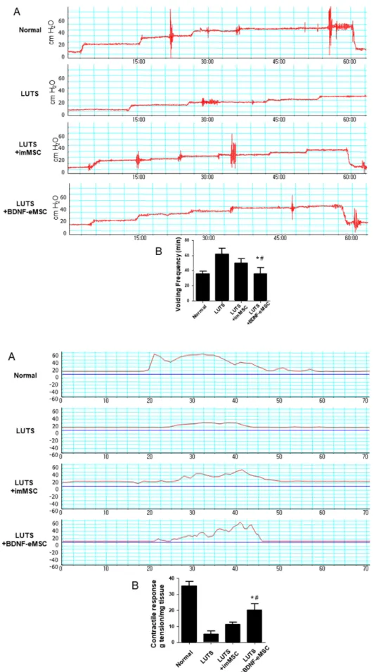
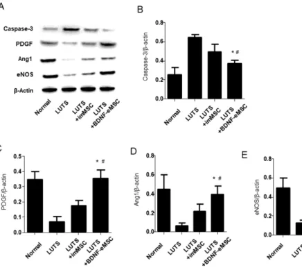
December 1, 2020This study aimed to explore the effects of brain-derived neurotrophic factor (BDNF), produced by engineered immortalized mesenchymal stem cells (imMSC), on lower urinary tract symptoms (LUTS) in a rat model with neurogenic bladder (NB). Forty-eight Sprague-Dawley (SD) rats were randomly divided into the following groups: Sham control, LUTS, LUTS+imMSC (treated with immortalized MSC), and LUTS+BDNF-eMSC (treated with BDNF-expressing MSC) groups. LUTS was induced by a crush injury to the major pelvic ganglion (MPG). Bladder function was tested under anesthesia, and bladder tissue strips were collected thereafter for contractility test and western blot analysis.
Western blot results showed that the expression of both Angiopoietin 1 (Ang 1) and platelet-derived growth factor (PDGF) increased with MSC injection. The effect of treatment with BDNF-eMSC on LUTS was also evaluated, and the results were found to be better than those with imMSC ( P <0.05).
BDNF-eMSC prevented fibrosis in the bladder tissue and significantly reduced caspase-3 levels. In conclusion, high expression of BDNF in vivo resulted in recovery of bladder function and contractility, along with the inhibition of apoptosis in a rat model.
Copyright Ⓒ 2020 The Korean Society for Clinical Laboratory Science. All rights reserved.
Key words Bladder function
Brain-derived neurotrophic factor Lower urinary tract symptoms
INTRODUCTION
Lower urinary tract symptoms (LUTS), including urinary tract damage, urinary tract infection, storage symptoms, voiding symptoms, and renal failure, may be related to nervous system injury. In a rat model of
neurogenic bladder (NB), such symptoms have been shown to be caused by diabetes, cerebrovascular accidents, brain injury, or spinal cord injury [1, 2].
Regenerative medicine, or stem cell therapy, which involves tissue formation and repair to restore the functionality of damaged organs or tissues, have often been employed for tissue regeneration [3-5].
Stem cell therapy has been used to treat various diseases till date [6, 7]. Mesenchymal stem cells (MSCs) are pluripotent stem cells that are capable of
Korean Society for Clinical Laboratory Science