Malignant Gastrointestinal Stromal Tumor of the Esophagus
-One case report-
Gastrointestinal stromal tumors (GISTs) are rare, but potentially aggressive tumors. GISTs are generally found in the stomach or small intestine and less commonly in the colon, rectum, or an intra-abdominal sites but have rarely been documented in the esophagus. GISTs were definded as the most common mesenchymal tumors of the gastrointestinal tract for which there is incomplete understanding of their lineage, while their relationship with differenciated. We reported a very rare case of GISTs of lower esophagus in a 66-year-old woman with relevant literature review.
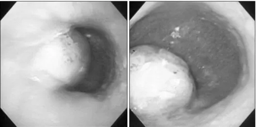
Fig. 1. The pre-operative esopha- goscophy found the about 5×6 cm sized oval shape-submucosal like mass at about 35 cm from upper incisor.
Fig. 2. The pre-operative chest CT shows the round shaped- lower esophageal mass about 4 cm above gastroesophageal junction but not infiltrating to aorta.
Fig. 3. Intra-operative field reveal the submucosal esophageal
mass.
(3)
Fig. 4. The microscopic finding show whorling and palisading patterns with spindle cell and epithelioid histology. The mitotic figures are present up to 10/50 high-power fields (H-E).
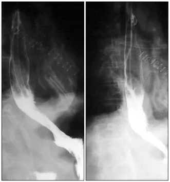
Fig. 5. The Post-operative esophagography revealed well pas-