Microarray Analysis for Genes Associated with Angiogenesis in Diabetic OLETF Keratocytes
The purpose of this study was to identify the differences in angiogenesis gene expression between normal and diabetic keratocytes stimulated with interleukin-1α (IL-1α) and tumor necrosis factor-α (TNF-α). Primarily cultured normal and diabetic keratocytes were treated with 20 ng/mL of IL-1a and TNF-α for 6 hr. cDNA was hybridized to an oligonucleotide microarray. Microarray analysis was used to identify differentially expressed genes that were further evaluated by real-time polymerase chain reaction (RT-PCR). Diabetes keratocytes overexpressed vital components of angiogenesis including Agtr1, and under- expressed components related to the blood vessel maturation, including Dcn. Cytokine- treated diabetic keratocytes differentially expressed components of angiogenesis. OLETF keratocytes after treatment with IL-1α and TNF-α showed the newly expressed 15 and 14 genes, respectively. Newly and commonly under-expressed five genes followed by treatment with both IL-1α and TNF-α were also evident. RT-PCR showed results similar to the microarray results. Agtr1 and Itga1 showed an increased expression in diabetic keratocytes compared with normal corneal keratocytes, especially after TNF-α treatment.
Il6 appeared strong expression after interleukin-1α treatment, but showed down expression after TNF-α treatment. Further studies to analyze and confirm the significance of the identified angiogenetic genes of diabetes are needed.
Keywords: Angiogenesis; Cytokines; Inflammation; Corneal Keratocyte; Microarray Analysis; OLETF
Jun-Mo Park,1 Young Min Park,2 Wook Jung,2 Ji-Eun Lee,2 and Jong-Soo Lee2
1Department of Ophthalmology, Busan St. Mary’s Hospital, Busan; 2Department of Ophthalmology, School of Medicine, Pusan National University and Medical Research Institute, Pusan National University Hospital, Busan, Korea Received: 19 August 2013 Accepted: 19 November 2013 Address for Correspondence:
Jong-Soo Lee, MD
Department of Ophthalmology, School of Medicine, Pusan National University, and Medical Research Institute, Pusan National University Hospital, 179 Gudeok-ro, Seo-gu, Busan 602-739, Korea
Tel: +82.51-240-7323, Fax: +82.51-242-7341 E-mail: [email protected]
This study was supported by Biomedical Research Institute, Pusan National University Hospital.
http://dx.doi.org/10.3346/jkms.2014.29.2.265 • J Korean Med Sci 2014; 29: 265-271
INTRODUCTION
Angiogenesis is essential for many physiological processes in
cluding wound healing, cell regeneration, tissue growth and de
velopment, and embryonic development, and plays a very im
portant role in the development of proliferative diabetic retino
pathy, a serious complication of diabetes (13). Angiogenesis or the formation of new blood vessels from existing vessels has re
cently been widely studied; it has been found that various types of cytokines and growth factors, particularly vascular endothe
lial growth factor (VEGF), are involved in angiogenesis (4, 5).
It has been observed that retinal capillary closure and retinal ischemia can result in spread of angiogenic factors such as VEGF into the ischemic retina, leading to the formation of abnormal blood vessels (68). The mechanism of angiogenesis is similar in both corneal and retinal tissues. It has been reported that the VEGF is also involved in the angiogenesis of the cornea (9, 10).
Therefore, it is very important to identify the factors associated with the angiogenesis, and also studies of genes related to an
giogenesis are expected to be very useful in the treatment of corneal neovascular diseases.
Accordingly, we investigated the differences in angiogenesis gene expression of keratocytes between the genetic model for
OLETF in diabetic rats and the control group of normal rats. In addition, an experiment was performed to observe the angio
genesisrelated genes in the inflamed cornea by treating with interleukin1α (IL1α) and tumor necrosis factorα (TNFα).
MATERIALS AND METHODS Cell culture and cytokine treatment
Fiftyweekold, genetically affected type 2 diabetic (OLETF, Ot
suka LongEvans Tokushima Fatty) and normal rats were pur
chased from Otsuka Pharmaceutical Company (Tokushima, Ja
pan). The corneal epithelium was removed; the corneal stroma was isolated, and washed several times with saline containing antibiotics. The corneal endothelial layer was removed, and then the corneal stromal tissue was cut into small pieces and cultur
ed serially in Dulbecco’s modified Eagle medium (DMEM, Gib
co BRL, Grand Island, NY, USA) containing 10% fetal bovine se
rum (FBS, Gibco BRL), 100 units/mL penicillin (Gibco BRL), and 100 mg/mL streptomycin (Gibco BRL). The culture medi
um was changed at 2 to 3day intervals. When the cells became confluent, the culture medium was removed, the cells were wash
ed once with Dulbecco’s phosphatebuffered saline (DPBS, Gibco BRL) and dissociated with 0.25% trypsin0.02% EDTA.
Ophthalmology
For the microarray experiments, cells between the third and fourth generation of serial culture were collected and stored at
70°C. The corneal stromal cells of diabetic and normal rats were treated with the cytokines, IL1α and TNFα (R&D Systems, Min
neapolis, MN, USA), at a concentration of 20 ng/mL for 6 hr.
RNA extraction
Total cellular RNAs were extracted from the corneal stromal cells of diabetic and normal rats in primary culture using the RNeasy mini kit (QIAGEN Inc, Valencia, CA, USA) for gene mi
croarrays. First, denaturing solution was added to cell pellets on ice for 5 min, and then phenol and chloroform were added to all samples. The samples were centrifuged, and the supernatant containing RNA was transferred to a new tube. The process was repeated. Subsequently, isopropanol at 20°C was added to precipitate the RNA, and then the mixture was centrifuged and the supernatant was discarded carefully. The pellet was washed with 80% ethanol at 20°C and then airdried. Extraction of RNA was quantitated and confirmed by electrophoresis.
DNA gene microarrays
Sixtymer oligonucleotides corresponding to each gene were synthesized and subsequently placed on a slide using a robotic gene microarray. The robotic gene microarray places 0.251 nL DNA samples on a slide sample in spots averaging 100150 nm.
cDNA was synthesized from the isolated RNA samples using RT primers (Genisphere Inc, Hatfield, PA, USA) and SuperScript II reverse transcriptase (Invitrogen, Grand Island, NY, USA). The purified cDNA was hybridized to an Agilent Rat oligo 22K chip (Agilent tech, Santa Clara, CA, USA). For hybridization, slides were placed in a dark hybridization chamber at 62°C for 16 hr.
The slides were removed, washed three times, and dried using a centrifuge. A fluorescent dye was added to DNA capture re
agents that bind RT primers. Thereafter, the second hybridiza
tion was performed for 4 hr. The slides were removed, washed, and dried using a centrifuge. Afterward, the fluorescence level was measured using an Axon Laser Fluorescence Scanner (Axon Instruments Inc, Foster City, CA, USA). The microarray analyz
er we used in our study has 105 rows and 215 columns. A total of 22,575 genes pertinent to angiogenesis were included.
Analysis
The hybridized slides were scanned by an Axon GenePix Laser Fluorescence Scanner and analyzed by GenePix Pro 5.1 (Axon Instruments) and GeneSpring 7.0 (Silicongenetics, Redwood City, CA, USA). Gene expression was considered as significant for genes showing more than a twofold difference in expression level between normal and diabetic rats. Genes showing similar patterns were distinguished by a Pearson’s correlation analysis.
The images thus obtained show that genes that are upregulat
ed are shown in red, whereas genes that are downregulated
are shown in green. First of all, we investigated the differences in angiogenesis gene expression in keratocytes between dia
betic and normal rats. In addition, the angiogenesisrelated genes that show different expressions between normal and dia
betic keratocytes after treatment with IL1α and TNFα were also studied.
Real-time polymerase chain reaction
Realtime polymerase chain reaction (RTPCR) was performed with SYBR Green I immunofluorescence dye and the HotStar
Taq DNA polymerase QuantiTect SYBR Green PCR Kit (QIA
GEN GmbH, Hamburg, Germany). To quantitate the control group, a standard curve for the genes of interest and glyceralde
hydes 3phosphate dehydrogenase (GAPDH) was prepared.
GAPDH was reacted to three genes: Il6, Itga1, and Agtr1. The reactions were carried out using the ABI PRISM 7900 Sequence Detection System version 1.6 software (PerkinElmer Biosys
tems, Foster City, CA, USA). Relative quantificational measure
ments were performed using the relative standard curve meth
od according to the manufacturer’s instructions.
RESULTS
The subjects used in our study were 50weekold diabetic OLETF rats. Mean blood glucose was 115.0 ± 10.8 mg/dL in the control group and 193.8 ± 18.9 mg/dL in the OLETF group, which dem
onstrated statistically significant differences between the two groups. Mean body weight was significantly higher in the OLETF group (control group: 518.0 ± 13.3 g; OLETF group: 628.5 ± 63.2 g). These are the typical characteristics of diabetic rats.
Diabetic keratocytes showed upward expression in 13 genes related to angiogenesis, compared to normal keratocytes, inclu
ding the Vegfc, Agtr1, Itga1, Ddah1, Adora2b, and Cspg4. Twen
tytwo genes including the Mmp3, Dcn, Amotl2, and Pdgfa were downregulated (Table 1).
Three genes in diabetic keratocytes, namely Itga1, Ddah1, and Pttg1, which promoted angiogenesis, showed an increased expression after both cytokine treatments, as well as compared to normal keratocytes. Genes that showed a decreased expres
sion after both cytokine treatments were Mmp3 and Pdgfa.
Some genes showed different patterns of expression in re
sponse to two cytokines. Itga1 showed no reaction to TNFα and responded to IL1α only. Conversely, Esm1 did not show a reaction to IL1α and indicated a decreased expression to TNFα compared to the control group (Table 1).
Newly expressed genes after IL1α treatment are shown in Table 2. After IL1α treatment, 15 genes showed different ex
pression in diabetic keratocytes as compared to control group.
Among them, eight genes showed an increased expression in
cluding Egr1, Cxcl12, and Tn. Seven genes showed a decreased expression including Il1b, Pdgfra, and Klf5.
Newly expressed genes after TNFα treatment are shown in Table 3. After TNFα treatment, 14 genes showed different ex
pression in diabetic keratocytes as compared to control group.
TNFα treatment increased gene expression in the Agtr1, Tnmd, and C1galt1. However, 11 genes including Edn1, Mmp9, and Prok1 showed a decreased expression compared to the control group.
Newly expressed genes after both IL1α and TNFα treatment are shown in Table 4. Five genes showed decreased gene expres
sion, including tissue inhibitor of Timp3 and Mmp14. Conver
sely, for Ctss, Hmox1, and Il6, which control and induce angio
genesis, expression was increased by IL1α stimulation and de
creased by TNFα treatment.
The RTPCR was performed on three genes: Il6, Itga1, and Agtr1 that showed significantly different expression between
normal and diabetic keratocytes, or were newly expressed after cytokine treatment. The primer preparation and annealing tem
perature for these genes are shown in Table 5.
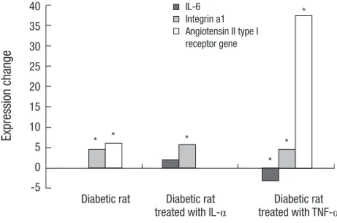
After cytokine treatment, the Itga1 significantly showed an increased expression in diabetic rats. The Agtr1 showed an in
creased expression compared to normal rats, but showed no expression by IL1α treatment and increased expression by TNFα treatment. Il6 showed no expression in the keratocytes of dia
betic rats, but increased expression by IL1α treatment and de
creased expression by TNFα treatment (Fig. 1).
DISCUSSION
Thirtyfive genes that showed different expressions in diabetic keratocytes compared to normal keratocytes, are related to VEGF.
Table 1. Up- and down-regulated genes in cultured diabetic rat stromal keratocytes, and angiogenesis related gene expression after cytokine treatment, compared with normal rat stromal keratocytes
Gene names Symbol Accession No. Fold change
I* II† III‡
Angiotensin II type 1 receptor Agtr1 NM_031009 6.08 - -
Immunoglobulin superfamily, member 4C (predicted) Plaur XM_344870 4.82 2.32 -
Integrin alpha 1 Itga1 NM_030994 4.67 5.77 4.62
Dimethylarginine dimethylaminohydrolase 1 Ddah1 NM_022297 4.56 3.63 3.41
Adenosine A2B receptor Adora2b NM_017161 3.16 - -
Pituitary tumor-transforming 1 Pttg1 NM_022391 3.16 3.14 2.54
Chemokine (C-X-C motif) ligand 12 Cxcl12 NM_022177 3.09 - -
Chondroitin sulfate proteoglycan 4 Cspg4 NM_031022 2.64 2.03 -
Vascular endothelial growth factor C Vegfc NM_053653 2.56 - -
Calponin 1 Cnn1 NM_031747 2.50 - -
Tenomodulin Tnmd NM_022290 2.14 - -
Stathmin 1 Stmn1 NM_017166 2.10 - -
Protein tyrosine phosphatase, receptor type, M Ptprm U66567 2.04 - -
Bone morphogenetic protein 4 Bmp4 NM_012827 -2.00 -100.00 -100.00
Angiopoietin 2 Agpt2 XM_344544 -2.04 - -
3-Phosphoinositide dependent protein kinase-1 Pdpk1 NM_031081 -2.04 - -
Endothelin 1 Edn1 NM_012548 -2.08 - -
Insulin-like growth factor binding protein 2 Igfbp2 NM_013122 -2.08 -2.22 -11.11
Neuropeptide Y receptor Y2 Npy2r NM_023968 -2.22 - -
Prostaglandin-endoperoxide synthase 2 Ptgs2 NM_017232 -2.22 - -2.04
Angiomotin like 2 Amotl2 XM_343457 -2.43 - -
Down syndrome critical region homolog 1 (human) Dscr1 NM_153724 -2.44 - -3.22
Prostaglandin-endoperoxide synthase 2 Ptgs2 L20085 -2.50 - -
Stanniocalcin 1 Stc1 NM_031123 -2.50 - -
Epiregulin Ereg NM_021689 -2.86 -2.22 -
Insulin-like growth factor 1 receptor Igf1r NM_052807 -3.13 -2.17 -
Runt-related transcription factor 1 Runx1 NM_017325 -3.23 - -2.77
Kinase insert domain protein receptor Kdr NM_013062 -3.33 - -
V-ets erythroblastosis virus E26 oncogene homolog 1 (avian) Ets1 NM_012555 -3.44 -7.14 -2.08
Histidine-rich glycoprotein Hrg NM_133428 -4.00 - -2.04
Endothelial cell-specific molecule 1 Esm1 NM_022604 -4.00 -3.70 -14.28
Matrix metallopeptidase 3 Mmp3 NM_133523 -4.76 -16.66 -25.00
Platelet-derived growth factor, alpha Pdgfa NM_012801 -5.26 -6.66 -9.09
Alanyl (membrane) aminopeptidase Anpep NM_031012 -33.33 -100.00 -25.00
Decorin Dcn NM_024129 -50.00 -5.26 -8.33
*The ratio of gene expression in diabetic rat stromal keratocytes compared with normal rat stromal keratocytes. †The ratio of gene expression following interleukin-1α (IL-1α) treatment. ‡The ratio of gene expression following tumor necrosis factor-α (TNF-α) treatment.
Table 2. Genes up- and down-regulated for newly expressed genes following interleukin-1α treatment in cultured diabetic rat stromal keratocytes compared with normal rat stromal keratocytes
Gene names Symbol Accession No. Fold change
Hypothetical gene supported by NM_053652 Flt4 NM_053652 3.01
Early growth response 1 Egr1 NM_012551 2.64
Chemokine (C-X-C motif) ligand 12 Cxcl12 NM_022177 2.50
G4 protein Tnf XM_579722 2.26
Peroxisome proliferator activated receptor, gamma Pparg NM_013124 2.20
Parathyroid hormone-like peptide Pthlh NM_012636 2.15
Secreted phosphoprotein 1 Spp1 NM_012881 2.04
Sphingosine kinase 1 Sphk1 NM_133386 2.04
Interleukin 1 beta Il1b NM_031512 -2.22
Platelet-derived growth factor receptor, alpha polypeptide Pdgfra XM_214030 -2.22
Mitogen-activated protein kinase 14 Mapk14 U73142 -2.44
CEA-related cell adhesion molecule 1 Ceacam1 NM_031755 -2.94
Glypican 1 Gpc1 NM_030828 -3.12
Platelet/endothelial cell adhesion molecule Pecam U77697 -3.12
Kruppel-like factor 5 Klf5 NM_053394 -3.33
Table 3. Genes up- and down-regulated for newly expressed genes following tumor necrosis factor-α treatment in cultured diabetic rat stromal keratocytes compared with nor- mal rat stromal keratocytes
Gene names Symbol Accession No. Fold change
Angiotensin II type 1 receptor Agtr1 NM_031009 37.44
Core 1 UDP-galactose:N-acetylgalactosamine-alpha-R beta 1,3-galactosyltransferase C1galt1 NM_022950 2.34
Tenomodulin Tnmd NM_022290 2.09
Serine (or cysteine) proteinase inhibitor, clade E, member 1 Serpine1 NM_012620 -2.00
Matrix metallopeptidase 9 Mmp9 NM_031055 -2.08
Prokineticin 1 Prok1 NM_138851 -2.22
Basigin Bsg NM_012783 -2.22
Secreted acidic cysteine-rich glycoprotein Sparc NM_012656 -2.27
Jagged 1 Jag1 NM_019147 -2.5
Hypothetical gene Cdh2 NM_031333 -2.63
Endothelin 1 Edn1 NM_012548 -2.94
WAP four-disulfide core domain 1 Wfdc1 NM_133581 -3.125
Corticotropin-releasing hormone receptor 2 Crhr2 NM_022714 -3.22
Interferon, beta 1 Ifnb1 NM_019127 -3.7
Table 4. Genes up- and down-regulated for newly expressed genes following treatment with both interleukin-1α and tumor necrosis factor-α in cultured diabetic rat stromal keratocytes compared with normal rat stromal keratocytes
Gene names Symbol Accession No. Fold change
I* II†
Cathepsin S Ctss NM_017320 2.53 -2.00
Heme oxygenase (decycling) 1 Hmox1 NM_012580 2.42 -2.27
Interleukin 6 Il6 NM_012589 2.02 -3.22
Cbp/p300-interacting transactivator, with Glu/Asp-rich carboxy-terminal domain, 2 Cited2 NM_053698 -2.04 -4.76
Tissue inhibitor of metalloproteinase 3 Timp3 NM_012886 -2.12 -2.85
Matrix metalloproteinase 14 Mmp14 NM_031056 -2.77 -2.04
Adrenomedullin Adm NM_012715 -5.55 -2.85
Neuropeptide Y Npy NM_012614 -33.33 -12.5
*A ratio of newly expressed genes associated with cell motility and communication in cultured diabetic rat stromal keratocytes compared with that of normal rat stromal kerato- cytes after treatment with interleukin-1α (IL-1α). †A ratio of newly expressed genes associated with cell motility and communication in cultured diabetic rat stromal keratocytes compared with that of normal rat stromal keratocytes after treatment with tumor necrosis factor-α (TNF-α).
The VEFG is one of the most important regulators of angiogen
esis, and is associated with endothelial cell proliferation of an
giogenesis process (11). Activated angiogenic process from de
creased blood supply to organs and tissue in diabetes, is related
to the increased gene expression related to VEGF.
The angiotensin II type 1 receptor gene (Agtr1) is observed to have the highest increased expression in diabetic rats compar
ed to normal rats. Angiotensin II upregulates mRNA expres
sion of VEGF and potentiates VEGFmediated angiogenic ac
tivity through an upregulation in kinase domain region in en
dothelial cells (12, 13). Therefore, upregulated expression of Agtr1 in corneal stromal cells results in the activation of angio
genesis through the reinforcement of the VEGF action. Ferna
dez et al. (14) also reported that angiotensin II induces angio
genesis in the rabbit cornea, which coincides with the result of our study.
Twentytwo genes show decreased expression in diabetic keratocytes compared to normal keratocytes, and the represen
tative gene is decroin (Dcn). This gene stabilizes the extracellu
lar matrix assembly to provide a template for endothelial cells to form capillary tubes by interacting with specific other angio
genesisassociated extracellular matrix molecules such as type I collagen and fibronectin. In addition, Dcn is also known to prevent apoptosis of endothelial cells and to be involved in mat
uration of the blood vessels (1518). Therefore, downregulated expression of Dcn in diabetes suggests that normal blood vessel growth and maturation is impossible in diabetic tissues (incom
plete angiogenesis). Furthermore, decorin is known to be in
volved in angiogenesis, particularly in condition in which the inflammatory component is dominant (19). This coincides with the result of our study, in which expression of Dcn increases when inflammation is induced by IL1α and TNFα treatment.
In addition, angiopoietin 2 (Agpt2) and plateletderived growth factor, alpha (PDGF-α, Pdgfa) show a decreased expression, which are related to stabilization and maturation of blood ves
sels through the recruitment of pericytes and vascular smooth muscle cells. The decreased expression of these genes is related to the fact that although neovascularization is stimulated in dia
betes, the newly formed blood vessels are immature and are easily damaged by external stimulation.
Genes that show a significant increased expression after IL
1α and TNFα treatment include Itga1, Ddah1, and Pttg1. Integ
rin alpha 1 (Itga1) is found to be directly involved in the process of angiogenesis by facilitating endothelial cell migration (20). In addition, dimethylarginine dimethylaminohydrolase 1 (Ddah1) is known to stimulate the process of angiogenesis (21) and that pituitary tumortransforming 1 (Pttg1) induces basic fibroblast growth factor expression and thereby promoting angiogenesis (22).
The genes that show a decreased expression, Mmp3 and Pdg- fa, exhibited greater action with cytokines. The decreased ex
pression of these genes implies that the recovery of diabetic tis
sues and blood vessels may be delayed or cause damage due to inflammation. PDGF-α (Pdgfa) plays a critical role in vessel ma
turation by recruiting vascular pericytes which aid in basal lam
ina assembly. And it also prevents vascular endothelial cell apo
ptosis after VEGF withdrawal (2325). An experiment on mice showed that 50% reduction of the pericyte density can cause retinopathy which is similar to diabetic retinopathy (26). De
creased expression of these genes in diabetic keratocyte might be related to the dysplasia of the vessel wall.
Among the eight angiogenesisrelated genes that show an increased expression after IL1α treatment, early growth respon
se 1 (Egr1) has a direct reaction on angiogenesis through the formation, proliferation, and migration of endotheliocytes in microblood vessels (27). Chemokine (CXC motif) ligand 12 (Cxcl12) is a potent promoter of angiogenesis that promotes re
cruitment of endothelial progenitor cells from the bone mar
row, and has an important role in the pathogenesis of chronic inflammatory disorders (28). Conversely, among the angiogen
esisrelated genes that are newly expressed after IL1α treatment, plateletderived growth factor receptor, alpha polypeptide (Pdg- fra) and kruppellike factor 5 (Klf5) exhibited a decreased ex
pression. If Pdgfra and Klf5 are insufficient during embryogen
esis, the incomplete formation of blood vessels or vascular mal
formation is induced by endothelial cell necrosis, significant re
duction in the numbers of vessel wall pericytes and smooth muscle cells, and decreased deposition of extracellular matrix (29). This result shows that the retinal neovascularization is ac
tivated in proliferative diabetic retinopathy, but the formation of immature blood vessels promotes complications of diabetes such as hemorrhage and necrosis of normal retinal tissues.
Fig. 1. Quantitative real-time PCR of three genes using GAPDH as an endogenous control. “*” indicates a significant difference between normal and diabetic rats stimu- lated with or without IL-1α or TNF-α. Itga1 and Agtr1 show a significant difference between untreated normal and diabetic rat, but the diabetic rat showed significant change in the expression of Il6. Agtr1 show an increased expression in the diabetic rat treated with TNF-α.
Expression change
Diabetic rat Diabetic rat Diabetic rat treated with IL-α treated with TNF-α 40
35 30 25 20 15 10 5 0 -5
* * *
*
* IL-6 *
Integrin a1 Angiotensin II type I receptor gene Table 5. Primers for real-time PCR
Genes Primer sequences (5´-3´) Annealing
temperature (°C) PCR product size (bp) Il6 F: CCTGGAGTTTGTGAAGAACAACT
R: GGAAGTTGGGGTAGGAAGGA
60 142
Itga1 F: AGGCTGATCTGCAGTACCG
R: GAGTGCCTGATGCATTCTG 60 130
Agtr1 F: GTGTTTTCATCATGCTGGCT R: CGTGAAGTGGCTGTTGATCT
60 115
F, Forward; R, Reverse.
After TNFα treatment, 14 angiogenesisrelated genes are new
ly expressed. As mentioned above, Agtr1, which clearly shows an increased expression, is known to increase the expression of VEGF and potentiate VEGFmediated angiogenic activity. The increased expression of tenomodulin (Tnmd) is also known as an angiogenesis inhibitor to interfere with blood vessel forma
tion. The reciprocal expression of these genes demonstrated that angiogenesis is induced but the formation of complete blood vessel is impossible in inflammationrelated diabetes cases.
Furthermore, the matrix metallopeptidase 9 (Mmp9) and en
dothelin 1 (Edn1), which are involved in angiogenesis, show a lower expression in diabetic keratocytes after TNFα treatment as compared to the control group. This downregulate neovas
cularization in diabetes, and is related to the pathological mech
anism of this disease.
Newly and decreased expressed genes by IL1 α and TNFα treatment are Timp3, Mmp14, Adm, Npy, and Cited2. Adreno
medullin (Adm) is recognized as a novel growth factor for en
dothelial cells and promoter of angiogenesis. Neuropeptide Y (Npy) is known to stimulate angiogenesis through endothelial cell migration, proliferation, and differentiation in vitro and acts on angiogenesis for ischemic stimulation (30). In particular, the expression of these genes decreases in diabetic rats after cyto
kine treatment, which induces inflammation and delays angio
genesis in diabetes. As a result, an ischemic change is caused in tissues that may lead to diabetic complications. Therefore, much more histopathologic research in this area is anticipated. More
over, cathepsin (Ctss) plays a functional role in angiogenesis through the production of type IV collagenderived antiangio
genic peptides and the generation of bioactive proangiogenic gamma 2 fragments from laminin5 (31); heme oxygenase 1 (Hmox1) promotes VEGFdriven noninflammatory angiogene
sis (32). In addition IL6 (Il6) shows an increased expression af
ter treatment with IL1α and decreased expression after treat
ment with TNFα. This result demonstrates that inflammation reactions can vary by the type of cytokine used. More studies on individual genes expressed differently after the cytokine treat
ment may be necessary.
In this study, we selected three genes that showed a signifi
cant difference of expression between normal and diabetic ker
atocytes to create a primer and performed an RTPCR to con
firm the result of microarray analysis. In other words, the RT
PCR was performed on Il6 that showed an increased and de
creased expression on cytokine treatment, Itga1 that showed an increased expression in diabetes compared to normal rats, and Agtr1 that clearly increased expression by TNFα treatment. The degree of expression was then examined. Consequently, the same expression pattern could be confirmed, as was the case with the microarray analysis.
The genes expressed more often in diabetic keratocytes than in normal keratocytes were found to stimulate the process of
angiogenesis while at the same time forming immature blood vessels. These effects are also stimulated by newly expressed genes after the IL1α and TNFα treatment which causes infla
mmation. Although much more time and effort are needed in this kind of studies, there will be an important opportunity to evaluate or identify new genes related with angiogenesis in dia
betics.
ACKNOWLEDGMENTS
The authors would like to thank GaHyun Lee, Department of Biological Science (cellular and molecular biology), California State University, Chico, California, USA for the support and as
sistance with this manuscript.
DISCLOSURE
The authors have no proprietary or commercial interest in any of the materials discussed in this article.
ORCID
JunMo Park http://orcid.org/0000-0002-3942-3989 Young Min Park http://orcid.org/0000-0001-8495-9224 Wook Jung http://orcid.org/0000-0003-2364-9526 JiEun Lee http://orcid.org/0000-0002-4927-2854 JongSoo Lee http://orcid.org/0000-0002-1052-2924
REFERENCES
1. Folkman J. Tumor angiogenesis. In: Mendelsohn J, Howley PM, Israel MA, Liotta LA, editors. The molecular basis of cancer. Philadephia: WB Saunders, 1995, p206-32.
2. Ma JJ, Adamis AP. Corneal angiogenesis. In: Foster CS, Azar DT, Dohlman CH, editors. Smolin and Thoft’s the cornea scientific foundation and clin- ical practice. 4th ed. Philadelphia: Williams & Wilkins, 2005, p141-9.
3. Kim SO, Lee HS, Ahn K, Park K. COMP-angiopoietin-1 promotes cav- ernous angiogenesis in a type 2 diabetic rat model. J Korean Med Sci 2013;
28: 725-30.
4. Folkman J, Klagsburn M. Angiogenic factors. Science 1987; 235: 442-7.
5. Risau W, Flamme I. Vasculogenesis. Annu Rev Cell Dev Biol 1995; 11:
73-91.
6. Casey R, Li WW. Factors controlling ocular angiogenesis. Am J Ophthal- mol 1997; 124: 521-9.
7. D’Amore PA. Mechanisms of retinal and choroidal neovascularization.
Invest Ophthalmol Vis Sci 1994; 35: 3974-9.
8. Kim JM, Kim JY, Lee YH, Choi GJ. Angiogenesis according to expressive change of angiogenic related factor in human RPE under oxidative stress.
J Korean Ophthalmol Soc 2005; 46: 366-76.
9. Kenyon BM, Voest EE, Chen CC, Flynn E, Folkman J, D’Amato RJ. A mo- del of angiogenesis in the mouse cornea. Invest Ophthalmol Vis Sci 1996;
37: 1625-32.
10. Lee JB, Jung SE, Lee HC, Kwon HM, Hong BK, Park HY, Kim EK, Oh JH.
Effect of vascular endothelial growth factor (VEGF) on the corneal neo- vascularization and expression of MMP-2, 9, TIMP-1, 2 and flk-1. J Ko- rean Ophthalmol Soc 2001; 42: 1053-62.
11. Nordlund ML, Pepose JS. Corneal response to infection. In: Krachmer JH, Mannis MJ, Holland EJ, editors. Cornea. 2nd ed. Philadelphia: Else- vier, 2005, p110.
12. Chou E, Suzuma I, Way KJ, Opland D, Clermont AC, Naruse K, Suzuma K, Bowling NL, Vlahos CJ, Aiello LP, et al. Decreased cardiac expression of vascular endothelial growth factor and its receptors in insulin-resis- tant and diabetic States: a possible explanation for impaired collateral formation in cardiac tissue. Circulation 2002; 105: 373-9.
13. Otani A, Takagi H, Suzuma K, Honda Y. Angiotensin II potentiates vas- cular endothelial growth factor-induced angiogenic activity in retinal microcapillary endothelial cells. Circ Res 1998; 82: 619-28.
14. Fernandez LA, Twickler J, Mead A. Neovascularization produced by an- giotensin II. J Lab Clin Med 1985; 105: 141-5.
15. Jackson CJ, Jenkins KL. Type I collagen fibrils promote rapid vascular tube formation upon contact with the apical side of cultured endotheli- um. Exp Cell Res 1991; 192: 319-23.
16. Schmidt G, Robenek H, Harrach B, Glössl J, Nolte V, Hörmann H, Rich
ter H, Kresse H. Interaction of small dermatan sulfate proteoglycan from fibroblasts with fibronectin. J Cell Biol 1987; 104: 1683-91.
17. Schönherr E, WitschPrehm P, Harrach B, Robenek H, Rauterberg J, Kre
sse H. Interaction of biglycan with type I collagen. J Biol Chem 1995; 270:
2776-83.
18. Schönherr E, O’Connell BC, Schittny J, Robenek H, Fastermann D, Fish
er LW, Plenz G, Vischer P, Young MF, Kresse H. Paracrine or virus-medi- ated induction of decorin expression by endothelial cells contributes to tube formation and prevention of apoptosis in collagen lattices. Eur J Cell Biol 1999; 78: 44-55.
19. Nelimarkka L, Salminen H, Kuopio T, Nikkari S, Ekfors T, Laine J, Pelli
niemi L, Järveläinen H. Decorin is produced by capillary endothelial cells in inflammation-associated angiogenesis. Am J Pathol 2001; 158: 345- 53.
20. Witmer AN, Vrensen GF, Van Noorden CJ, Schlingemann RO. Vascular endothelial growth factors and angiogenesis in eye disease. Prog Retin Eye Res 2003; 22: 1-29.
21. Jacobi J, Sydow K, von Degenfeld G, Zhang Y, Dayoub H, Wang B, Pat
terson AJ, Kimoto M, Blau HM, Cooke JP. Overexpression of dimethylar- ginine dimethylaminohydrolase reduces tissue asymmetric dimethylar- ginine levels and enhances angiogenesis. Circulation 2005; 111: 1431-8.
22. Ishikawa H, Heaney AP, Yu R, Horwitz GA, Melmed S. Human pituitary tumor-transforming gene induces angiogenesis. J Clin Endocrinol Metab 2001; 86: 867-74.
23. Benjamin LE, Golijanin D, Itin A, Pode D, Keshet E. Selective ablation of immature blood vessels in established human tumors follows vascular endothelial growth factor withdrawal. J Clin Invest 1999; 103: 159-65.
24. Benjamin LE, Hemo I, Keshet E. A plasticity window for blood vessel re- modelling is defined by pericyte coverage of the preformed endothelial network and is regulated by PDGF-B and VEGF. Development 1998; 125:
1591-8.
25. Lindahl P, Johansson BR, Levéen P, Betsholtz C. Pericyte loss and micro- aneurysm formation in PDGF-B-deficient mice. Science 1997; 277: 242-5.
26. Enge M, Bjarnegård M, Gerhardt H, Gustafsson E, Kalén M, Asker N, Hammes HP, Shani M, Fässler R, Betsholtz C. Endothelium-specific plate- let-derived growth factor-B ablation mimics diabetic retinopathy. EMBO J 2002; 21: 4307-16.
27. Khachigian LM. Early growth response-1 in cardiovascular pathobiolo- gy. Circ Res 2006; 98: 186-91.
28. Belperio JA, Keane MP, Arenberg DA, Addison CL, Ehlert JE, Burdick MD, Strieter RM. CXC chemokines in angiogenesis. J Leukoc Biol 2000;
68: 1-8.
29. Kuo CT, Veselits ML, Barton KP, Lu MM, Clendenin C, Leiden JM. The LKLF transcription factor is required for normal tunica media forma- tion and blood vessel stabilization during murine embryogenesis. Genes Dev 1997; 11: 2996-3006.
30. Lee EW, Michalkiewicz M, Kitlinska J, Kalezic I, Switalska H, Yoo P, Sang
kharat A, Ji H, Li L, Michalkiewicz T, et al. Neuropeptide Y induces isch- emic angiogenesis and restores function of ischemic skeletal muscles. J Clin Invest 2003; 111: 1853-62.
31. Wang B, Sun J, Kitamoto S, Yang M, Grubb A, Chapman HA, Kalluri R, Shi GP. Cathepsin S controls angiogenesis and tumor growth via matrix- derived angiogenic factors. J Biol Chem 2006; 281: 6020-9.
32. Bussolati B, Ahmed A, Pemberton H, Landis RC, Di Carlo F, Haskard DO, Mason JC. Bifunctional role for VEGF-induced heme oxygenase-1 in vivo: induction of angiogenesis and inhibition of leukocytic infiltra- tion. Blood 2004; 103: 761-6.