Pulmonary Adenofibroma Manifesting as Two Nodules in Different Lobes of
the Lung: A Case Report
서로 다른 폐엽에 두 개의 결절로 발현된 폐의 선섬유종: 증례 보고
Minsu Kim, MD1 , Young-A Bae, MD1* , Sun-Ju Byeon, MD2 , Jung-Ah Choi, MD1
Departments of 1Radiology, 2Pathology, Hallym University Dongtan Sacred Heart Hospital, Hwaseong, Korea
Pulmonary adenofibroma is a rare tumor, with a few reported cases in the literature. Radiologi- cally, the lesion appears as a solitary pulmonary nodule in most cases, and the multiplicity of this disease entity is extremely rare. We present an unusual case of pulmonary adenofibroma in a 71-year-old woman manifested as two nodules in different lobes of the lung on CT. Histo- pathological and immunohistochemical examinations are needed to establish the definitive di- agnosis of pulmonary adenofibroma.
Index terms Adenofibroma; Lung; Computed Tomography, X-Ray
INTRODUCTION
Pulmonary adenofibroma is a rare benign biphasic tumor composed of epithelial and stromal components, which histologically resembles adenofibroma of the woman genital tract and fibroadenoma of the breast (1). Only limited number of case reports are available on this disease entity due to its rarity. Clinical manifestations are usually nonspecific. Radiologically, the lesion appears as a solitary pulmonary nodule and needs to be distinguished from other benign and malignant lesions. The diagnosis of pulmonary adenofibroma is based entirely on histopathological and immunohisto- chemical evaluations (2). The multiplicity of pulmonary adenofibroma is extremely rare, and to our knowledge, only one case was reported (3).
Herein, we present an extremely rare case of pulmonary adenofibromas manifesting
Received May 11, 2019 Revised June 17, 2019 Accepted June 19, 2019
*Corresponding author Young-A Bae, MD Department of Radiology, Hallym University Dongtan Sacred Heart Hospital, 7 Keunjaebong-gil, Hwaseong 18450, Korea.
Tel 82-31-8086-2588 Fax 82-31-8086-2582 E-mail [email protected] This is an Open Access article distributed under the terms of the Creative Commons Attribu- tion Non-Commercial License (https://creativecommons.org/
licenses/by-nc/4.0) which permits unrestricted non-commercial use, distribution, and reproduc- tion in any medium, provided the original work is properly cited.
ORCID iDs Minsu Kim https://
orcid.org/0000-0002-7228-1839 Young-A Bae
https://
orcid.org/0000-0001-7719-0154 Sun-Ju Byeon
https://
orcid.org/0000-0002-9599-4970 Jung-Ah Choi
https://
orcid.org/0000-0002-0896-4766
as slowly growing pulmonary nodules on serial chest CT, which was difficult to diagnose be- fore surgical resection due to its rarity and multiplicity.
CASE REPORT
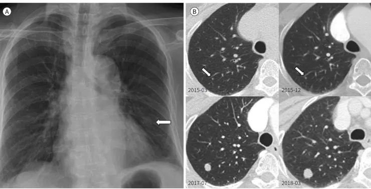
A 71-year-old woman visited our outpatient pulmonary clinic with cough and sputum for 10 days. The patient had been previously healthy but had a history of total hysterectomy due to leiomyomas and had undergone hormone therapy. On chest radiograph, there was a well- circumscribed nodular opacity in the left lower lung zone, measuring about 1.7 cm in size (Fig. 1A).
For further evaluation, low dose chest CT was performed (Fig. 1B, C) using a 128-row de- tector CT scanner (Somatom Definition AS, Siemens Healthcare, Erlangen, Germany). On CT image, it showed a 1.7 cm-sized well-circumscribed, round, low-density nodule in left lower lobe (LLL). Another smaller nodule was seen in right upper lobe (RUL), measuring less than 0.3 cm in size.
Follow-up chest CT was performed with contrast enhancement after 9 months (Fig. 1B, C).
Contrast-enhanced CT showed slight increase in size of preexisting nodule in LLL, 1.7 cm to 1.9 cm. The lesion showed no contrast enhancement (Fig. 1D). Another smaller nodule in RUL revealed no change in size. The enhancement of this lesion was difficult to evaluate due to its small size. We suspected benign conditions such as benign metastasizing leiomyomas considering patient’s hysterectomy history or hamartomas but malignancy could not be ex- cluded because of its gradual increase in size. For pathologic confirmation, fluoroscopy- guided percutaneous needle aspiration of the LLL nodule was performed and the result was negative for malignancy.
The patient was lost to follow-up for one and a half years and returned to the outpatient clinic. Contrast-enhanced chest CT was then performed for follow-up of the nodules. The nodule in LLL grew in size, from 1.9 cm to 3.6 cm. The smaller nodule in RUL increased in size, from 0.3 cm to 0.9 cm (Fig. 1B, C). Subsequent CT-guided percutaneous needle biopsy was done at LLL nodule. The pathologic result showed a possibility of benign mesenchymal tumor, but there was a difficulty of getting a definite diagnosis due to insufficient amount of biopsied tissue. The pulmonologist decided to do a 6-month follow-up. Follow-up contrast- enhanced chest CT showed that the size of nodules in LLL and RUL increased even more, from 3.6 cm to 4.0 cm and from 0.9 cm to 1.1 cm, respectively (Fig. 1B, C). Wedge resection of LLL and RUL nodules by video-assisted thoracoscopic surgery (VATS) was performed for ac- curate diagnosis.
Grossly, the nodules were well-circumscribed, intraparenchymal lesions. Cut surface was firm, rubbery, homogenous, and grayish white. The tumor showed leaf-like (phyllodes) fibro- epithelial pattern with stromal and epithelial components (Fig. 1E, upper). The epithelial component composed of gland like structures with simple cuboidal to columnar epithelium and the stromal component composed of spindle cell fibroblastic proliferation. The epitheli- al component showed immunopositivity for cytokeratin 7 (Fig. 1E, lower left). The stromal component showed immunopositivity for smooth muscle actin (SMA), estrongen receptor (ER) (Fig. 1E, lower middle), CD34 and Bcl-2. Both epithelial and stromal component showed
immunopositivity for signal transducer and activator of transcription 6 (STAT6) (Fig. 1E, low- er right).
Considering these morphologic and immunohistochemical results, the tumor was compat- ible with the diagnosis of adenofibromas of the lung. After 10 months, the patient underwent regular follow-up radiograph, and there was no evidence of recurrence.
DISCUSSION
Pulmonary adenofibroma is a rare pathologic entity, first described by Scarff and Gowar (4) in 1944 as fibroadenoma of the lung, given its architectural similarity to breast fibroadeno- ma. Since then, very few cases have been reported. The histogenesis of this rare tumor re- mains a subject of debate with some authors maintaining the hypothesis of hamartomatous origin and others suggesting that it should be regarded as a true neoplastic lesion (1, 2, 5-7).
Pulmonary adenofibroma is histologically characterized by biphasic proliferations of stro- mal and epithelial components, arranged in a distinctive phyllodes-like architecture. Stromal component resembles that of solitary fibrous tumor (SFT) whereas the glands are composed of respiratory epithelium, typically distributed throughout the entire lesion. Epithelial cells are positive for cytokeratin, thyroid transcription factor-1, epithelial membrane antigen, whereas stromal cells show variable degrees of positive immunostainings for vimentin, des- min, CD34, Bcl-2, CD99, SMA (1, 3, 5, 8, 9).
Recently, Fusco et al. (5) reported highly recurrent NAB2-STAT6 fusion variant (exon 4-exon 2) in the stromal element, suggesting that pulmonary adenofibromas are benign tumors that belong to the spectrum of SFTs. Furthermore, they found ER to be overexpressed in the stro- mal cells, suggesting that a subset of pulmonary adenofibromas are likely to be hormone- sensitive.
Clinical manifestations of pulmonary adenofibroma are nonspecific (9). It is usually de- tected incidentally on radiologic examination (1). It recognized in adults, mostly in 5th and 6th decade, without gender and race predominance (9). Radiologically, pulmonary adenofi- broma is usually known to be a well-circumscribed solitary pulmonary nodule that is com- pletely surrounded by pulmonary parenchyma, usually peripherally located. The tumor has a variable size ranging from 0.8 cm to 9.5 cm (9) and does not show enhancement after con- trast injection (1, 10). On PET-CT previously reported, pulmonary adenofibroma showed no or minimal fluorodeoxyglucose FDG hypermetabolism (1, 2, 10).
As mentioned above, pulmonary adenofibroma usually manifests as a solitary pulmonary nodule. In contrast, two adenofibromas were revealed in our case, which showed interval size increase on serial chest CT scans.
Initially, we suspected benign metastasizing leiomyomas considering her past history of hysterectomy for leiomyomas. Histologically, benign metastasizing leiomyoma usually shows higher stromal cellularity than adenofibroma. Entrapped pulmonary epithelial struc- ture can also occasionally be seen in peripheral portion of leiomyoma, while in adenofibro- ma, it shows a diffuse homogeneous distribution (1). Furthermore, unlike leiomyoma, ade- nofibroma shows phyllodes-like structure. But radiologically, there are no significant differences between benign metastasizing leiomyoma and adenofibromas in the cases of
Fig. 1. Adenofibromas of the lung in a 71-year-old woman.
A. Initial chest radiograph shows an approximately 1.7 cm large, well-circumscribed nodular opacity (arrow) in the left lower lung.
B. Axial images of the initial low-dose chest CT show a nodule, approximately 0.3 cm in size, in the right upper lobe (arrows). Follow-up CT im- ages show the nodule gradually enlarging and measuring 1.1 cm on the last follow-up image.
multiple nodules.
Pulmonary adenofibroma can be distinguished from other tumors showing biphasic pat- tern by several features as shown below.
Histologically, pulmonary hamartoma is composed of varying amounts of at least two mesenchymal elements such as cartilage, fat, connective tissue, and smooth muscle. En- trapped respiratory epithelium is also found in addition to the mesenchymal elements. This can be distinguished from pulmonary adenofibroma by the presence of two or more mesen- chymal components (3).
Intrapulmonary SFT is another tumor that can be confused with pulmonary adenofibro- ma. It is characterized by a combination of hypo- and hypercellular areas separated by thick bands with thin-walled branching vessels. When this occurs in the lung, it may entrap nor- mal respiratory epithelium at the periphery of the lesion in a haphazard arrangement as op- posed to the diffuse distribution and complex arrangement of the glandular and epithelial el- ement in pulmonary adenofibroma (3). Stromal overexpression of ER and progesterone receptor (PR) is observed in majority of cases of pulmonary adenofibroma whereas SFT and pulmonary hamartoma are ER/PR-negative (5).
Pulmonary adenofibroma poses a diagnostic challenge, particularly in small diagnostic specimen and on frozen section owing to its rarity. Therefore, most appropriate diagnostic and therapeutic modality is surgical resection using VATS rather than biopsy (1, 5). Pulmo- nary adenofiroma shows favorable prognosis. To the best of our knowledge, there are no re- current or metastatic cases in any studies previously reported with variable durations of fol-
A B
2015-03 2015-12
2017-07 2018-03
low-up, upto 8 years (7, 8).
In summary, we report a rare case of a 71-year-old woman with pulmonary adenofibromas of different lobes of the lung, mimicking other disease entities such as benign metastasizing leiomyomas or hamartomas. In such case as ours, it is difficult to diagnose on imaging alone and histopathological examination is necessary for confirmation.
Author Contributions
Conceptualization, B.Y.; investigation, K.M., B.Y.; project administration, B.Y.; supervision, B.Y.; writ- ing—original draft, all authors; and writing—review & editing, all authors.
Conflicts of Interest
The authors have no potential conflicts of interest to disclose.
REFERENCES
1. Kumar R, Desai S, Pai T, Pramesh CS, Jambhekar NA. Pulmonary adenofibroma: clinicopathological study of 3 cases of a rare benign lung lesion and review of the literature. Ann Diagn Pathol 2014;18:238-243 Fig. 1. Adenofibromas of the lung in a 71-year-old woman.
C. Axial images of the initial low-dose chest CT show a well-circumscribed nodule, sized about 1.7 cm, in the LLL. Follow-up CT images show the nodule gradually enlarging and measuring 4.0 cm on the last follow-up image.
D. Axial images of contrast-enhanced chest CT show a well-circumscribed, round, low-density nodule in LLL (left), with no enhancement after contrast injection (right, 22 → 25 HU).
E. Microscopic sections of the LLL nodule show a leaf-like (phyllodes) fibroepithelial pattern (upper left; H&E, × 10), with diffuse, homogeneously distributed epithelial (glandular) and stromal components (upper right; H&E, × 100). On immunohistochemical staining, the epithelial compo- nent is positive for cytokeratin 7 (lower left, × 100), and the stromal component is positive for the estrogen receptor (lower middle, × 100). Both epithelial and stromal components are positive for signal transducer and activator of transcription 6 (lower right, × 100).
H&E = hematoxylin-eosin stain, LLL = left lower lobe
C D
2015-03 2015-12
2017-07 2018-03
E
2. Vitkovski T, Zeltsman D, Esposito M, Morgenstern N. Pulmonary adenofibroma: cytologic and clinicopatho- logic features of a rare benign primary lung lesion. Diagn Cytopathol 2013;41:991-996
3. Hao J, Zhang C, Cao Q, Zou J, Wang C. Pulmonary adenofibroma: report of a case with multiple masses.
Ann Clin Lab Sci 2016;46:691-695
4. Scarff RW, Gowar FJ. Fibroadenoma of the lung. J Pathol 1944;56:257-258
5. Fusco N, Guerini-Rocco E, Augello C, Terrasi A, Ercoli G, Fumagalli C, et al. Recurrent NAB2-STAT6 gene fu- sions and oestrogen receptor-α expression in pulmonary adenofibromas. Histopathology 2017;70:906-917 6. Cavazza A, Rossi G, De Marco L, Putrino I, Pellegrino S, Piana S. Solitary fibrous pseudopapillary tumor of
the lung: pulmonary fibroadenoma and adenofibroma revisited. Pathologica 2003;95:162-166
7. Suster S, Moran CA. Pulmonary adenofibroma: report of two cases of an unusual type of hamartomatous lesion of the lung. Histopathology 1993;23:547-551
8. Al-Amer M, Abdeen Y, Shaaban H, Alderink C. Solitary pulmonary adenofibroma in a middle-aged man with bladder cancer. Lung India 2017;34:570-572
9. Esmaeili H, Azimpouran M, Shokohi B, Mostafidi E, Karbasi M. Pulmonary adenofibroma; a rare finding.
Am J Med Case Rep 2016;4:101-107
10. Corzani R, Bellan C, Luzzi L, Ghisalberti M, Ligabue T, Meniconi F, et al. A rare pulmonary adenofibroma mimicking a metastatic lesion. Clin Surg 2017;2:1691
서로 다른 폐엽에 두 개의 결절로 발현된 폐의 선섬유종: 증례 보고
김민수1 · 배영아1* · 변선주2 · 최정아1
폐에서 발생한 선섬유종은 드물게 발생하는 종양으로 이 양성 병변에 대해 발표된 보고는 매 우 적다. 영상의학적으로 대부분의 이 병변은 단일 폐결절로 나타나며, 다발성은 극히 드물 다. 저자는 71세 여자 환자에서, 전산화단층촬영 검사상 서로 다른 엽에 두 개의 결절로 보인 폐의 선섬유종에 대한 흔치 않은 증례를 보고하고자 한다. 폐의 선섬유종의 확실한 진단을 위해서는 조직병리학적 검사와 면역조직화학 검사가 필요하다.
한림대학교 동탄성심병원 1영상의학과, 2병리과