Osteoradionecrosis
of the Anterior Thoracic Wall after Radiation Therapy
for Breast Cancer
유방암으로 방사선 치료를 받은 후 흉벽에 발생한 방사선 골괴사
Young Seon Kim, MD1 , Jung-Hee Yoon, MD2*
1Department of Radiology, Yeungnam University Medical Center, College of Medicine, Yeungnam University, Daegu, Korea
2Department of Radiology, Haeundae Paik Hospital, Inje University College of Medicine, Busan, Korea
Although osteoradionecrosis of the thoracic wall is rare, severe complications following radia- tion therapy for breast cancer can occur. The authors report the case of a 65-year-old woman who developed osteoradionecrosis in the left thoracic wall 17 years after undergoing radiation therapy for breast cancer. Chest CT revealed fractures in the left third and fourth ribs as well as in the sternal body, with severe sclerotic and lytic changes and cortical irregularity. Ultrasonog- raphy of the left chest wall revealed diffuse edematous changes in the overlying skin layer. Ra- diologists should be familiar with the imaging findings of osteoradionecrosis to differentiate osteoradionecrosis from bony metastasis.
Index terms Radiotherapy; Osteoradionecrosis; Breast Cancer; Thoracic Wall
INTRODUCTION
Osteoradionecrosis of the thoracic wall is rare; nevertheless, severe complications of radiation therapy (RT) can occur in patients with breast and lung cancers. Blood vessels that supply the bone with nutrients can be damaged by RT. Vascular compromise with obliterative endarteritis and damage to osteoblasts and osteoclasts is a possible cause of osteoradionecrosis (1). It usually occurs > 1 year after completion of RT in patients treated with doses > 6 Gy (1). Radiological findings of osteoradionecrosis include focal
Received February 23, 2018 Revised August 9, 2018 Accepted December 18, 2018
*Corresponding author Jung-Hee Yoon, MD Department of Radiology, Haeundae Paik Hospital, Inje University College of Medicine, 875 Haeun-daero, Haeundae-gu, Busan 48108, Korea.
Tel 82-51-797-0355 Fax 82-51-797-0379 E-mail radyjh@hanmail.net This is an Open Access article distributed under the terms of the Creative Commons Attribu- tion Non-Commercial License (https://creativecommons.org/
licenses/by-nc/4.0) which permits unrestricted non-commercial use, distribution, and reproduc- tion in any medium, provided the original work is properly cited.
ORCID iDs Jung-Hee Yoon https://
orcid.org/0000-0001-5152-6668 Young Seon Kim
https://
orcid.org/0000-0002-9168-8204
A
C
B
D
was approved by the author’s Institutional Review Board, and the requirement for informed consent was waived.
CASE REPORT
A 65-year-old woman presented with an open wound and pain in her left chest wall. She underwent left radical mastectomy for breast cancer 17 years previously. She subsequently underwent adjuvant chest wall RT with 5000 cGy in 25 fractions, and adjuvant chemotherapy with 8 cycles of 5-florourasil, epirubicin, and cyclophosphamide after surgery. Subsequently, tamoxifen treatment of 20 mg/day for 5 years was continued. The wound on her left chest wall was developed a few days previous to presentation. She denied a history of trauma to her left chest wall. On physical examination, an ulcer approximately 1 cm in size was ob- served on the left mastectomy site. CT of the chest revealed fractures of the left third and
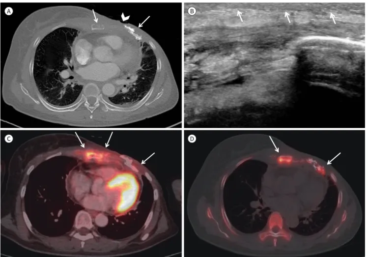
Fig. 1. Radiologic findings of osteoradionecrosis of the ribs and sternum after radiation therapy for breast cancer in a 65-year-old woman.
A. CT of the chest wall revealing fractures with sclerotic change and cortical irregularity of left third and fourth ribs, and sternal body (arrows).
Ulceration at overlying skin layer is also noted (arrowhead).
B. Ultrasonography of the left chest wall reveals diffuse thickening of skin (arrows) and edematous change of subcutaneous fat layer.
C. PET/CT demonstrating increased uptake of radioisotope in the sternal body and left ribs, and surrounding soft tissue (arrows).
D. Two years later, follow-up PET/CT demonstrating continued hot uptake in the sternal body and left ribs, with progressed sclerotic changes and non-union state of rib fractures (arrows).
fourth ribs and the sternal body, with severe lytic and sclerotic changes, and cortical irregu- larity (Fig. 1A). Ultrasonography of the left chest wall revealed diffuse thickening of the skin and increased echogenicity and edematous changes in the subcutaneous fat layer (Fig. 1B).
PET/CT of the chest wall demonstrated increased uptake in the sternal body, left ribs, and overlying soft tissue (Fig. 1C). Thus, osteomyelitis with associated cellulitis was initially sus- pected. However, bone metastasis could not be excluded based on these imaging findings. A skin punch biopsy of the ulcer lesion at the left chest wall was performed and revealed fibri- noid materials and calcifications without viable cells. Bone biopsy was recommended to dif- ferentiate it from skeletal metastasis, however, the patient refused to undergo the procedure.
Conservative treatment with pain control and management of the open wound was imple- mented. Two years later, follow-up PET/CT was performed and continued to demonstrate in- creased uptake of radioisotope in the sternal body and ribs, with progressed sclerotic chang- es and non-union state of fractures (Fig. 1D). Therefore, the possibility of radiation-related osteonecrosis was raised. Conservative treatment, including analgesia, was performed for the patient. Over the 5-year follow-up period, the patient’s anterior rib pain resolved; howev- er, her wound persisted with repeated improvement and deterioration.
DISCUSSION
The primary goal of RT is the eradication of microscopic residual disease adjacent to the tumor site and to eliminate the possibility of multicentric disease. Usually, external beam ra- diation is used for the treatment of breast cancer; it affects the adjacent structures as well as the original site of the tumor. Several complications associated with RT may occur. Early complications (during the weeks to months after completion of RT) include skin changes, breast edema, dystrophic calcifications, fat necrosis, radiation-induced pneumonia and pleu- ral effusion. Months to years after the completion of RT, fibrotic changes in the breast(s), glandular atrophy, lactational difficulty, lymphedema, brachial plexopathy, bone fracture, pulmonary fibrosis and pericardial disease can occur (1). Late complications (> 10 years after completion of RT) include cardiomyopathy and secondary malignancies (1, 2). Radiation-in- duced rib fracture is a very rare, late complication of conventionally fractionated RT for breast and lung cancer. The previously reported incidence is between 0.1% and 5% (2, 3). Se- vere osteoradionecrosis of the ribs is extremely rare, with only a few case reports published over the past decades (4, 5). While osteoradionecrosis of the ribs is rare, the mandible is a rel- atively common site of osteoradionecrosis due to the higher radiation doses administered for head and neck cancers, and the poor vascular system of mandible (6).
Radiologically, osteoradionecrosis may present as focal lucent area in bone, periostitis, sclerosis, cortical thinning and irregularity, fatty marrow change, and insufficiency fractures (2). Bone scan will demonstrate decreased uptake of radioisotope in the early stages and, lat- er, increased uptake of radioisotope with fracture will appear. On PET scan, osteoradionecro- sis may appear as a false-positive finding. Multiple previous studies have reported the relative
ing bone metastasis from osteoradionecrosis. For example, a discrete, associated solid or cys- tic mass is a significant diagnostic indication for the presence of skeletal metastasis (7). In one previous study, a permeative pattern of bone loss (< 75% loss of total bone trabecula) was more commonly seen in osteoradionecrosis, and lucent pattern (> 75% loss of total bone tra- becula) is more often observed in metastasis (7). Because, irradiation leads to relatively hy- poxic, hypocellular, hypovascular substrate with an inconsistent ability to remodel tissue loss, this would lead to relatively less bony loss than would be evident when tissue is being actively destroyed and replaced by tumor. Additionally, intraosseous gas may be evident in osteoradionecrosis because of superimposed osteomyelitis, but not in metastasis (7).
Irradiated bone is susceptible to infection and is associated with a high risk for bone sarco- mas. The differentiation of osteoradionecrosis from bone metastasis is very important for ra- diologists to plan appropriate treatment. Predisposing factors for osteoradionecrosis include trauma, infection, inflammation, overdose radiation, bony invasion by tumor, tumor loca- tion around the bone, and individual sensitivity of the patient (8). The most frequent symp- toms of osteoradionecrosis include pain, infection, and pathological fractures. Osteoradione- crosis can be diagnosed through joint evaluation of clinical findings, pathology, and radiology (5). Biologically, radiation-induced fibroatrophic changes, chronic inflammation, and tumor necrosis factor-α have been suggested to be potential causes of osteoradionecrosis (9). Various treatments such as pentoxifylline, vitamin E and clodronate for inhibiting tumor necrosis factor-α have been developed (10). Hyperbaric oxygen has been proposed as an ad- junctive therapy and may improve outcomes (8). Surgical debridement with reconstruction of the chest wall can be an option for patients who fail treatment with conservative management (5). However, there are no established treatment guidelines for osteoradionecrosis. The man- agement of radiation-induced rib fracture is to optimize analgesia, exclude recurrent disease, and trialling established treatments for osteoradionecrosis (5).
In summary, we reported a case of osteoradionecrosis affecting the left ribs following adju- vant chest wall RT, and presented a brief review of the literature. Radiologists should be aware of imaging findings of osteoradionecrosis to differentiate it from bony metastasis.
Conflicts of Interest
The authors have no potential conflicts of interest to disclose.
REFERENCES
1. Yi A, Kim HH, Shin HJ, Huh MO, Ahn SD, Seo BK. Radiation-induced complications after breast cancer radi- ation therapy: a pictorial review of multimodality imaging findings. Korean J Radiol 2009;10:496-507 2. Meric F, Buchholz TA, Mirza NQ, Vlastos G, Ames FC, Ross MI, et al. Long-term complications associated
with breast-conservation surgery and radiotherapy. Ann Surg Oncol 2002;9:543-549
3. Whelan T, MacKenzie R, Julian J, Levine M, Shelley W, Grimard L, et al. Randomized trial of breast irradia- tion schedules after lumpectomy for women with lymph node-negative breast cancer. J Natl Cancer Inst 2002;94:1143-1150
4. Makboul M, Salama Ayyad MA. Is myocutaneous flap alone sufficient for reconstruction of chest wall os- teoradionecrosis? Interact Cardiovasc Thorac Surg 2012;15:447-451
5. Nicholls L, Gorayski P, Harvey J. Osteoradionecrosis of the ribs following breast radiotherapy. Case Rep Oncol 2015;8:332-338
6. Reuther T, Schuster T, Mende U, Kübler A. Osteoradionecrosis of the jaws as a side effect of radiotherapy of
head and neck tumour patients--a report of a thirty year retrospective review. Int J Oral Maxillofac Surg 2003;32:289-295
7. Alhilali L, Reynolds AR, Fakhran S. Osteoradionecrosis after radiation therapy for head and neck cancer:
differentiation from recurrent disease with CT and PET/CT imaging. AJNR Am J Neuroradiol 2014;35:1405- 1411
8. Bennett MH, Feldmeier J, Hampson NB, Smee R, Milross C. Hyperbaric oxygen therapy for late radiation tissue injury. Cochrane Database Syst Rev 2016;4:CD005005
9. Rice N, Polyzois I, Ekanayake K, Omer O, Stassen LF. The management of osteoradionecrosis of the jaws--a review. Surgeon 2015;13:101-109
10. Delanian S, Chatel C, Porcher R, Depondt J, Lefaix JL. Complete restoration of refractory mandibular os- teoradionecrosis by prolonged treatment with a pentoxifylline-tocopherol-clodronate combination (PEN- TOCLO): a phase II trial. Int J Radiat Oncol Biol Phys 2011;80:832-839
유방암으로 방사선 치료를 받은 후 흉벽에 발생한 방사선 골괴사
김영선1 · 윤정희2*
유방암으로 방사선 치료를 받은 후 흉벽에 발생한 방사선 골괴사는 매우 드물지만 위중한 합 병증이다. 저자들은 유방암 치료를 위해 방사선 치료를 받은 후 17년 뒤 흉골과 왼쪽 늑골에 방사선 골괴사가 발생한 65세 환자의 증례를 보고하고자 한다. 환자는 흉부 단층촬영사진에 서 왼쪽 3번째, 4번째 늑골과 흉골에 경화성과 용해성 변화, 불규칙한 피질을 동반한 골절 소 견을 보였다. 또한, 초음파 검사에서 왼쪽 흉벽의 피부와 피하층의 전반적인 부종 소견이 동 반되어 있었다. 영상의학과 의사들은 골전이와 감별하기 위해 방사선 골괴사의 영상의학적