J Korean Surg Soc 2012;82:365-369 http://dx.doi.org/10.4174/jkss.2012.82.6.365
ORIGINAL ARTICLE
Journal of the Korean Surgical Society
JKSS
pISSN 2233-7903ㆍeISSN 2093-0488
Received February 2, 2012, Revised April 17, 2012, Accepted April 30, 2012 Correspondence to: Dong-Ik Kim
Division of Vascular Surgery, Department of Surgery, Samsung Medical Center, Sungkyunkwan University School of Medicine, 81 Irwon-ro, Gangnam-gu, Seoul 135-710, Korea
Tel: +82-2-3410-3467, Fax: +82-2-3410-0040, E-mail: [email protected]
cc Journal of the Korean Surgical Society is an Open Access Journal. All articles are distributed under the terms of the Creative Commons Attribution Non-Commercial License (http://creativecommons.org/licenses/by-nc/3.0/) which permits unrestricted non-commercial use, distribution, and reproduction in any medium, provided the original work is properly cited.
Ten year outcomes after bypass surgery in aortoiliac occlusive disease
Gwan-Chul Lee, Shin-Seok Yang, Keun-Myoung Park, Yangjin Park, Young-Wook Kim, Kwang Bo Park
1, Hong Suk Park
1, Young-Soo Do
1, Dong-Ik Kim
Division of Vascular Surgery, Department of Surgery, Samsung Medical Center, Sungkyunkwan University School of Medicine, Seoul, 1Department of Radiology, Samsung Medical Center, Sungkyunkwan University School of Medicine, Seoul, Korea
Purpose: Most outcome studies of bypass surgery are limited to five years of follow-up. However, as human life expectancy has increased, analyses of more long-term outcomes are needed. The aim of this study is to evaluate 10-year outcomes of ana- tomical bypasses in aortoiliac occlusive disease. Methods: From 1996 to 2009, 92 patients (82 males and 10 females) under- went aortic anatomical bypasses to treat aortoiliac occlusive disease at Samsung Medical Center. The patients were reviewed retrospectively. Kaplan-Meier survival analyses were performed using PASW ver. 18.0 (IBM Co). Results: A total of 72 pa- tients (78.3%) underwent aorto-femoral bypasses (uni- or bi-femoral), 15 patients (16.3%) underwent aorto-iliac bypasses (uni- or bi-iliac), and 5 patients (5.4%) underwent aorto-iliac and aorto-femoral bypasses. The overall primary patency rates of the 92 patients were 86.2% over 5 years and 77.6% over 10 years. The 10-year limb salvage rate and overall survival rate were 97.7% and 91.7%, respectively. Conclusion: The overall patency rates of bypass graft and limb salvage rates decreased as time passed. The analysis of results after bypass surgery to treat arterial occlusive disease will be needed to extend for 10 years of follow-up.
Key Words: Aortoiliac occlusive disease, Leriche syndrome, Bypass
INTRODUCTION
Aortoiliac occlusive disease is a relatively rare artery oc- clusive disease compared to infrainguinal arterial occlu- sive disease [1]. Leriche and Morel [2] first described this disease in 1948. The main treatment is surgical revascu- larization. In the past, endarterectomy was the only treat- ment of choice, but with the development of artificial vas- cular graft materials, anatomical bypass graft surgery has
now become common. Axillo-femoral bypass should be considered in patients at high risk for laparotomy surgery or in whom the aortic approach is difficult due to previous abdominal surgery [3]. Endovascular treatment for athe- rosclerotic occlusive disease has recently emerged as a non-surgical option, but this procedure may be applied in relatively few patients. Thus, aorto-iliac bypass remains the most popular treatment for aortoiliac occlusive disease.
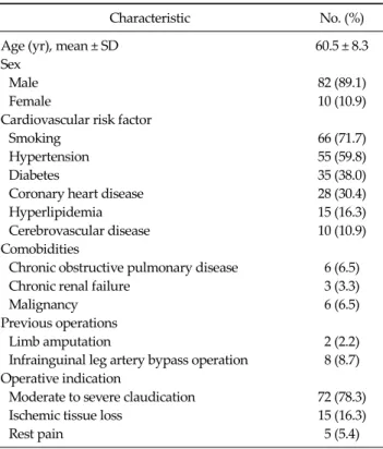
Table 1. Patient characteristics
Characteristic No. (%)
Age (yr), mean ± SD 60.5 ± 8.3
Sex
Male 82 (89.1)
Female 10 (10.9)
Cardiovascular risk factor
Smoking 66 (71.7)
Hypertension 55 (59.8)
Diabetes 35 (38.0)
Coronary heart disease 28 (30.4)
Hyperlipidemia 15 (16.3)
Cerebrovascular disease 10 (10.9)
Comobidities
Chronic obstructive pulmonary disease 6 (6.5)
Chronic renal failure 3 (3.3)
Malignancy 6 (6.5)
Previous operations
Limb amputation 2 (2.2)
Infrainguinal leg artery bypass operation 8 (8.7) Operative indication
Moderate to severe claudication 72 (78.3)
Ischemic tissue loss 15 (16.3)
Rest pain 5 (5.4)
Table 2.Operative procedure
No. (%) Occlusion level
Infrarenal 73 (79.3)
Juxtarenal 19 (20.7)
Reconstruction type
Aorto-uni or bi-femoral 72 (78.3)
Aorto-uni or bi-iliac 15 (16.3)
Aorto-iliac + aorto-femoral 5 (5.4) Graft material
Polytetrafluoroethylene (Gore-tex) 79 (85.9)
Dacron (hemasheild) 13 (14.1)
Approach of aorta
Transperitoneal 90 (97.8)
Retroperitoneal 2 (2.2)
Ten-year long-term outcomes of this disease are rarely reported in the literature [4]. The aim of this study is to evaluate the 10-year long-term outcomes of anatomical bypass in aortoiliac occlusive disease.
METHODS
From 1996 to 2009, 111 patients were surgically treated for aortoiliac occlusive disease at Samsung Medical Cen- ter. Of this group, 92 patients underwent aortic anatomical bypasses, including 82 male and 10 female patients.
Nineteen patients who underwent extra-anatomical by- passes were excluded from this study. Types of prosthetic graft were selected according to surgeon preference. Pa- tient charts were reviewed retrospectively. All patients were examined for patency of graft by duplex ultrasound at 1, 3, and 6 months postoperatively. Thereafter, duplex scan and/or CT angiography were undertaken every 6 to 12 months.
Statistical analyses were performed using PASW ver.
18.0 (IBM Co., Armonk, NY, USA). The patency rates of
grafts, limb salvage rates and overall survival rates were analyzed using Kaplan-Meier survival analysis.
RESULTS
The median follow-up period was 41.5 months (range, 1 to 155.8 months). Twenty-eight patients were receiving medical therapy due to co-existing coronary heart disease, nine patients had coronary artery stenting, and three pa- tients received coronary artery bypass grafts (CABGs) be- fore the anatomical bypass surgery. Eight patients had un- dergone distal lower extremity bypass surgeries and two patients had undergone distal limb amputations due to is- chemic tissue loss (Table 1).
The indications for bypass surgery were clinically clas- sified into three categories. There were 72 patients with moderate to severe claudication, 15 patients with ischemic tissue loss, and 5 patients with rest pain (Table 1).
Seventy-three patients had infrarenal occlusions and 19 patients had juxtarenal occlusions. The surgical approach was transperitoneal in 90 patients and retroperitoneal in 2 patients. Median hospital stays were 13 days, and there were no immediate postoperative mortalities. The by- passes were aorto-femoral (uni- or bi-femoral) in 72 pa- tients (78.3%), aorto-iliac (uni- or bi-iliac) in 15 patients (16.3%), and aorto-iliac and aorto-femoral in 5 patients (5.4%) (Table 2).
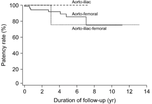
In the aorto-femoral bypass cases, the 5-year and
Fig. 1. Comparison of primary patency rates between recon- struction types.
Fig. 2. Comparison of primary patency rates between types of graft material. PTFE, polytetrafluoroethylene.
Fig. 3. Overall primary and secondary patency rates.
Fig. 4. Overall limb salvage rates.
10-year primary patency rate were 85.3% and 74.6%, res- pectively. In the aorto-iliac bypass cases, the 5-year and 10-year primary patency rates were both 100%. In the aor- to-iliac and aorto-femoral bypass cases, the 5-year and 10-year primary patency rates were both 75.0%. There were no significant differences among the three groups (P
= 0.512) (Fig. 1).
Two graft materials were used, polytetrafluoroethylene (PTFE) in 79 (85.9%) and Dacron in 13 (14.1%). The 5-year primary patency rates for PTFE and Dacron were 86.4%
and 82.1%, respectively, and the 10-year primary patency rates were 75.6% and 82.1%, respectively. However, these differences were not significant (P = 0.698) (Fig. 2).
During follow-up, grafts were occluded in nine pa- tients. Recurrent symptoms were 4 patients with claudica- tion, 3 rest pain, 1 paralysis and 1 skin color change in foot.
There were 8 procedures to rescue of graft-failure; 2 thrombolysis, 2 stent insertion, 1 thrombectomy with patch angioplasty, 2 thrombectomy with bypass graft and 1 interposition graft. In a patient undergone aorto-bife- moral bypass and below the knee amputation in right leg, conservative management was performed due to long seg- mental occlusion from descending thoracic aorta to left femoral artery. The 5-year and 10-year overall primary pa- tency rates were 86.2% and 77.6%, respectively, and the secondary patency rates at 5 years and 10 years were 91.6%
and 83.3%, respectively (Fig. 3). Statistical analysis of the variables such as age, gender and comorbidities failed to detect predictors of graft failure. Two patients underwent lower extremity amputations after bypass surgery. The 10-year overall limb salvage rate was therefore 97.7% (Fig.
4).
Four patients died due to acute myocardial infarction (one patient) or malignancy (three patients: stomach can-
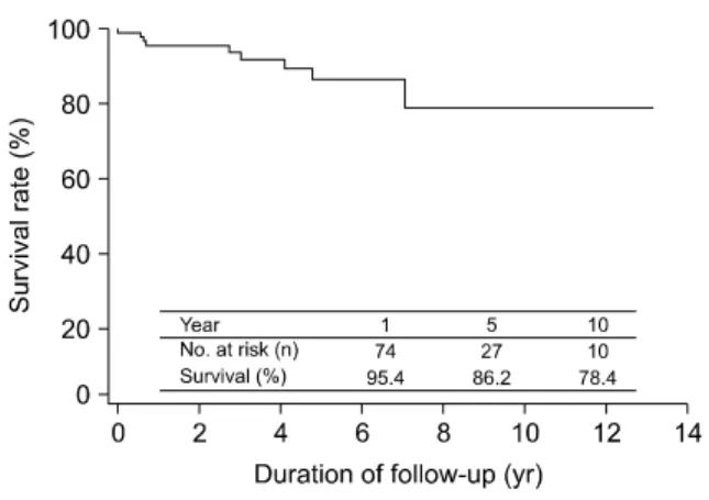
Fig. 5. Overall survival rates.
cer, lung cancer, and pancreatic cancer). The 10-year over- all survival rate was 91.7% (Fig. 5).
DISCUSSION
The management options for aortoiliac occlusive dis- ease are surgical or non-surgical management. The surgi- cal treatment of aortoiliac occlusive disease can be divided into two categories, direct anatomical bypass vs. ex- tra-anatomical bypass [5]. Direct anatomical bypasses are aorto-iliac bypasses or aorto-femoral bypasses, while ex- tra-anatomical bypasses are axillo-femoral bypasses.
Some high-risk patients underwent extra-anatomical by- pass surgeries (e.g., axillo-femoral bypass) or endovas- cular revascularization [3,6]. Because patency outcomes after extra-anatomical bypass were less favorable than af- ter anatomical bypass, the treatment of choice in aortoiliac occlusive disease is anatomical bypass surgery [7].
Recently, a totally laparoscopic approach and robotic-as- sisted laparoscopic approach were used for the treatment of aortoiliac occlusive disease, but the indications for lapa- roscopy are limited [8,9]. In any case, this study analyzed the long-term outcomes of direct anatomical bypasses alone, not in comparison with extra-anatomical bypass surgery.
The primary patency rates of aortic bypass graft re- construction are good compared to infrainguinal arterial bypass graft reconstruction [4]. We observed good results (5-year primary patency rate of 86.2% and 10-year primary patency rate of 77.6%) in the present study, but these out-
comes were not compared directly to infrainguinal leg by- pass outcomes.
We observed no significant differences in patency rates between PTFE and Dacron in this study (P = 0.698). This re- sult agrees with previous studies that did not detect sig- nificant differences between artificial graft materials in pa- tients who underwent aorto-femoral bypass and femo- ropopliteal bypass [10,11].
Some patients experienced symptoms of sexual dys- function due to aortoiliac occlusive disease [12-14]. Im- provement of sexual function is expected after revascu- larization. However, we did not evaluate pre- and post-op- erative male sexual function in our study. Due to the limi- tations of retrospective study design, further evaluation of sexual function is needed.
The most common cause of death in peripheral artery occlusive disease is cardiovascular disease [15]. In our study, 28 patients (30.4%) also had coronary artery disease, but only one patient died of acute myocardial infarction.
This low rate of death was probably due to coronary inter- vention and the management of coronary artery disease.
In conclusion, most outcome studies of bypass surgery are limited to five years of follow-up. However, as human life expectancy has increased, analyses of more long-term outcomes are needed. A total of 72 patients (78.3%) under- went aorto-femoral bypasses (uni- or bi-femoral), 15 pa- tients (16.3%) underwent aorto-iliac bypasses (uni- or bi-iliac), and 5 patients (5.4%) underwent aorto-iliac and aorto-femoral bypasses. The overall primary patency rates of 92 patients were 86.2% over 5 years and 77.6% over 10 years. The 10-year limb salvage rate and overall survival rate were 97.7% and 91.7%.
CONFLICTS OF INTEREST
No potential conflict of interest relevant to this article was reported.
REFERENCES
1. Szilagyi DE, Elliott JP Jr, Smith RF, Reddy DJ, McPharlin
M. A thirty-year survey of the reconstructive surgical treatment of aortoiliac occlusive disease. J Vasc Surg 1986;3:421-36.
2. Leriche R, Morel A. The syndrome of thrombotic ob- literation of the aortic bifurcation. Ann Surg 1948;127:193- 206.
3. Park UJ, Kim DI. Thromoboagiitis obliterans (TAO). Int J Stem Cells 2010;3:1-7.
4. de Vries SO, Hunink MG. Results of aortic bifurcation grafts for aortoiliac occlusive disease: a meta-analysis. J Vasc Surg 1997;26:558-69.
5. Brewster DC. Current controversies in the management of aortoiliac occlusive disease. J Vasc Surg 1997;25:365-79.
6. Kashyap VS, Pavkov ML, Bena JF, Sarac TP, O'Hara PJ, Lyden SP, et al. The management of severe aortoiliac occlu- sive disease: endovascular therapy rivals open reconstruc- tion. J Vasc Surg 2008;48:1451-7.e3.
7. Kim IH, Kim DI, Huh SH, Lee BB, Kim DK, Do YS, et al.
Clinical experiences of the arterial bypass in aortoiliac oc- clusive disease. J Korean Surg Soc 2001;61:600-3.
8. Di Centa I, Coggia M, Cerceau P, Javerliat I, Alfonsi P, Beauchet A, et al. Total laparoscopic aortobifemoral by- pass: short- and middle-term results. Ann Vasc Surg 2008;22:227-32.
9. Novotny T, Dvorak M, Staffa R. The learning curve of ro- bot-assisted laparoscopic aortofemoral bypass grafting for aortoiliac occlusive disease. J Vasc Surg 2011;53:414-20.
10. Davidovic L, Vasic D, Maksimovic R, Kostic D, Markovic D, Markovic M. Aortobifemoral grafting: factors influenc- ing long-term results. Vascular 2004;12:171-8.
11. Takagi H, Goto SN, Matsui M, Manabe H, Umemoto T. A contemporary meta-analysis of Dacron versus polytetra- fluoroethylene grafts for femoropopliteal bypass grafting. J Vasc Surg 2010;52:232-6.
12. Miles JR Jr, Miles DG, Johnson G Jr. Aortoiliac operations and sexual dysfunction. Arch Surg 1982;117:1177-81.
13. Cormio L, Edgren J, Lepantalo M, Lindfors O, Nisen H, Saarinen O, et al. Aortofemoral surgery and sexual function. Eur J Vasc Endovasc Surg 1996;11:453-7.
14. Flanigan DP, Schuler JJ, Keifer T, Schwartz JA, Lim LT.
Elimination of iatrogenic impotence and improvement of sexual function after aortoiliac revascularization. Arch Surg 1982;117:544-50.
15. Hertzer NR, Beven EG, Young JR, O'Hara PJ, Ruschhaupt WF 3rd, Graor RA, et al. Coronary artery disease in periph- eral vascular patients: a classification of 1000 coronary an- giograms and results of surgical management. Ann Surg 1984;199:223-33.