ISSN 2234-3806 • eISSN 2234-3814
466 www.annlabmed.org http://dx.doi.org/10.3343/alm.2015.35.4.466 Ann Lab Med 2015;35:466-468
http://dx.doi.org/10.3343/alm.2015.35.4.466
Letter to the Editor
Diagnostic Hematology
An Unusual Case of Myeloperoxidase-Positive Acute Megakaryoblastic Leukemia
Hyeyoung Lee, M.D.1, Yonggoo Kim, M.D.1, Yoo-Jin Kim, M.D.2, and Kyungja Han, M.D.1
Departments of Laboratory Medicine1 and Internal Medicine2, College of Medicine, The Catholic University of Korea, Seoul, Korea
Dear Editor
We report an unusual case of myeloperoxidase (MPO)-positive acute megakaryoblastic leukemia (AMKL). The patient was a 63-yr-old woman referred to our hospital with anemia and dys- pnea. General physical examination revealed no lymphadenopa- thy or hepatosplenomegaly. An initial complete blood count indi- cated white blood cell counts of 26.78 ×10⁹/L, including 47%
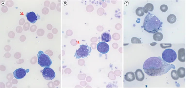
blasts, hemoglobin of 7.2 g/dL, and platelets of 519×109/L. A pe- ripheral blood smear showed blasts with prominent nucleoli and blue cytoplasm. Some giant platelets were also observed. A bone marrow (BM) biopsy showed hypercellularity with no fibrosis, packed with blasts, and revealed an increase in megakaryocyte number. BM aspirates showed that blasts accounted for 70% of all nucleated elements. Most blasts had medium-to-large, finely chromatinated nuclei with distinct nucleoli. Some showed multi- ple clear cytoplasmic projections (Fig. 1A, B) or Auer rods. No dysplastic features of hematologic precursors were observed. Cy- tochemical stain showed some blasts were MPO-positive (Fig.
1C) but negative for periodic acid–Schiff (PAS) stain and non- specific esterase (NSE, α-naphthyl butyrate).
Immunophenotyping showed a distinct population in the CD45 vs. SSC plot that was positive for CD7, CD11c, CD13, CD33, CD41a, CD117, cytoplasmic MPO, and HLA-DR and negative for CD2, CD3, CD5, CD10, CD14, CD19, CD20, CD22,
CD34, CD56, CD64, and cytoplasmic CD79a. About 57.0% of blasts were CD41a-positive, and 57.7% were cytoplasmic MPO- positive. A blast population co-expressing CD41a and cytoplas- mic-MPO was also observed (Fig. 2). Conventional karyotyping revealed a 46,XX karyotype. Molecular studies did not reveal any genetic abnormalities. As the patient refused treatment, only supportive care was provided, and the patient died from pneu- monia four months later.
Diagnosis of AMKL relies on multiple criteria including mor- phology, immunophenotyping, cytochemical stain, and ultra- structural studies [1]. The standard for diagnosis is demonstra- tion of platelet glycoprotein (GP)-CD41 (GP IIb/IIIa) and/or CD61 (GP IIIa) by immunophenotyping. Cytoplasmic expression is more specific owing to possible contamination of platelets [2]. In cytochemical stain, megakaryoblasts are not reactive with MPO or Sudan Black B. Cells of the megakaryocytic lineage are usu- ally positive in PAS stain owing to glycogen granules in the cyto- plasm, and they are typically present in the periphery on stain, with prominent cytoplasmic blebs [3]. Reactivity with α-naphthyl acetate, but not with α-naphthyl butyrate, a different substrate for NSE, is characteristic of megakaryoblasts [4]. An ultrastruc- tural platelet peroxidase reaction by cytochemistry, while difficult to perform, is also diagnostic for megakaryoblasts [1]. Mega- karyoblasts also show acid phosphatase reactivity localizing to
Received: October 7, 2014
Revision received: February 16, 2015 Accepted: April 30, 2015
Corresponding author: Kyungja Han
Department of Laboratory Medicine, College of Medicine, The Catholic University of Korea, Seoul St. Mary’s Hospital, 222 Banpo-daero, Seocho-gu, Seoul 137-701, Korea
Tel: +82-2-2258-1644, Fax: +82-2-2258-1719 E-mail: hankja@catholic.ac.kr
© The Korean Society for Laboratory Medicine.
This is an Open Access article distributed under the terms of the Creative Commons Attribution Non-Commercial License (http://creativecommons.org/licenses/by-nc/3.0) which permits unrestricted non-commercial use, distribution, and reproduction in any medium, provided the original work is properly cited.
Lee H, et al.
MPO-positive acute megakaryoblastic leukemia
http://dx.doi.org/10.3343/alm.2015.35.4.466 www.annlabmed.org 467
the Golgi [4]. There are no distinct chromosomal abnormalities, but inv(3)(q21q26.2) and t(3:3)(q21q26.2) are associated with
megakaryocytic/megakaryoblastic differentiation [2].
Although MPO is an exclusive marker for leukemia of the Fig. 1. (A, B) Bone marrow aspirates showing megakaryoblasts with cytoplasmic blebs (red arrows) (Wright-Giemsa stain, ×1,000). (C) Bone marrow megakaryoblasts positive for myeloperoxidase stain (×1,000).
A B C
Fig. 2. Flow cytometry of the bone marrow sample. (A) CD45/SSC dot plot with the blast population highlighted. (B) FSC/SSC plot of the sample. Blasts are positive for (C) CD41a (57.0%) and (D) cytoplasmic myeloperoxidase (MPO) (57.7%). (E) Back-gating of CD41a.
C D E
A B
Lee H, et al.
MPO-positive acute megakaryoblastic leukemia
468 www.annlabmed.org http://dx.doi.org/10.3343/alm.2015.35.4.466 megakaryoblastic lineage, few MPO-positive AMKL cases have
been reported [5-7]. Park et al. [5] reported one Korean case in 1996, where blasts showed morphology typical of megakaryo- blasts, and CD61 expression was confirmed by flow cytometry.
The blasts also were weakly positive for MPO in cytochemical stain, and some Auer rods were observed. The sample was PAS- negative, but positive foci in the Golgi were observed with NSE stain (α-naphthyl acetate). Tallman et al. [6] reviewed 20 patients previously diagnosed as having AMKL using morphologic evi- dence and found two MPO-positive patients. One patient ex- pressed factor VIII, a megakaryocytic lineage marker, and karyo- typing revealed t(3:3)(q21;q26). The other patient had a normal karyotype and no evidence of the megakaryocytic lineage. In an- other study, AMKL was diagnosed by using another platelet marker, CD31, with MPO [7]. CD31, also known as PECAM-1 (platelet endothelial cell adhesion molecule 1), is a 130-kDa transmembrane glycoprotein on the surface of platelets, mono- cytes, macrophages, and neutrophils [8]. Immunolocalization of CD31 is limited to megakaryocytes in normal BM or in cases of myelofibrosis [9]. A failed BM aspiration prevented analysis by flow cytometry. The sample was negative for factor VIII on immu- nohistochemistry. However, the authors diagnosed the case as AMKL on the basis of typical blast morphology and positive im- munohistochemical reactivity for CD31, CD43, and MPO in the BM biopsy [7].
In this case, blasts showed typical megakaryoblastic morphol- ogy with some Auer rods. Unlike most cases, the blasts co-ex- pressed CD41a and cytoplasmic-MPO, were MPO-positive on cytochemical stain, and PAS-negative. According to the 2008 WHO classification, megakaryocytic lineage markers are not in- cluded in the diagnosis of mixed phenotype acute leukemia [10].
On the basis of previous reports, we conclude that this was an MPO-positive AMKL. AMKL is rare, and its diagnosis is not clearly defined compared with other types of AML. Morphologic evidence is still important, and comprehensive analyses are re- quired when diagnosing acute leukemia, especially with a mega-
karyocytic lineage.
Authors’ Disclosures of Potential Conflicts of Interest
No potential conflicts of interest relevant to this article were re- ported.
REFERENCES
1. Mathur NB, Joshi N, Singh T, Singh M. Congenital acute megakaryo- cytic leukemia. Indian J Med Paediatr Oncol 2011;32:165-7.
2. Arber DA, Brunning RD, Orazi A, Porwit A, Peterson L, Thiele J, et al.
Acute myeloid leukemia, not otherwise specified. In: Swerdlow SH, Campo E, Harris NL, Jaffe ES, Pileri SA, Stein H, et al., eds. WHO classi- fication of tumours of haematopoietic and lymphoid tissues. 4th ed.
Lyon: IARC, 2008:130-9.
3. Sun T, ed. Flow cytometry, immunohistochemistry, and molecular ge- netics for hematologic neoplasms. 2nd ed. Philadelphia: Lippincott Wil- liams & Wilkins, 2012:150-2.
4. Pombo De Oliveira MS, Gregory C, Matutes E, Parreira A, Catovsky D.
Cytochemical profile of megakaryoblastic leukaemia: a study with cyto- chemical methods, monoclonal antibodies, and ultrastructural cyto- chemistry. J Clin Pathol 1987;40:663-9.
5. Park CJ, Cho HC, Park YS. A case of acute megakaryoblastic leukemia showing Aure rods. Korean J Hematol 1996;31;161-5.
6. Tallman MS, Neuberg D, Bennett JM, Francois CJ, Paietta E, Wiernik PH, et al. Acute megakaryocytic leukemia: the Eastern Cooperative On- cology Group experience. Blood 2000;96:2405-11.
7. Majhi U, Murhekar K, Sundersingh S, Rajalekshmi KR. Megakaryoblas- tic leukemia presenting as pancytopenia and extensive myelofibrosis in a child diagnosed by myeloid markers and CD 31. Indian J Med Paedi- atr Oncol 2012;33:59-61.
8. Pusztaszeri MP, Seelentag W, Bosman FT. Immunohistochemical ex- pression of endothelial markers CD31, CD34, von Willebrand factor, and Fli-1 in normal human tissues. J Histochem Cytochem 2006;54:385- 95.
9. Calapso P, Vitarelli E, Crisafulli C, Tuccari G. Immunocytochemical de- tection of megakaryocytes by endothelial markers: a comparative study.
Pathologica 1992;84:215-23.
10. Borowitz MJ, Bene MC, Harris NL, Porwit A, Matutes E. Acute leuke- mias of ambiguous lineage. In: Swerdlow SH, Campo E, Harris NL, Jaffe ES, Pileri SA, Stein H, et al., eds. WHO classification of tumours of hae- matopoietic and lymphoid tissues. 4th ed. Lyon: IARC, 2008:150-5.