https://doi.org/10.4174/astr.2020.99.1.37 Annals of Surgical Treatment and Research
Clinical characteristics and oncologic outcomes in
patients with preoperative clinical T3 and T4 colon cancer who were staged as pathologic T3
Jeong-Min Choo, Se-Jin Baek, Jung-Myun Kwak, Jin Kim, Seon-Hahn Kim
Department of Surgery, Korea University College of Medicine, Seoul, Korea
INTRODUCTION
Colorectal cancer is the third most malignant cancer in the world and it is the second most common cancer after stomach cancer in Korea according to the National Cancer Registry in 2015 [1,2]. Therefore, diagnosis and management of colorectal cancer are very important. Precise staging should be a basic tool for evaluating cancer, and the American Joint Committee on Cancer (AJCC) guideline for TNM staging is most widely
used in colorectal cancer as well as in many other cancers. This staging system can predict cancer prognosis and can be used as a guideline for determining postoperative management policy. Therefore, accurate staging is clinically important. TNM staging is based on pathologic examination, and the tumor is similarly evaluated using TNM staging in preoperative imaging.
In general, the accuracy of imaging tests is lower than the accuracy of histologic examination, especially for N stage [3- 5]. To increase the accuracy of evaluation, various tests are
Received November 11, 2019, Revised March 18, 2020, Accepted April 28, 2020
Corresponding Author: Se-Jin Baek
Department of Surgery, Korea University College of Medicine, 73 Goryeodae-ro, Seongbuk-gu, Seoul 02841, Korea
Tel: +82-2-920-6412, Fax: +82-2-928-1631 E-mail: [email protected]
ORCID: https://orcid.org/0000-0002-3185-8777
Copyright ⓒ 2020, the Korean Surgical Society
cc Annals of Surgical Treatment and Research is an Open Access Journal. All articles are distributed under the terms of the Creative Commons Attribution Non- Commercial License (http://creativecommons.org/licenses/by-nc/4.0/) which permits unrestricted non-commercial use, distribution, and reproduction in any medium, provided the original work is properly cited.
Purpose: Clinically suspected T4 stage colon cancer from a preoperative exam is often diagnosed as T3 stage colon cancer pathologically after surgery, raising concerns about understaging. The aims of this study were to compare the survival of clinical T3 and T4 colon cancer patients who had received a pathologic T3 stage diagnosis postoperatively.
Methods: Patients who were diagnosed with pathologic T3 stage colon cancer postoperatively were reviewed. Patients with clinically suspected T3 or T4 stage cancer on preoperative exam were enrolled in the study. We compared patient demographics and survival of the cT3 and cT4 groups.
Results: Out of the 536 patients with pT3 colon cancer, 503 patients were cT3 (93.8%) and 33 patients were cT4 (6.2%) preoperatively. The most common reason for suspected clinical T4 stage cancer was free perforation (78.8%). There were no statistically significant differences between the 5-year overall survival and the total 5-year disease-free survival (DFS) between the cT3 and cT4 groups; however, local recurrence was significantly higher in the cT4 group (local 5-year DFS: 98.6% vs. 84.0%, P < 0.001). Multivariate analysis showed cT stage was associated with local recurrence, but the association was not statistically significant (P = 0.056).
Conclusion: Preoperative clinically suspected T4 stage colon cancer showed inferior local recurrence despite a postoperative pathologic diagnosis of T3 stage cancer. It is necessary to address the shortcomings of pathologic exams in the matter of the understaging of T4 colon cancer, and to reinforce the treatment for local control in patients with cT4 colon cancer.
[Ann Surg Treat Res 2020;99(1):37-43]
Key Words: Colonic neoplasms, Diagnosis, Survival, Tumor Staging
performed before surgery and evaluated comprehensively [6-10].
Nevertheless, current examinations are limited since there are some differences between staging before and after surgery.
The postoperative stage is occasionally different from the stage predicted before surgery, which makes it difficult for the clinician to trust the diagnosis and define a treatment plan. In colon cancer, a preoperative suspected cT4 stage can often be diagnosed as pT3 after surgery. In these cases, preoperative clinical and imaging staging were very reliable, leading to challenges in determining which results to trust [11- 13]. Since there are concerns around whether pathologic T3 is understaged, there are also concerns around undertreatment.
However, there are few studies on the accuracy of pathologic diagnosis, especially for T stage. Therefore, this study was planned to confirm the diagnostic accuracy of the postoperative pathologic results by comparing the survival of patients who were suspected of cT3 and cT4 before surgery and who were diagnosed with pT3 after surgery.
METHODS
We reviewed the data of patients who underwent surgical resection for colon cancer from September 2006 to September 2016, and who were diagnosed with pathologic T3 stage cancer with or without lymph node metastasis.
Patients with preoperative clinical T3 or T4 stage cancer were selected. Clinical T4 was defined as free perforation, localized peritumoral abscess, and direct invasion of an adjacent organ [14]. Patients diagnosed with primary colon cancer with pathologically proven adenocarcinoma from the cecum to the sigmoid colon were included in this study. The exclusion criteria were as follows: patients with appendiceal or rectosigmoid colon cancer, patients with multiple colon cancers, patients with distant metastasis, or patients who had undergone neoadjuvant treatment. This study was approved by the Institutional Review Board (IRB) of Korea University Anam Hospital (IRB No. 2019AN0071), and all patients had provided informed consent.
All patients with colon cancer had been evaluated preoperatively by physical examination, total colonoscopy, abdominopelvic CT, chest CT, and routine laboratory, which included tests for tumor markers. In cases of postoperative stage 2 disease with risk factors or stage 3 disease, oxaliplatin- based adjuvant chemotherapy was performed for 8–12 cycles.
In patients who had undergone chemotherapy, laboratory tests, which included CEA, and abdominopelvic CT were performed at 3–4 cycle intervals during chemotherapy. Other examinations such as chest CT, colonoscopy, and PET-CT were added when necessary. Radiation therapy was determined after a multidisciplinary team discussion for cases where there was suspicion of peritoneum or abdominal wall involvement, or
if there was a high risk of recurrence in a locally limited area before or during surgery. After adjuvant treatment, follow-up examinations were carried out at 3-month intervals during the first 2 years postoperatively, at 6-month intervals until 5 years after surgery, and then annually if there was no evidence of recurrence.
Data for cT3 and cT4 groups were compared in terms of patient demographics, tumor characteristics, operative and postoperative outcomes, pathologic results, and oncologic outcomes. Descriptive results are presented as a mean with standard deviation or median with interquartile range for continuous outcomes and as frequency and percentage for categorical outcomes. Student t-tests were used to compare continuous variables, and the chi-square test or Fisher exact test was applied for categorical variables. Five-year overall survival (OS) and recurrence-free survival were analyzed using the Kaplan-Meier method. Comparison of survival between groups was performed by log-rank test. Univariate and multivariate analyses of clinicopathologic factors were performed by the Cox regression analysis in order to determine prognostic factors of survival. Statistical analysis was performed using IBM SPSS Statistics ver. 22.0 (IBM Co., Armonk, NY, USA). A P-value <0.05 was considered statistically significant.
RESULTS
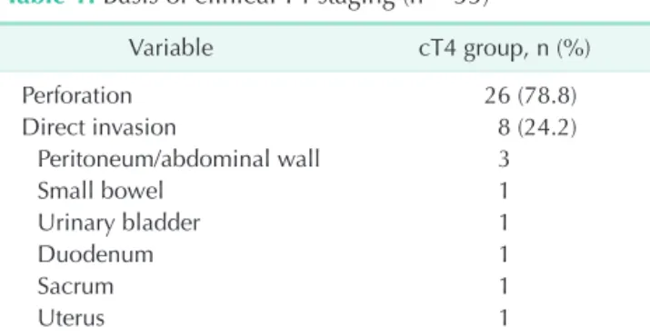
A total of 536 patients were included in the study. In the patient population, 503 had clinical T3 (93.8%), and 33 had clinical T4 (6.2%). Table 1 summarizes the reasons for the stage 4 diagnoses in the cT4 group. The most common finding was free perforation (26 patients, 78.8%), followed by direct invasion of adjacent organs (24.2%). The most commonly invaded organ was the peritoneum or abdominal wall, followed by the small bowel or urogenital organ. Four patients had suspected localized peritumoral abscess (12.1%).
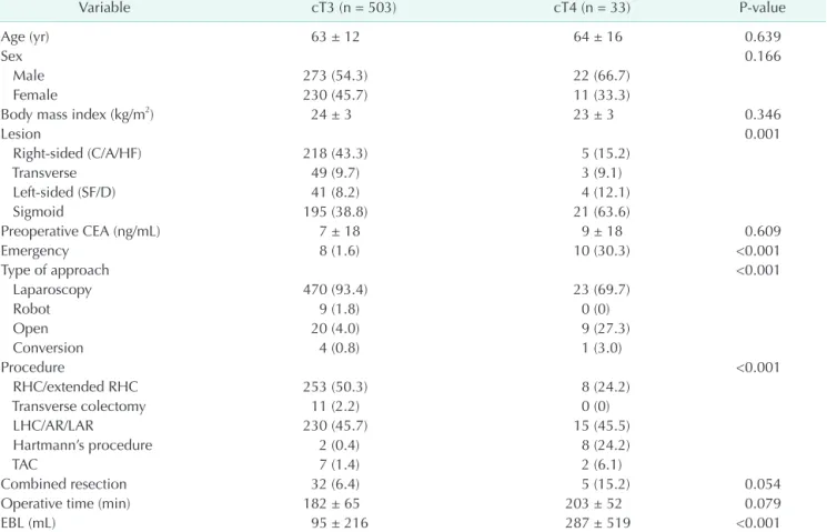
In the cT4 group, the proportion of sigmoid colon cancer was high (38.8% vs. 63.6%), whereas the proportion of right colon
Table 1. Basis of clinical T4 staging (n = 33)a)
Variable cT4 group, n (%)
Perforation 26 (78.8)
Direct invasion 8 (24.2)
Peritoneum/abdominal wall 3
Small bowel 1
Urinary bladder 1
Duodenum 1
Sacrum 1
Uterus 1
Peritumoral abscess 4 (12.1)
a)Patients are duplicated.
cancer was high in the cT3 group (43.3% vs. 15.2%, P < 0.001) (Table 2). In the cT4 group, the rate of emergency surgery was high (1.6% vs. 30.3%, P < 0.001), and the rate of open surgery was high (4.0% vs. 27.3%, P < 0.001). In terms of operative procedure, the cT3 group was more likely to undergo right colonic resection (50.3% vs. 24.2%), while the cT4 group was significantly more likely to undergo Hartmann's procedure (0.4% vs. 24.2%, P < 0.001). Estimated blood loss was also higher in the cT4 group (95 mL vs. 287 mL, P < 0.001). The rates of combined resection and operation time were higher in the cT4 group, but the results were not statistically significant (6.4%
vs. 15.2%, P = 0.054; 182 minutes vs. 203 minutes, P = 0.079, respectively). There were no significant differences between the 2 groups in terms of pathologic results (Table 3). There were no differences in postoperative stay, complication, and adjuvant chemotherapy, but the rate of adjuvant radiotherapy in the cT4 group was high (0.6% vs. 6.1%, P = 0.002).
Median follow-up periods for the cT3 and cT4 groups were 48.3 months and 24 months, respectively, and the shorter follow-up period for the cT4 group was significant (P = 0.037).
Table 2. Patient demographics and operative outcomes
Variable cT3 (n = 503) cT4 (n = 33) P-value
Age (yr) 63 ± 12 64 ± 16 0.639
Sex 0.166
Male 273 (54.3) 22 (66.7)
Female 230 (45.7) 11 (33.3)
Body mass index (kg/m2) 24 ± 3 23 ± 3 0.346
Lesion 0.001
Right-sided (C/A/HF) 218 (43.3) 5 (15.2)
Transverse 49 (9.7) 3 (9.1)
Left-sided (SF/D) 41 (8.2) 4 (12.1)
Sigmoid 195 (38.8) 21 (63.6)
Preoperative CEA (ng/mL) 7 ± 18 9 ± 18 0.609
Emergency 8 (1.6) 10 (30.3) <0.001
Type of approach <0.001
Laparoscopy 470 (93.4) 23 (69.7)
Robot 9 (1.8) 0 (0)
Open 20 (4.0) 9 (27.3)
Conversion 4 (0.8) 1 (3.0)
Procedure <0.001
RHC/extended RHC 253 (50.3) 8 (24.2)
Transverse colectomy 11 (2.2) 0 (0)
LHC/AR/LAR 230 (45.7) 15 (45.5)
Hartmann’s procedure 2 (0.4) 8 (24.2)
TAC 7 (1.4) 2 (6.1)
Combined resection 32 (6.4) 5 (15.2) 0.054
Operative time (min) 182 ± 65 203 ± 52 0.079
EBL (mL) 95 ± 216 287 ± 519 <0.001
Values are presented as mean ± standard deviation or number (%).
BMI, body mass index; C, cecal; A, ascending; HF, hepatic flexure; SF, splenic flexure; D, descending; RHC, right hemicolectomy;
LHC, left hemicolectomy; AR, anterior resection; LAR, low anterior resection; TAC, total abdominal colectomy; EBL, estimated blood loss.
Table 3. Pathologic results
Variable cT3 (n = 503) cT4 (n = 33) P-value
pN 0.333
pN0 261 (51.9) 20 (60.6)
pN1 179 (35.6) 10 (30.3)
pN2 63 (12.5) 3 (9.1)
Positive LN 1 ± 3 1 ± 2 0.410
Retrieved LN 30 ± 17 29 ± 18 0.568
pTNM stage 0.332
Stage 2 261 (51.9) 20 (60.6)
Stage 3 242 (48.1) 13 (39.4)
Tumor size (cm) 6 ± 2 6 ± 3 0.360
Differentiation 0.215
WD/MD/mucinous 479 (95.2) 33 (100) PD/signet ring cell 22 (4.4) 0 (0)
Etc. 2 (0.4) 0 (0)
PRM (cm) 15 ± 11 16 ± 14 0.671
DRM (cm) 12 ± 9 9 ± 7 0.084
Values are presented as number (%) or mean ± standard deviation.
LN, lymph node; WD, well differentiation; MD, moderate differentiation; PD, poor differentiation; PRM, proximal resection margin; DRM, distal resection margin.
On the other hand, local recurrence was significantly higher in the cT4 group (9.1%) than in the cT3 group (1.4%) (P = 0.002) (Table 4). The 5-year OS was 87.7% in the cT3 group and
79.6% in the cT4 group; the difference was not statistically significant (P = 0.470). There was no difference in 5-year total disease-free survival (DFS) between the cT3 and the cT4 groups (82.7% vs 71.5%, P = 0.286). However, the 5-year local DFS was significantly worse in the cT4 group than in the cT3 group (98.6% vs. 84.0%, P < 0.001) (Fig. 1). When performing subgroup analysis according to stage, in stage 2, only 5-year local DFS varied between the 2 groups (99.6% vs. 88.1%, P <
0.001). On the other hand, in stage 3, no measures of survival differed between the 2 groups. In the univariate analysis, cT4, emergency surgery, and operative procedure were risk factors for local recurrence. However, multivariate analysis showed no statistically significant risk factors (P = 0.056) (Table 5).
DISCUSSION
In our study, patients who had suspected cT4 before surgery but had been diagnosed with pT3 after surgery were more likely to have perforation, and local recurrence was significantly higher in these patients. This implies that preoperative cT4 patients have a worse prognosis. Therefore, the cT4 group should be treated more aggressively after surgery. However, Table 4. Postoperative outcomes
Variable cT3 (n = 503) cT4 (n = 33) P-value
POD (day) 8 (7–12) 11 (7–15) 0.506
Complication 76 (15.1) 8 (24.2) 0.163
Leakage 10 (2.0) 2 (6.1) 0.126
Adjuvant chemotherapy 353 (70.2) 20 (60.6) 0.165
FOLFOX 226 (44.9) 15 (45.5)
IV 5-FU 25 (5.0) 1 (3.0)
Oral 5-FU 93 (18.5) 4 (12.1)
Capecitabine 9 (1.8) 0 (0)
Adjuvant radiotherapy 3 (0.6) 2 (6.1) 0.002 Follow-up (mo) 48.3 (23.9–69.1) 24 (15–55) 0.037
Recurrence 71 (14.1) 6 (18.2) 0.520
Local 7 (1.4) 3 (9.1) 0.002
Systemic 65 (12.9) 5 (15.2) 0.713
Death 54 (10.7) 2 (6.1) 0.804
Values are presented as median (interquartile range) or number (%).
POD, postoperative day; FOLFOX, oxaliplatin-based chemo- therapy; IV, intravenous; FU, fluorouracil.
0
5-yearoverallsurvival
Time (mo) 100
80
60
40
20
0
12 24 36 48 60 0
5-yeardisease-freesurvival(total)
Time (mo) 100
80
60
40
20
0
12 24 36 48 60
5-yeardisease-freesurvival(systemic) 5-yeardisease-freesurvival(local)
0
Time (mo) 100
80
60
40
20
0
12 24 36 48 60 0
Time (mo) 100
80
60
40
20
0
12 24 36 48 60
cT3 cT4
87.7%
79.6%
P = 0.470
cT3 cT4
82.7%
71.5%
P = 0.286
cT3 cT4
83.6%
74.6%
P = 0.435
cT3 cT4
98.6%
84.0%
P < 0.001
A B
C D
Fig. 1. Five-year overall (A), total disease-free (B), systemic disease-free (C), and local disease-free (D) survivals in the cT3 and cT4 groups.
according to our results there was no difference in adjuvant chemotherapy between the cT3 and cT4 groups. Following the National Comprehensive Cancer Network guidelines [15], we recommended adjuvant chemotherapy for either stage 2 cancer that is associated with preoperative obstruction or perforation or stage 3 disease; however, these principles were not always applicable.
This phenomenon seems to be due primarily to the influence of pathologic staging. Because there is high regard for pathologic staging as an errorless and immutable result, we are dependent on pathologic staging in determining a treatment plan. As compared to clinical staging, which is generally dependent on the preoperative imaging tests, postoperative pathologic results are thought to be highly accurate and have been the standard for treatment plans. However, actual results may be inaccurate because the opinion of the pathologist is reflected in the postoperative pathologic results. In particular, gross examination is very important and requires a discussion between the surgeon and the pathologist with regard to the surgical specimen [16]. For example, in the case of a cancer with perforation, this can be diagnosed as T4 due to the identification of a nonconsecutive serosal lining through finding an accurate perforation site, otherwise a T4 diagnosis is difficult pathologically.
In our study, the proportion of patients with suspected cT4 due to perforation was relatively high. Perforation often cannot be detected during pathologic examination, and thus understaging as pT3 can occur in these patients. In these patients, clinical stage may have been more important in determining the postoperative treatment plan because the reliability of the clinical or imaging results was relatively very high [11-13]. These patients are likely to have been treated with an adjuvant treatment based on the postoperative pT3. In addition, the condition of the patient after surgery may have influenced the adjuvant treatment. For this reason, there was no difference in adjuvant chemotherapy between the cT3 and cT4 groups.
Therefore, it should be noted that the diagnostic accuracy of pathologic results may also be inaccurate, and a discussion
between the surgeon and pathologist would be needed for accurate oncological evaluation. Fortunately, the 8th edition of the AJCC guideline specifies the definition of pT4a [14].
Unlike the previous version, which defines T4 as simply “tumor penetration to the surface of the visceral peritoneum,” T4 is specifically defined in the 8th edition as “tumor invades through the visceral peritoneum (including gross perforation of the bowel through tumor and continuous invasion of tumor through areas of inflammation to the surface of the visceral peritoneum).” Although it remains controversial, it seems to reflect a willingness to diagnose more actively than before.
In our study, postoperative local recurrence of cT4 patients was similar to those of other studies [17-22]. If the tumor invades the serosa, seeding tumor cells may remain in the peritoneal cavity.
This is probably due to the presence of seeding tumor cells in the peritoneal cavity when the tumor invades the serosa.
Considering these results, it is highly suggestive that a pT3 diagnosis after surgery is likely to be understaged. Therefore, if cT4 is clinically suspected before surgery, aggressive adjuvant treatment is required regardless of pathologic staging. If necessary, aggressive treatments, such as hyperthermic intraperitoneal chemotherapy or early postoperative intraperitoneal chemotherapy, may be considered. In cases of suspected abdominal wall or retroperitoneal invasion, intraoperative marking, and postoperative radiotherapy may be performed to reduce local recurrence.
A limitation of our study is that the analysis included various colon cancers. The colon has large differences in luminal diameter depending on whether it is right or left, and there are many differences in clinical features and surgical methods.
Recently, there have been many studies by tumor sideness, in which there are differences in tumor biology between the midgut and hindgut [23-25]. The basis for suspecting cT4 or the pattern of recurrence may be different between retroperitoneal colon (ascending or descending colon) and intraperitoneal colon (transverse or sigmoid colon). Therefore, a subgroup analysis can also be considered for accurate analysis of survival, especially local recurrence. Nevertheless, to our knowledge there have been no survival analyses according to preoperative staging Table 5. Risk factors for local recurrence
Variable (Reference)
Univariate analysis Multivariate analysis
OR P-value OR P-value
cT4 group 7.791 0.003 2.939 0.304
Age 1.022 0.437 1.008 0.755
Male sex 0.813 0.744 0.818 0.759
Emergency, yes 15.734 <0.001 5.046 0.222
Procedure - 0.001 - 0.306
OR, odds ratio.
in patients with pathologic T3 until this current study. Our study is important since it questions the accuracy of current pathologic examinations and confirms a correlation between clinical staging and oncologic outcome.
In conclusion, patients with colon cancer who were suspected of cT4 before surgery had a high local recurrence even after a T3 diagnosis on final postoperative pathology. Considering that the current pathologic exam has limitations in accurately diagnosing the T4 stage, the clinician should perform more aggressive adjuvant treatment in patients with colon cancer who have suspected preoperative T4 stage cancer. In addition, a discussion between the surgeon and the pathologist is needed for accurate T staging after surgery.
ACKNOWLEDGEMENTS
This study was presented as a poster at the Annual Scientific Meeting of The American Society of Colon & Rectal Surgeons (ASCRS) June 10-14, 2017, Seattle, WA, USA.
Conflict of Interest
No potential conflict of interest relevant to this article was reported.
ORCID iD
Jeong-Min Choo: https://orcid.org/0000-0002-6059-5064 Se-Jin Baek: https://orcid.org/0000-0002-3185-8777 Jung-Myun Kwak: https://orcid.org/0000-0002-2181-4279 Jin Kim: https://orcid.org/0000-0001-6479-9673
Seon-Hahn Kim: https://orcid.org/0000-0002-4526-5147
Auth or Contribution
Conceptualization: SJB Formal Analysis: JMC, SJB Investigation: JMC, SJB Methodology: JMC, SJB Project Administration: SJB Writing – Original Draft: JMC
Writing – Review & Editing: SJB, JMK, JK, SHK
REFERENCES
1. Ferlay J, Soerjomataram I, Ervik M, Dikshit R, Eser S, Mathers C, et al. GLOBOCAN 2012: estimated cancer incidence, mortality and prevalence worldwide in 2012. Lyon: International Agency for Research on Cancer; 2015.
2. National Cancer Center. Annual report of cancer statistics in Korea in 2015. Goyang (Korea): National Cancer Center; 2017.
3. Sjovall A, Blomqvist L, Egenvall M, Johansson H, Martling A. Accuracy of preoperative T and N staging in colon cancer--a national population-based study.
Colorectal Dis 2016;18:73-9.
4. Elmas N, Killi RM, Sever A. Colorectal carcinoma: radiological diagnosis and staging. Eur J Radiol 2002;42:206-23.
5. Thoeni RF. Colorectal cancer. Radiologic staging. Radiol Clin North Am 1997;35:
457-85.
6. Kijima S, Sasaki T, Nagata K, Utano K, Lefor AT, Sugimoto H. Preoperative evaluation of colorectal cancer using CT colonography, MRI, and PET/CT. World J Gastroenterol 2014;20:16964-75.
7. de Haan MC, van Gelder RE, Graser A,
Bipat S, Stoker J. Diagnostic value of CT- colonography as compared to colonoscopy in an asymptomatic screening population:
a meta-analysis. Eur Radiol 2011;21:1747- 63.
8. Pham TT, Liney GP, Wong K, Barton MB.
Functional MRI for quantitative treatment response prediction in locally advanced rectal cancer. Br J Radiol 2017;90:20151078.
9. Will O, Purkayastha S, Chan C, Athanasiou T, Darzi AW, Gedroyc W, et al. Diagnostic precision of nanoparticle-enhanced MRI for lymph-node metastases: a meta- analysis. Lancet Oncol 2006;7:52-60.
10. Figueiras RG, Goh V, Padhani AR, Naveira AB, Caamano AG, Martin CV. The role of functional imaging in colorectal cancer.
AJR Am J Roentgenol 2010;195:54-66.
11. Utano K, Endo K, Togashi K, Sasaki J, Kawamura HJ, Horie H, et al. Preoperative T staging of colorectal cancer by CT colonography. Dis Colon Rectum 2008;51:
875-81.
12. Nerad E, Lahaye MJ, Maas M, Nelemans P, Bakers FC, Beets GL, et al. Diagnostic accuracy of CT for local staging of colon
cancer: a systematic review and meta- analysis. AJR Am J Roentgenol 2016;207:
984-95.
13. Kothari K, Friedman B, Grimaldi GM, Hines JJ. Nontraumatic large bowel perforation: spectrum of etiologies and CT findings. Abdom Radiol (NY) 2017;42:2597-608.
14. Amin MB, Edge S, Greene F, Byrd DR, Brookland RK, Washington MK, et al.
AJCC Cancer Staging Manual. 8th ed. New York: Springer International Publishing;
2017.
15. Nat ion a l Comprehensive C a ncer Network. Clinical Practice Guidelines in Oncology (NCCN Guidelines®) Colon Cancer (Version 1. 2018, October 19, 2018). Plymouth Meeting (PA); National Comprehensive Cancer Network; 2018.
Available from: https://www.nccn.org/
patients/guidelines/content/PDF/colon- patient.pdf.
16. White VA, Trotter MJ. Intraoperative consultation/final diagnosis correlation:
relationship to tissue type and pathologic process. Arch Pathol Lab Med 2008;132:29-
36.
17. Asano H, Kojima K, Ogino N, Fukano H, Ohara Y, Shinozuka N. Postoperative recurrence and risk factors of colorectal cancer perforation. Int J Colorectal Dis 2017;32:419-24.
18. Lee KY, Park JW, Song I, Lee KY, Cho S, Kwon YH, et al. Prognostic significance of sealed-off perforation in colon cancer:
a prospective cohort study. World J Surg Oncol 2018;16:232.
19. Belt EJ, Stockmann HB, Abis GS, de Boer JM, de Lange-de Klerk ES, van Egmond M, et al. Peri-operative bowel perforation in early stage colon cancer is associated with an adverse oncological outcome. J Gastrointest Surg 2012;16:2260-6.
20. Biondo S, Kreisler E, Millan M, Fraccalvieri D, Golda T, Marti Rague J, et al. Diffe- rences in patient postoperative and long- zterm outcomes between obstruc tive and perforated colonic cancer. Am J Surg 2008;195:427-32.
21. Abdelrazeq AS, Scott N, Thorn C, Verbeke CS, Ambrose NS, Botterill ID, et al. The impact of spontaneous tumour perforation on outcome following colon cancer surgery.
Colorectal Dis 2008;10:775-80.
22. Chen HS, Sheen-Chen SM. Obstruction and perforation in colorectal adenocarcinoma:
an analysis of prognosis and current trends. Surgery 2000;127:370-6.
23. Petrelli F, Tomasello G, Borgonovo K, Ghidini M, Turati L, Dallera P, et al.
Prognostic survival associated with left-sided vs right-sided colon cancer: a systematic review and meta-analysis.
JAMA Oncol 2017;3:211-9.
24. Boeckx N, Koukakis R, Op de Beeck K, Rolfo C, Van Camp G, Siena S, et al.
Primary tumor sidedness has an impact on prognosis and treatment outcome in metastatic colorectal cancer: results from two randomized first-line panitumumab studies. Ann Oncol 2017;28:1862-8.
25. Kim SR, Song N, Yothers G, Gavin PG, Allegra CJ, Paik S, et al. Tumour sidedness and intrinsic subtypes in patients with stage II/III colon cancer: analysis of NSABP C-07 (NRG Oncology). Br J Cancer 2018;118:629-33.