Introduction
Menopause is a status characterized with decrease in estrogen and increase in ferritin levels. As serum estrogen is known to prevent the prevalence of atherosclerosis, postmenopausal women are at high risk of developing cardiovascular disease (CVD). Consequently, CVD including coronary artery disease (CAD) and strokes contribute to have higher morbidity and mortality rates in postmenopausal
women.1
Prevalence of high levels of thyroid stimulating hormone (TSH) increases with age, especially after menopause.2 Thyroid hormonal level abnormally increases in this group because of decrease in serum estrogen levels. Moreover, symptoms occurring due to thyroid disease are similar to postmenopausal symptoms, that differentiating these two diseases is difficult. On top of that, missing thyroid dysfunction in postmenopausal women would more increase
Received: June 23, 2016 Revised: August 8, 2016 Accepted: August 16, 2016
Address for Correspondence: Seok Kyo Seo, Department of Obstetrics and Gynecology, Severance Hospital, Yonsei University College of Medicine, 50-1 Yonsei-ro, Seodaemun-gu, Seoul 03722, Korea
Tel: +82-2-2228-2230, Fax: +82-2-313-8357, E-mail: tudeolseo@yuhs.ac
Original Article
pISSN: 2288-6478, eISSN: 2288-6761 https://doi.org/10.6118/jmm.2016.22.3.146 Journal of Menopausal Medicine 2016;22:146-153
J MM
Serum Thyroid Stimulating Hormone Levels Are Associated with the Presence of Coronary Atherosclerosis in Healthy Postmenopausal Women
Seung Joo Chon1, Jin Young Heo2,3, Bo Hyon Yun2,3, Yeon Soo Jung4, Seok Kyo Seo2,3
1Department of Obstetrics and Gynecology, Gil Hospital, Gachon University College of Medicine, Incheon, 2Department of Obstetrics and Gynecology, Severance Hospital, Yonsei University College of Medicine, 3Institute of Women’s Life Medical Science, Yonsei University College of Medicine, Seoul, 4Department of Obstetrics and Gynecology, Wonju Severance Christian Hospital, Yonsei University Wonju College of Medicine, Wonju, Korea
Objectives: Menopause is a natural aging process causing estrogen deficiency, accelerating atherogenic processes including dyslipidemia. Prevalence of thyroid dysfunction is also high in postmenopausal women, and it is known to elevate the risk of cardiovascular disease (CVD). Therefore, we are to study on the associations in between serum thyroid stimulating hormone (TSH) and prevalence of CVD in postmenopausal women who have normal thyroid function.
Methods: We performed a retrospective review of 247 Korean postmenopausal women who visited the health promotion center from January, 2007 to December, 2009. Postmenopausal women with normal serum TSH were included in the study. Coronary atherosclerosis was assessed by 64-row multidetector computed tomography.
Results: In multiple linear regression analysis, serum TSH was associated with serum triglyceride (TG) (β = 0.146, P = 0.023).
In multiple logistic regression analysis, increasing age and serum TSH were associated with an increased risk of coronary atherosclerosis in euthyroid postmenopausal women (odds ratio [OR] = 1.107 [1.024-1.197], P = 0.011 and OR = 1.303 [1.024-1.658], P = 0.031, respectively).
Conclusions: It revealed that significant predictor of serum TSH was serum TG, and increasing age and TSH were found to have associations with an increased risk of coronary atherosclerosis in euthyroid postmenopausal women. Screening and assessing risks for CVD in healthy postmenopausal women would be helpful before atherosclerosis develops. (J Menopausal Med 2016;22:146-153) Key Words: Cardiovascular diseases · Coronary artery disease · Postmenopause · Thyrotropin
Seung Joo Chon, et al. TSH, Atherosclerosis in Postmenopausal Woman
the risk of CVD. Therefore, routine screening of thyroid function in postmenopausal women would be crucial.
Thyroid hormone has both direct and indirect actions on heart and vascular systems. Both hypothyroidism and hyperthyroidism have been studied to cause detrimental effects in cardiovascular system. Also, from previous studies, invasive and noninvasive measurements of thyroid function in women having thyroid disease are known to be closely linked to cardiac functions such as heart rate, cardiac output and systemic vascular resistance.2 Serum TSH has also been to have positive associations with serum total cholesterol (TC), low-density lipoprotein cholesterol (LDL-C), non-high- density lipoprotein cholesterol (non-HDL-C), triglycerides (TG) and negative association with serum HDL-C.3
Based on these facts, a question raises whether associations exist in between serum TSH and prevalence of CVD in postmenopausal women who have normal thyroid function. However, not many studies on this field have been investigated. Therefore, this study was undertaken to determine whether serum TSH levels are associated with coronary atherosclerosis in euthyroid postmenopausal women.
Materials and Methods
1. Participants
This is a retrospective study of postmenopausal women, who attended health care center at Severance Hospital, Seoul, Korea for routine check-up from January, 2007 to December, 2009. We included postmenopausal women over 40 years old, who are at menopause, defined as 12 months of amenorrhea. Before recruitment, their postmenopausal status were once more confirmed with serum follicle stimulating hormone (FSH) levels ≥ 40 IU/L, and all of them required to have normal thyroid function with normal serum free T4 (fT4). Exclusion criteria included participants who are currently smoking, and those who were diagnosed with hypertension (HTN), diabetes mellitus (DM), hypercholesterolemia, and CVD. Women who were receiving menopausal hormone therapy (MHT) were also excluded. A total of 247 participants who satisfied above criteria were included in the study. The study was carried
out in accordance with the ethical standards of the Helsinki Declaration, and was approved by the Yonsei University Health System, Severance Hospital, Institutional Review Board.
Height and body weight were measured with light clothing, and body mass index (BMI) was calculated as weight (kg) divided by square meters of height (m2). Blood pressure (BP) was measured using an automated device (TM-2665P; A&D Co., Ltd., Tokyo, Japan) in a sitting position, after 10 minutes of rest. All participants had serum blood test after 8 hours of fasting, including glucose, TC, TG, HDL-C, LDL-C, glucose, white blood cell (WBC), C-reactive protein (CRP), uric acid, gamma-glutamyl transferase (γGT), alkaline phosphatase (ALP), bilirubin, estradiol (E2), FSH, TSH, and fT4.
2. Coronary artery assessment
Cardiac computed tomography (CT) was performed using a 64-multidetector-row CT (MDCT) scanner (Philips Brilliance 64; Philips Medical System, Best, the Netherlands). In women with a heart rate of ≥ 70 beats/
minute, a β-blocker (40-80 mg of propranolol hydrochloride;
Pranol, Dae Woong, Seoul, Korea) was administered orally 1 hour before the scan. Images were reconstructed on the scanner’s work station using commercially available software (Extended Brilliance Workstation, Philips Medical System).
Coronary atherosclerosis was defined if there is any size of calcified, or non-calcified, atherosclerotic plaque with luminal narrowing.
3. Statistical analysis
Baseline characteristic of the participants were expressed as the mean ± standard deviation (SD). In order to find clinical variables that are related to serum TSH, bivariate correlation analysis was done for Pearson’s correlation coefficients. Based on the findings, multiple regression analysis was performed. To find independent parameters to the presence of coronary atherosclerosis, stepwise multiple logistic regression analysis was conducted.
All statistical analyses were conducted using SPSS ver.
15.0 (SPSS Inc., Chicago, IL, USA). A P value less than 0.05 was considered to be statistically significant.
Journal of Menopausal Medicine 2016;22:146-153
J MM
Results
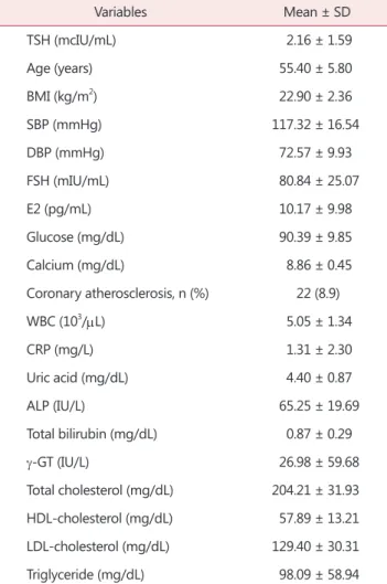
Baseline characteristics of the subjects are displayed on Table 1. The mean age was 55.40 ± 5.80 years old and the mean serum TSH was 2.16 ± 1.59 mcIU/mL. The mean serum FSH and E2 were 80.84 ± 25.07 mIU/mL and 10.17
± 9.98 pg/mL, respectively. The mean BMI was 22.90 ± 2.36 kg/m2, and mean lipid profiles were as follows; TC 204.21 ± 31.93 mg/dL, HDL-C 57.89 ± 13.21 mg/dL, LDL-C 129.40 ± 30.31 mg/dL, and TG 98.09 ± 58.94 mg/
dL. Among 247 postmenopausal women, 22 people (8.90%)
were found to have coronary atherosclerosis.
We were to investigate on association in between serum TSH level and clinical variables using simple correlation analysis. Age, TG were weakly positively correlated (r = 0.148, P = 0.020 and r = 0.169, P = 0.008, respectively), and HDL-C was weakly negatively correlated (r = -0.145, P
= 0.023) with serum TSH (Table 2). Based on these findings, multiple linear regression analysis was done, and it revealed that serum TSH was associated with serum TG (β = 0.146, P = 0.023). Although they were not significant, serum TSH also had tendency to have associations with age and serum HDL-C (Table 3).
To find factors which independently affect on presence of coronary atherosclerosis, stepwise multiple logistic regression Table 1. Baseline characteristics of 247 euthyroid postmenopausal
women
Variables Mean ± SD
TSH (mcIU/mL) 2.16 ± 1.59
Age (years) 55.40 ± 5.80
BMI (kg/m2) 22.90 ± 2.36
SBP (mmHg) 117.32 ± 16.54
DBP (mmHg) 72.57 ± 9.93
FSH (mIU/mL) 80.84 ± 25.07
E2 (pg/mL) 10.17 ± 9.98
Glucose (mg/dL) 90.39 ± 9.85
Calcium (mg/dL) 8.86 ± 0.45
Coronary atherosclerosis, n (%) 22 (8.9)
WBC (103/μL) 5.05 ± 1.34
CRP (mg/L) 1.31 ± 2.30
Uric acid (mg/dL) 4.40 ± 0.87
ALP (IU/L) 65.25 ± 19.69
Total bilirubin (mg/dL) 0.87 ± 0.29
γ-GT (IU/L) 26.98 ± 59.68
Total cholesterol (mg/dL) 204.21 ± 31.93 HDL-cholesterol (mg/dL) 57.89 ± 13.21 LDL-cholesterol (mg/dL) 129.40 ± 30.31
Triglyceride (mg/dL) 98.09 ± 58.94
SD: standard deviation, TSH: thyroid stimulating hormone, BMI:
body mass index, SBP: systolic blood pressure, DBP: diastolic blood pressure, FSH: follicle-stimulating hormone, E2: estradiol, WBC: white blood cell, CRP: C-reactive protein, ALP: alkaline phos- phatase, γ-GT: gamma-glutamyl transferase, HDL: high-density lipoprotein, LDL: low-density lipoprotein
Table 2. Pearson’s correlation coefficients between serum thyroid stimulating hormone and clinical variables
Variables r P value
Age (years) 0.148 0.020
BMI (kg/m2) 0.034 0.596
SBP (mmHg) 0.033 0.602
DBP (mmHg) 0.016 0.806
FSH (mIU/mL) 0.040 0.533
E2 (pg/mL) -0.041 0.521
Glucose (mg/dL) 0.061 0.340
Calcium (mg/dL) -0.079 0.218
WBC (103/μL) 0.015 0.811
CRP (mg/L) -0.043 0.496
Uric acid (mg/dL) -0.003 0.958
ALP (IU/L) 0.060 0.346
Total bilirubin (mg/dL) 0.043 0.500
γ-GT (IU/L) 0.053 0.411
Total cholesterol (mg/dL) -0.028 0.660 HDL-cholesterol (mg/dL) -0.145 0.023 LDL-cholesterol (mg/dL) -0.033 0.650
Triglyceride (mg/dL) 0.169 0.008
BMI: body mass index, SBP: systolic blood pressure, DBP: diastolic blood pressure, FSH: follicle-stimulating hormone, E2: estradiol, WBC: white blood cell, CRP: C-reactive protein, ALP: alkaline phos- phatase, γ-GT: gamma-glutamyl transferase, HDL: high-density lipoprotein, LDL: low-density lipoprotein
Journal of Menopausal Medicine 2016;22:146-153 Seung Joo Chon, et al. TSH, Atherosclerosis in Postmenopausal Woman
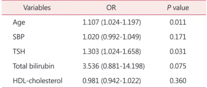
analysis was conducted. Firstly, in simple logistic regression analysis, age, systolic BP (SBP), serum TSH, total bilirubin, and HDL-C were statistically significantly associated with presence of coronary atherosclerosis in postmenopausal
women (Table 4). Based on these data, multiple logistic regression analysis was done. It revealed that increasing age and serum TSH were associated with an increased risk of coronary atherosclerosis in postmenopausal woman (odds ratio [OR] = 1.107 [1.024-1.197], P = 0.011 and OR = 1.303 [1.024-1.658], P = 0.031, respectively). On the other hand, SBP, serum total bilirubin, and HDL-C did not have any association with prevalence of coronary atherosclerosis (Table 5).
Discussion
The findings of the present study indicate that serum TSH levels could be considered as an independent predictive marker of the presence of coronary atherosclerosis in euthyroid postmenopausal women. Also, serum TSH levels within a normal range may be associated with age and some lipid profiles in these women. These findings suggest that increase in age, TG and decrease in HDL-C levels may be related to TSH elevation, and these changes would lead to higher risk of having coronary atherosclerosis in healthy postmenopausal women.
Menopause is a natural aging process causing estrogen deficiency. In premenopausal period, women have a significantly lower risk for CVD compared to postmenopausal women or age matched men.4 However, after menopause, decline in serum estrogen accelerates atherogenic processes, including dyslipidemia, endothelial dysfunction, arterial stiffness, and increase the risk for CVD, which threaten women’s health.5 This disparity between prevalence of CVD Table 3. Multiple regression analysis for serum thyroid stimulating
hormone
Variables β P value β P value
Age (years) 0.128 0.058 0.121 0.060
HDL-cholesterol (mg/dL) -0.118 0.068
Triglyceride (mg/dL) 0.146 0.023
HDL: high-density lipoprotein
Table 5. Multiple logistic regression analysis for the presence of coronary atherosclerosis in postmenopausal women
Variables OR P value
Age 1.107 (1.024-1.197) 0.011
SBP 1.020 (0.992-1.049) 0.171
TSH 1.303 (1.024-1.658) 0.031
Total bilirubin 3.536 (0.881-14.198) 0.075 HDL-cholesterol 0.981 (0.942-1.022) 0.360 OR: odds ratio, SBP: systolic blood pressure, TSH: thyroid stimu- lating hormone, HDL: high-density lipoprotein
Table 4. Simple logistic regression analysis for the presence of coronary atherosclerosis in euthyroid postmenopausal women
Variables OR P value
Age 1.139 (1.061-1.222) < 0.001
BMI 0.962 (0.794-1.166) 0.691
SBP 1.027 (1.002-1.051) 0.032
DBP 1.041 (0.998-1.086) 0.064
FSH 1.002 (0.984-1.019) 0.863
E2 1.012 (0.979-1.047) 0.473
Glucose 0.993 (0.949-1.040) 0.776
TSH 1.379 (1.134-1.721) 0.002
WBC 1.059 (0.765-1.467) 0.729
CRP 0.627 (0.326-1.207) 0.162
Uric acid 1.028 (0.614-1.721) 0.916
ALP 1.001 (0.979-1.024) 0.910
Total bilirubin 4.184 (1.133-15.451) 0.032
γ-GT 1.004 (0.990-1.010) 0.108
Total cholesterol 0.993 (0.979-1.007) 0.300 HDL-cholesterol 0.959 (0.923-0.995) 0.027 LDL-cholesterol 0.989 (0.974-1.004) 0.146 Triglyceride 1.004 (0.998-1.010) 0.179 OR: odds ratio, BMI: body mass index, SBP: systolic blood pres- sure, DBP: diastolic blood pressure, FSH: follicle-stimulating hormone, E2: estradiol, TSH: thyroid stimulating hormone, WBC:
white blood cell, CRP: C-reactive protein, ALP: alkaline phospha- tase, γ-GT: gamma-glutamyl transferase, HDL: high-density lipo- protein, LDL: low-density lipoprotein
Journal of Menopausal Medicine 2016;22:146-153
J MM
in premenopausal and postmenopausal women has been elucidated to the actions of estrogens on cardiovascular system, particularly on vascular endothelium. Previous studies have reported that both endogenous and exogenous estrogens have cardio-protective effects in premenopausal women and women starting estrogen therapy within the first few years after menopause, whereas initiating estrogen therapy many years after menopause may have hazardous effects on cardiovascular systems.6~8 Like this, actions of estrogen depends on the time of initiation of estrogen therapy in postmenopausal women, and it may be due to endothelial injury, changes in vascular estrogen receptor expression, intracellular signaling which could alter the cardiovascular effects of this steroid hormone.9 Although risk and benefit of HRT since the first announcement of Women's Health Initiative study in 2002 were controversial, hormone replacement therapy in young menopausal women could decrease the morbidity and mortality related to coronary heart disease and stroke.10 Also in one previous study, early menopausal women are known to face more physiological problems than the late menopausal women on their quality of life.11
In an aspect of lipid profiles, as women go through menopause, TC, LDL-C, TG, very LDC-C (VLDL-C) increase and HDL-C decreases contributing to form atherogenesis.1 In these women, small dense LDL particles also increase, which would further increase the coronary atherosclerosis in euthyroid postmenopausal women. In this study, in simple logistic regression analysis, increasing HDL-C seemed to have associations with a decrease in presence of coronary atherosclerosis (OR = 0.959 [0.923- 0.995], P = 0.027) in euthyroid postmenopausal women, although it was not valid in multiple logistic regression analysis (OR = 0.981 [0.942-1.022], P = 0.360). These negative effects on women’s lipid profiles increase and positive effects on women’s lipid profiles tend to decrease risks of CVD that screening for CVD is essential and should also include healthy postmenopausal women.
Prevalence of thyroid dysfunction appears to be high in postmenopausal woman.12 Thyroid dysfunction is diagnosed by measuring of the serum thyrotropin which is known as TSH, and it is known as the best and the most reliable test to diagnose thyroid disease.13,14 Thyroid dysfunction
is defined based on clinical signs, symptoms, and serum TSH level.15 In the Study of Women’s Health Across the Nation (SWAN), serum TSH levels outside the normal range were found to be up to 9.4% in women aged from 42 to 52 years old. Also, serum TSH statistically increased in 3.5%, for each 5 years as age increases.16 Elevated serum TSH is common in postmenopausal women, and clinical symptoms caused by abnormal thyroid function could be masked by postmenopausal symptoms. It is known that estrogen influences on serum thyroid hormone by increasing the level of thyroxine binding globulin, with the decrease of its clearance.17 Therefore, routine screening of thyroid function in menopausal period to determine thyroid disease is required.
Thyroid dysfunction is associated with diverse aspects of metabolic abnormalities and conditions such as obesity, hypercholesterolemia, osteoporosis, and CVD.18~20 Many studies have investigated on mechanisms of thyroid hormone on cardiovascular system. Thyroid hormone is known to cause cardiac contractility both directly and indirectly, by increasing peripheral oxygen consumption and substrate requirements.21~23 Triiodothyronine, the active cellular form of thyroid hormone, is known to decrease systemic vascular resistance through vasodilation by direct effecting on vascular smooth muscle cells promoting relaxations.24,25 Thyroid hormone also increases blood volume.26 As systemic vascular resistance decreases, the effective arterial filling volume falls, which causes increase in renin secretion and activation of the angiotensin-aldosterone axis. Then, it eventually stimulates renal sodium reabsorption and finally increases plasma volume.27
Patients with hyperthyroidism mainly have decrease in systemic vascular resistance, and increase in resting heart rate, left ventricular contractility, ejection fraction, and blood volume, which overall increase cardiac output, higher than in normal population.26,28 On the other hand, hemodynamic changes in hypothyroidism are opposite to the ones in hyperthyroidism. The usual signs are bradycardia, decrease in ventricular filling with decrease in cardiac contractility causing low cardiac output.29 Although hypothyroidism presents milder clinical symptoms than hyperthyroidism, it may cause accelerated atherosclerosis and CAD.30 In one population-based cross-sectional study, subclinical
Journal of Menopausal Medicine 2016;22:146-153 Seung Joo Chon, et al. TSH, Atherosclerosis in Postmenopausal Woman
hypothyroidism was found to be a strong risk factor for atherosclerosis and myocardial infarction in postmenopausal women.31
It is clear that abnormal thyroid function is a risk for CVD, but whether thyroid function within the reference range would also be associated with cardiovascular risk has to be evaluated. In one cross-sectional study with 2,205 postmenopausal women, upper normal range of serum TSH (2.48-4.00 mIU/L) showed 1.95 of adjusted OR for prevalence of metabolic syndrome.3 Our study revealed that serum TSH was associated with serum TG (β = 0.146, P
= 0.023). Like our study, one cross-sectional study with 1333 people with euthyroidism, people with serum TSH in upper normal range (2.5-4.5 mIU/L) had higher TG, with 1.7-fold increase in prevalence of metabolic syndrome.32 There were also studies on associations in between serum TSH and BP in euthyroid people. One study concluded that people with higher levels of TSH within a normal range, had more elevated BP than people with lower normal serum TSH range, whereas other study on 1,319 euthyroid people concluded that there is no association in between serum TSH and BP.33~35
Among many factors associated with cardiovascular risk factors, recent studies report on a direct effect of serum TSH in upper normal range to increase in arterial stiffness in euthyroid dialysis patients.36 Serum TSH is known to increase circulating inflammatory molecules and to inhibit nitric oxide production due to oxidative stress.37,38 Like previous study, our study found that increasing age and TSH level were associated with an increased risk of coronary atherosclerosis in euthyroid postmenopausal women, having OR = 1.107 (1.024-1.197), P = 0.011 and OR = 1.303 (1.024- 1.658), P = 0.031, respectively. Since serum TSH is easy to evaluate with blood sampling, measuring serum TSH would be an economical and convenient method of screening high risk group for CVD.
This study bears certain limitations. First, it is a retrospective study based on chart review, that causal associations cannot be demonstrated. Second, the studied subjects are from a single institution with restricted sample size. Korean people are known to have higher intake of iodine than people in other countries. Iodine is a substrate requiring for synthesis of thyroid hormones. It induces
thyroid hormone secretion by inhibiting the proteolytic release of iodothyronines from thyroglobulin, and also reduces thyroid cellularity and vascularity affecting on serum thyroid hormones. Therefore, applying our study’s result to people with other ethnicity having different lifestyles may not be appropriate. Third, we only included people who visited health care center in a tertiary hospital which would not fully present general population of Korean people. Therefore, applying our results to everyone would not be possible, but to certain group of postmenopausal women would be meaningful.
In conclusion, it revealed that significant predictor of serum TSH was serum TG, and increasing age and TSH were found to have associations with an increased risk of coronary atherosclerosis in euthyroid postmenopausal women. As life span is extended in these days, for even in healthy postmenopausal women, screening and risk assessment for CVD and proper management of modifiable risk factors would be required before atherosclerosis develops.
Conflict of Interest
No potential conflict of interest relevant to this article was reported.
References
1. Creatsas G, Christodoulakos G, Lambrinoudaki I. Car- diovascular disease: screening and management of the a-symptomatic high-risk post-menopausal woman.
Maturitas 2005; 52 Suppl 1: S32-7.
2. Klein I, Ojamaa K. Thyroid hormone and the cardiovascular system. N Engl J Med 2001; 344: 501-9.
3. Park HT, Cho GJ, Ahn KH, Shin JH, Hong SC, Kim T, et al. Thyroid stimulating hormone is associated with metabolic syndrome in euthyroid postmenopausal women.
Maturitas 2009; 62: 301-5.
4. Witteman JC, Grobbee DE, Kok FJ, Hofman A, Valkenburg HA. Increased risk of atherosclerosis in women after the menopause. BMJ 1989; 298: 642-4.
5. Yi SS, Hwang E, Baek HK, Kim TH, Lee HH, Jun HS, et al. Application of bioactive natural materials-based
Journal of Menopausal Medicine 2016;22:146-153
J MM
products on five women's diseases. J Menopausal Med 2015;
21: 121-5.
6. Salpeter SR, Walsh JM, Greyber E, Salpeter EE. Brief report: Coronary heart disease events associated with hormone therapy in younger and older women. A meta- analysis. J Gen Intern Med 2006; 21: 363-6.
7. Rossouw JE, Prentice RL, Manson JE, Wu L, Barad D, Barnabei VM, et al. Postmenopausal hormone therapy and risk of cardiovascular disease by age and years since menopause. JAMA 2007; 297: 1465-77.
8. Barton M, Meyer MR, Haas E. Hormone replacement therapy and atherosclerosis in postmenopausal women:
does aging limit therapeutic benefits? Arterioscler Thromb Vasc Biol 2007; 27: 1669-72.
9. Dworatzek E, Mahmoodzadeh S, Schubert C, Westphal C, Leber J, Kusch A, et al. Sex differences in exercise-induced physiological myocardial hypertrophy are modulated by oestrogen receptor beta. Cardiovasc Res 2014; 102: 418-28.
10. Koo YH, Song YJ, Na YJ. Mortality associated with hormone replacement therapy in postmenopausal women. J Korean Soc Menopause 2012; 18: 133-8.
11. Ahmed K, Jahan P, Nadia I, Ahmed F, Abdullah Al E.
Assessment of menopausal symptoms among early and late menopausal midlife Bangladeshi women and their impact on the quality of life. J Menopausal Med 2016; 22: 39-46.
12. Lambrinoudaki I, Armeni E, Rizos D, Georgiopoulos G, Kazani M, Alexandrou A, et al. High normal thyroid- stimulating hormone is associated with arterial stiffness in healthy postmenopausal women. J Hypertens 2012; 30:
592-9.
13. Ladenson PW, Singer PA, Ain KB, Bagchi N, Bigos ST, Levy EG, et al. American thyroid association guidelines for detection of thyroid dysfunction. Arch Intern Med 2000;
160: 1573-5.
14. Baskin HJ, Cobin RH, Duick DS, Gharib H, Guttler RB, Kaplan MM, et al. American association of clinical endocrinologists medical guidelines for clinical practice for the evaluation and treatment of hyperthyroidism and hypothyroidism. Endocr Pract 2002; 8: 457-69.
15. González-Rodríguez LA, Felici-Giovanini ME, Haddock L. Thyroid dysfunction in an adult female population:
A population-based study of Latin American Vertebral Osteoporosis Study (LAVOS) - Puerto Rico site. P R Health Sci J 2013; 32: 57-62.
16. Sowers M, Luborsky J, Perdue C, Araujo KL, Goldman MB, Harlow SD. Thyroid stimulating hormone (TSH) concentrations and menopausal status in women at the mid-life: SWAN. Clin Endocrinol (Oxf) 2003; 58: 340-7.
17. Redmond GP. Thyroid dysfunction and women's
reproductive health. Thyroid 2004; 14 Suppl 1: S5-15.
18. Rotondi M, Magri F, Chiovato L. Thyroid and obesity: not a one-way interaction. J Clin Endocrinol Metab 2011; 96:
344-6.
19. Walsh JP, Bremner AP, Bulsara MK, O'Leary P, Leedman PJ, Feddema P, et al. Subclinical thyroid dysfunction as a risk factor for cardiovascular disease. Arch Intern Med 2005; 165: 2467-72.
20. Razvi S, Weaver JU, Vanderpump MP, Pearce SH. The incidence of ischemic heart disease and mortality in people with subclinical hypothyroidism: reanalysis of the Whickham Survey cohort. J Clin Endocrinol Metab 2010;
95: 1734-40.
21. Klein I. Thyroid hormone and the cardiovascular system.
Am J Med 1990; 88: 631-7.
22. Dillmann WH. Biochemical basis of thyroid hormone action in the heart. Am J Med 1990; 88: 626-30.
23. Polikar R, Burger AG, Scherrer U, Nicod P. The thyroid and the heart. Circulation 1993; 87: 1435-41.
24. Park KW, Dai HB, Ojamaa K, Lowenstein E, Klein I, Sellke FW. The direct vasomotor effect of thyroid hormones on rat skeletal muscle resistance arteries. Anesth Analg 1997; 85:
734-8.
25. Ojamaa K, Klemperer JD, Klein I. Acute effects of thyroid hormone on vascular smooth muscle. Thyroid 1996; 6: 505- 12.
26. Braverman LE, Utiger RD, editors. Werner & Ingbar's the thyroid: a fundamental and clinical text. 8th ed.
Philadelphia, PA: Lippincott Williams & Wilkins; 2000.
27. Resnick LM, Laragh JH. PLasma renin activity in syndromes of thyroid hormone excess and deficiency. Life Sci 1982; 30: 585-6.
28. Kiss E, Jakab G, Kranias EG, Edes I. Thyroid hormone- induced alterations in phospholamban protein expression.
Regulatory effects on sarcoplasmic reticulum Ca2+
transport and myocardial relaxation. Circ Res 1994; 75:
245-51.
29. Crowley WF, Jr., Ridgway EC, Bough EW, Francis GS, Daniels GH, Kourides IA, et al. Noninvasive evaluation of cardiac function in hypothyroidism. Response to gradual thyroxine replacement. N Engl J Med 1977; 296: 1-6.
30. Bengel FM, Nekolla SG, Ibrahim T, Weniger C, Ziegler SI, Schwaiger M. Effect of thyroid hormones on cardiac function, geometry, and oxidative metabolism assessed noninvasively by positron emission tomography and magnetic resonance imaging. J Clin Endocrinol Metab 2000;
85: 1822-7.
31. Hak AE, Pols HA, Visser TJ, Drexhage HA, Hofman A, Witteman JC. Subclinical hypothyroidism is an independent
Journal of Menopausal Medicine 2016;22:146-153 Seung Joo Chon, et al. TSH, Atherosclerosis in Postmenopausal Woman
risk factor for atherosclerosis and myocardial infarction in elderly women: the Rotterdam Study. Ann Intern Med 2000; 132: 270-8.
32. Ruhla S, Weickert MO, Arafat AM, Osterhoff M, Isken F, Spranger J, et al. A high normal TSH is associated with the metabolic syndrome. Clin Endocrinol (Oxf) 2010; 72:
696-701.
33. Gumieniak O, Perlstein TS, Hopkins PN, Brown NJ, Murphey LJ, Jeunemaitre X, et al. Thyroid function and blood pressure homeostasis in euthyroid subjects. J Clin Endocrinol Metab 2004; 89: 3455-61.
34. Asvold BO, Bjoro T, Nilsen TI, Vatten LJ. Association between blood pressure and serum thyroid-stimulating hormone concentration within the reference range: a population-based study. J Clin Endocrinol Metab 2007; 92:
841-5.
35. Liu D, Jiang F, Shan Z, Wang B, Wang J, Lai Y, et al. A
cross-sectional survey of relationship between serum TSH level and blood pressure. J Hum Hypertens 2010; 24: 134- 8.
36. Tatar E, Sezis Demirci M, Kircelli F, Gungor O, Yaprak M, Asci G, et al. The association between thyroid hormones and arterial stiffness in peritoneal dialysis patients. Int Urol Nephrol 2012; 44: 601-6.
37. Desideri G, Bocale R, Milardi D, Ghiadoni L, Grassi D, Necozione S, et al. Enhanced proatherogenic inflammation after recombinant human TSH administration in patients monitored for thyroid cancer remnant. Clin Endocrinol (Oxf) 2009; 71: 429-33.
38. Dardano A, Ghiadoni L, Plantinga Y, Caraccio N, Bemi A, Duranti E, et al. Recombinant human thyrotropin reduces endothelium-dependent vasodilation in patients monitored for differentiated thyroid carcinoma. J Clin Endocrinol Metab 2006; 91: 4175-8.

