Department of Oral & Maxillofacial Surgery, School of Dentistry, Chosun University
국문초록
본 연구의 목적은 상악동 점막 거상술에 대한 동물과 사람에 대한 연구의 차이와 성공률에 대한 비교 및 향후 연구의 전망에 대해 논하고자 한다.
1997년부터 2006년까지 임프란트 식립을 위해 시행된 상악동저 점막 거상술에 대해 보고된 문헌을 Medline을 통해 검색하 였다. 검색시 사용한 key word는 sinus augmentation, sinus graft, sinus lift, sinus elevation, animal이었다.
상악동 점막 거상술에 관한 동물실험은 주로 양과 미니돼지가 실험대상이었으며, 골-임프란트 접촉률을 평가하는 논문이 대 부분이었고, 사람을 대상으로 하는 논문은 주로 임상적 성공률(survival rate)에 관한 논문이 주를 이루었다.
상악동 점막 거상술의 동물실험시 임프란트의 식립시기, 희생 시기, 조직학적 평가 기준 등에 관한 근거를 바탕으로 시행하 여야 할 것으로 보이며, 양, 돼지, 개 등을 이용한 상악동 점막 거상술의 실험은 새로운 임프란트나 골대체제 등에 관한 연구에 있어 임상실험 전에 중요한 과정으로 여겨진다.
종 설
Review of Human and Animal Studies on
Maxillary Sinus Grafting for Implant Placement
Yeol-Su Park, Su-Gwan Kim, Ji-Su Oh, Hak-Kyun Kim, Seong-Yong Moon
임프란트 식립을 위한 상악동 점막 거상술의 문헌고찰:
사람과 동물 연구
박열수, 김수관, 오지수, 김학균, 문성용 조선대학교 치과대학 구강악안면외과학교실
Methods to overcome insufficient bone due to poor bone quality, the pneumatization of a maxillary sinus and other anatomical limitations of implant placement in the maxillary molar area include the augmentation of residual alveolar bone using bone grafting with onlay grafts1or veneer grafting and bone grafting methods involving a Le Fort I osteotomy.2Of these, the maxillary sinus lift is a simple procedure, has fewer side effects than other techniques, and can be widely applied. After it was introduced by Tatum3and Bonye,4 Tatum3reported a procedure including implant place- ment and simultaneous maxillary sinus bone grafting using a lateral wall approach. Despite numerous sub- sequent studies examining various clinical techniques and bone graft materials, it is still recognized as the most reliable method. Both animal studies and clinical studies on patients have examined the maxillary sinus lifting. In this study, the success rate and the differ- ences between animal and human studies from the lit- erature study were compared and then the future per- spectives of studies on the maxillary sinus lifting were discussed.
Papers published between 1997 and 2006 on the maxillary sinus grafting for implant placement were surveyed through the Medline. The keywords used for this survey were sinus augmentation, sinus graft, sinus lift, sinus elevation, and animal.
In the literature study, 38 papers dealt with animal experiments on maxillary sinus floor lifting were found. Among the animal studies, sheep and minipig were used mostly as the experimental animal, in terms of the evaluation of the bone-implant contact ratio (BIC). However, the most clinical studies on humans were reported the clinical survival rate. The results of the literature review are presented in Tables I~III.
Ⅲ
종설
Table Ⅰ. List of recent animal studies on maxillary sinus augmentation R
Reeffeerreenncceess AAnniimmaall NNoo.. MMaatteerriiaallss RReessuullttss
Kirker-Head et al. (1997)5 goat 6 rhBMP-2.ACS no complications
Hass et al. (1998)6 sheep 27 Bio-Oss, autologous bone Autologous bone had significantly more BIC than the non-graft group.
Margolin et al. (1998)7 chimpanzee collagen matrix OP-1 Bone formation was superior to that in the collagen matrix-alone group.
Terheydenet al. (1999)8 minipig 5 Bio-Oss, OP-1 The combination group (BIC 80%) was better than the Bio-Oss group (36%).
Haas et al. (2002)9 sheep 36 DFDB The DFDB group required a slightly higher force than the non-augmented group in the pullout test.
Haas et al. (2002)10 sheep 27 HA autogenous bone HA & autogenous bone group had more BIC than the non-grafted group.
Fu¨rst et al. (2003)11 minipig 12 HA, PRP PRP combined with HA was not supe- rior to HA alone in terms of BIC.
Haas et al. (2003)12 sheep 27 HA, autogenous bone The HA group had significantly more BIC than the non-grafted group.
Jakse et al. (2003)13 sheep 12 autogenous bone The PRP group did not differ signifi- cantly in terms of BIC.
Butterfield et al. (2005)14 rabbit 12 autogenous bone pQ-CT, fail
*BIC: bone-implant contact; HA: hydroxyapatite; PRP: platelet-rich plasma; DFDB: demineralized freeze-dried
Blomqvist et al. (1996)15 49 171 iliac crest Nobel Biocare AB 82.5 Khoury (1999)16 216 467 autograft IMZ, Nobel Biocare, 10-15 94
Bra˚nemark II Frialit-2
Johansson et al. (1999)17 39 131 iliac graft Nobel Biocare AB 4 10, 13, 15, 18 95
Chanavaz (2000)18 982 Autograft, demineralized 99.34, 95.78
Artzi et al. (2002)19 10 36 Bio-Oss Steri-Oss, Spline 3.7, 4.5 10-15 Success
Sartori et al. (2003)20 1 Bio-Oss IMZ 86.7
Andreana et al. (2004)21 6 14 Capset Paragon 3.75-4.7 10 No failure Hatano et al. (2004)22 191 361 Bio-Oss, Nobel Biocare 3.75-6 8.5-15 94
autogenous bone
Engelke & 6 21 Cerasorb Friadent, Xive 3.4-4.5 13-15 95.2
Capobianco (2005)23
Simunek et al. (2005)24 24 45 algipore Friatlit-2 3.8 13 97.8
*A: animal study; Imp: Implant; D: diameter; L: Length.
Yeol-Su Park et al: Review of Human and Animal Studies on Maxillary Sinus Grafting for Implant Placement. Implantology 2009
종설
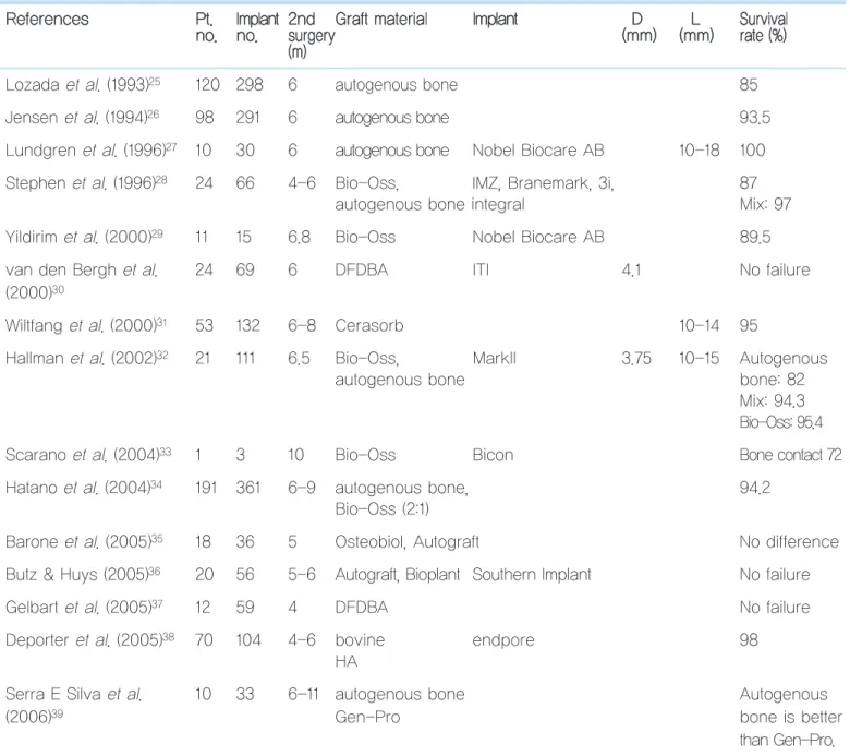
Table III. Human studies of maxillary sinus augmentation (two stage) R
Reeffeerreenncceess PPtt.. IImmppllaanntt 22nndd GGrraafftt mmaatteerriiaall IImmppllaanntt DD LL SSuurrvviivvaall n
noo.. nnoo.. ssuurrggeerryy ((mmmm)) ((mmmm)) rraattee ((%%)) ((mm))
Lozada et al. (1993)25 120 298 6 autogenous bone 85
Jensen et al. (1994)26 98 291 6 autogenous bone 93.5
Lundgren et al. (1996)27 10 30 6 autogenous bone Nobel Biocare AB 10-18 100 Stephen et al. (1996)28 24 66 4-6 Bio-Oss, IMZ, Branemark, 3i, 87
autogenous bone integral Mix: 97
Yildirim et al. (2000)29 11 15 6.8 Bio-Oss Nobel Biocare AB 89.5
van den Bergh et al. 24 69 6 DFDBA ITI 4.1 No failure
(2000)30
Wiltfang et al. (2000)31 53 132 6-8 Cerasorb 10-14 95
Hallman et al. (2002)32 21 111 6.5 Bio-Oss, MarkII 3.75 10-15 Autogenous
autogenous bone bone: 82
Mix: 94.3 Bio-Oss: 95.4
Scarano et al. (2004)33 1 3 10 Bio-Oss Bicon Bone contact 72
Hatano et al. (2004)34 191 361 6-9 autogenous bone, 94.2
Bio-Oss (2:1)
Barone et al. (2005)35 18 36 5 Osteobiol, Autograft No difference
Butz & Huys (2005)36 20 56 5-6 Autograft, Bioplant Southern Implant No failure
Gelbart et al. (2005)37 12 59 4 DFDBA No failure
Deporter et al. (2005)38 70 104 4-6 bovine endpore 98
HA
Serra E Silva et al. 10 33 6-11 autogenous bone Autogenous
(2006)39 Gen-Pro bone is better
than Gen-Pro.
*A: animal study; DFDBA: demineralized freeze-dried bone allograft; m: month; D: diameter; L: length
Bone resorption and regeneration are continuously ongoing in the skeletal system due to the interaction of osteoblasts and osteoclasts. When a bone defect devel- ops following trauma, surgery, or bone grafting, bone regeneration and resorption occur via this interaction of osteoblasts and osteoclasts.
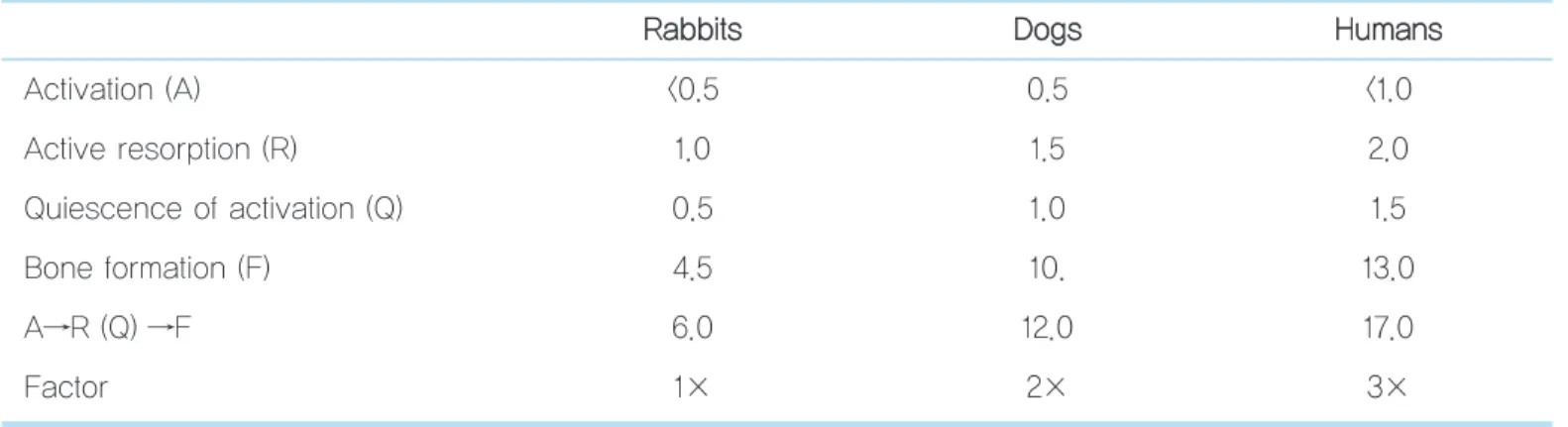
Bone remodeling involves the activation (A) of osseous precursor cells, leading to increased active resorption (R), quiescence or reversal of the activation (Q), and bone formation (F). This series of processes is referred to as the bone remodeling cycle. Each species, including humans, has a different cycle period. The cycle period is approximately 6 weeks in rabbits, 12 weeks in dogs, and 17 weeks in humans. The various stages are summarized in Table IV for rabbits, dogs, and humans.
bits, 1 week in dogs, and 1-2 weeks in humans. In the bone remodeling process, the bone formation period varies the most across species. In rabbits, dogs, and humans, it takes approximately 4.5, 10, and 13 weeks, respectively, and the time required for bone remodel- ing cycle is proportional to the size of animal.
Assigning rabbits a relative size of 1, dogs require approximately twice the relative corresponding period, and humans require three times the corresponding period.40
In rabbits, woven bone appears on the implant sur- face, which becomes sufficiently dense within approxi- mately 6 weeks; bone remodeling occurs, and it can sustain normal loading. Little quantitative data are available on the early healing process in humans, although based on the bone remodeling cycle period, it is estimated that the formation of the primary callus is similar to that in rabbits. Nevertheless, the maturation
Table IV. Parameters of the bone remodeling cycle in rabbits, dogs, and humans R
Raabbbbiittss DDooggss HHuummaannss
Activation (A) <0.5 0.5 <1.0
Active resorption (R) 1.0 1.5 2.0
Quiescence of activation (Q) 0.5 1.0 1.5
종설
process in humans bone modeling takes approximately three times longer (approximately 18 weeks) compared to rabbits (Table V).
In animal experiments, it is difficult to evaluate the implant success rate, and the BIC rate is primarily measured, although radiological, histological, and his- tomorphometric analyses are also performed. Many reports have evaluated the bone formation rate with bone grafts using xenogenic or synthetic bone, and numerous recent reports have evaluated the use of platelet concentrated plasma. Regarding the time of animal death, minipigs are killed after 3, 6, 12, and 26 weeks,8,11 rabbits after 2, 3, and 8 weeks,14 and sheep after 12, 16, and 26 weeks.6,9,10,12,13
The initial studies of implants involved animal experiments. Linder and Lundskog implanted a titani-
the clinical success rate of implants, usually based on long-term follow-up. Boyne and James4 performed the maxillary sinus floor lift procedure using autoge- nous bone; the implants were placed 6 months later, and they reported the first clinical results after a fol- low-up of 4 years.4In 1987, Misch43grafted a mixture of decalcified bone, blood, and tricalcium phosphate in the maxillary sinus and obtained a 98% success rate in 179 cases. In 1996, Blomqvist et al.15 studied the suc- cess rate of immediate implant placement in 49 patients with 2 to 4 mm of residual bone and reported an 82% success rate with 314 implants after the 32 months follow-up. Unlike animal studies, many clini- cal studies have investigated the use of autogenous bones in maxillary sinus floor elevation. Autogenous bone is the best graft material for bone defects, and Table V. Differences in the bone remodeling process in rabbits and humans
C
Chhaannggee iinn tthhee bboonnee RRaabbbbiittss HHuummaannss ((33××))
First step: Callus formation (weeks) 2 6
Second step: The deposition of lamellar bone (weeks) 6 18
Third step: Interface remodeling (weeks) 6 18
Fourth step: Bone maturation (weeks) 18 54
Yeol-Su Park et al: Review of Human and Animal Studies on Maxillary Sinus Grafting for Implant Placement. Implantology 2009
year follow-up, 98.9% of the implants were retained for a 90.3% success rate.
Most papers6,9,10,12on animal study had significantly more BIC than the non-grafted group.
Animal studies of maxillary sinus floor elevation appear to be standardized in terms of the time of implant placement, time of animal death, and criteria used for the histological evaluation. Experimental studies of maxillary sinus floor elevation in sheep, pigs, and dogs are important precursors of clinical experiments.
1. Marx RE, Wong ME. A technique for the compression and cartilage of autogenous bone bone during bone grafting procedures. J Oral Maxillofac Surg 1987; 45: 988-989.
2. Weingart D, Joos U, Hürzeler MB, et al. Restoration of maxillary resid- ual ridge atrophy using Le Fort I osteotomy with simultaneous endosseous implant placement: Technical report. Int J Oral Maxillofac Implants 1992; 7: 529-535.
3. Tatum H Jr. Maxillary and sinus implant reconstructions. Dent Clin North Am 1986; 30: 207-209.
4. Boyne PJ, James RA. Grafting of the maxillary sinus floor with autoge-
study between recombinant human osteogenic protein-1 and natural bone mineral. J Periodontol 1998; 69: 911-919.
8. Terheyden H, Jepsen S, Möller B, et al. Sinus floor augmentation with simultaneous placement of dental implants using a combination of deproteinized bone xenografts and recombinant human osteogenic pro- tein-1. A histometric study in miniature pigs. Clin Oral Implants Res 1999; 10: 510-521.
9. Haas R, Haidvogl D, D?rtbudak O, et al. Freeze-dried bone for maxillary sinus augmentation in sheep. Part II: biomechanical findings. Clin Oral Implants Res 2002; 13: 581-586.
10. Haas R, Baron M, Donath K, et al. Porous hydroxyapatite for grafting the maxillary sinus: a comparative histomorphometric study in sheep.
Int J Oral Maxillofac Implant 2002; 17: 337-346.
11. Fürst G, Gruber R, Tangl S, et al. Sinus grafting with autogenous platelet-rich plasma and bovine hydroxyapatitie. A histomorphometric study in minipigs. Clin Oral Implnats Res 2003; 14: 500-508.
12. Haas R, Baron M, Zechner W, et al. Porous hydroxyapatite for grafting the maxillary sinus in sheep: comparative pullout study of dental implants; Int J Oral Maxillofac Implants 2003; 18: 691-696.
13. Jakse N, Tangl S, Gilli R, et al. Influence of PRP on autogenous sinus grafts. An experimental study on sheep. Clin Oral Implants Res 2003;
14: 578-583.
14. Butterfield KJ, Bennett J, Gronowicz G, et al. Effect of platelet-rich plas- ma with autogenous bone graft for maxillary sinus augmentation in a rabbit model. J Oral Maxillofac Surg 2005; 63: 370-376.
15. Blomqvist JE, Alberius P, Isaksson S. Retrospective analysis of one- stage maxillary sinus augmentation with endosseous implants. Int J Oral Maxillofac Implants 1996; 11: 512-521.
16. Khoury F. Augmentation of the sinus floor with mandibular bone block and simultaneous implantation: A 6-year clinical investigation. Int J
종설
18. Chanavaz M. Sinus graft procedures and implant dentistry: A review of 21 years of surgical experience(1979-2000). Impalnt Dentistry 2000; 9:
197-203.
19. Artzi Z, Nemcovsky CE, Dayan D. Bovine-HA spongiosa blocks and immediate implant placement in sinus augmentation procedures.
Histopathological and histomorphometric observations on different his- tological stainings in 10 consecutive patients. Clin Oral Implants Res 2002; 13: 420-427.
20. Sartori S, Silvestri M, Forni F, et al. Ten-year follow-up in a maxillary sinus augmentation using anorganic bovine bone(Bio-Oss). A case report with histomorphometric evaluation. Clin Oral Implants Res 2003;
14: 369-372.
21. Andreana S, Cornelini R, Edsberg LE, et al. Maxillary sinus elevation for implant placement using calcium sulfate with and without DFDBA: six cases. Implant Dent 2004; 13: 270-277.
22. Hatano N, Shimizu Y, Ooya K. A clinical long-term radiographic evalu- ation of graft height changes after maxillary sinus floor augmentation with a 2:1 autogenous bone/xenograft mixture and simultaneous place- ment of dental implants. Clin Oral Implants Res 2004; 15: 339-345.
23. Engelke W, Capobianco M. Flapless sinus floor augmentation using endoscopy combined with CT scan-designed surgical templates:
method and report of 6 consecutive cases. Int J Oral Maxillofac Implants 2005; 20: 891-897.
24. Simunek A, Cierny M, Kopecka D, et al. The sinus lift with phycogenic bone substitute. Clin Oral Implants Res 2005; 16: 342-348.
25. Lozada JL, Emanuelli S, James RA, et al. Root-form implants placed in subantral grafted sites. CDA J 1993; 21: 31-35.
26. Jensen J, Sindet-Pedersen S, Oliver AJ. Varying treatment strategies for reconstruction of maxillary atrophy with implants: results in 98 patients.
J Oral Maxillofac Surg 1994; 52: 210-216.
27. Lundgren S, Moy P, Johansson C, et al. Augmentation of the maxillary
30. van den Bergh JP, ten Bruggenkate CM, Krekeler G, et al. Maxillary sinusfloor elevation and grafting with human demineralized freeze dried bone. Clin Oral Implants Res 2000; 11: 487-493.
31. Wiltfang J, Schultze-Mosgau S, Merten HA, et al. Endoscopic and ultra- sonographic evaluation of the maxillary sinus after combined sinus floor augmentation and implant insertion. Oral Surg Oral Med Oral Pathol Oral Radiol Endod 2000; 89: 288-291.
32. Hallman M, Sennerby L, Lundgren S. A clinical and histologic evalua- tion of implant integration in the posterior maxilla after sinus floor aug- mentation with autogenous bone, bovine hydroxyapatite, or a 20:80 mixture. Int J Oral Maxillofac Implant 2002; 17: 635-643.
33. Scarano A, Pecora G, Piattelli M, et al. Osseointegration in a sinus aug- mented with bovine porous bone mineral: Histological results in an implant retrieved 4 years after insertion. A case report. J Periodontol 2004; 75: 1161-1166.
34. Hatano N, Shimizu Y, Ooya K. A clinical long-term radiographic evalu- ation of graft height changes after maxillary sinus floor augmentation with 2:1 autogenous bone/xenograft mixture and simultaneous place- ment of dental implants. Clin Oral Implants Res 2004; 15: 339-345.
35. Barone A, Crespi R, Aldini NN, et al. Maxillary sinus augmentation: his- tologic and histomorphometric analysis. Int J Oral Maxillofac Implants 2005; 20: 519-525.
36. Butz SJ, Huys LW. Long-term success of sinus augmentation using a synthetic alloplast: a 20 patients, 7 years clinical report. Implant Dent 2005; 14: 36-42.
37. Gelbart M, Friedman R, Burlui V, et al. Maxillary sinus augmentation using a peptide-modified graft material in three mixtures: A prospective human case series of histologic and histomorphometric results. Implant Dent 2005; 14: 185-193.
38. Deporter DA, Caudry S, Kermalli J, et al. Further Data on the Predictability of the indirect sinus elevation procedure used with short,
43. Misch CE. Maxillary sinus augmentation for endosteal implants:
Organized alternative treatment plan. Int J Oral Implantol 1987; 4: 49- 58.
44. Wheeler SL. Sinus augmentation for dental implants: the use of alloplas- tic materials. J Oral Maxillofac Surg 1997; 55: 1287-1298.
45. Hürzeler MB, Kirsch A, Ackermann KL, et al. Reconstruction of the severly resorbed maxilla with dental implants with augmented maxillary sinus: A 5 year clinical investigation. Int J Oral Maxillofac implants 1996; 11: 466-475.