Tenebrio molitor (Mealworm) Extract Improves Insulin Sensitivity and Alleviates Hyperglycemia in C57BL/Ksj-db/db Mice
Seon Young Kim, Jae Eun Park and Ji Sook Han*
Department of Food Science and Nutrition, Pusan National University, Busan 46241, Korea Received April 24, 2019 /Revised May 10, 2019 /Accepted May 13, 2019
Diabetes is one of the serious chronic metabolic diseases caused by Westernized eating habits, and the goal of diabetes treatment is to keep blood glucose at a normal level and prevent diabetic complications. This study was designed to investigate the anti-diabetic effects of a mealworm (Tenebrio
molitor larva) extract (MWE) on hyperglycemia in an animal model with type 2 diabetes. DiabeticC57BL/Ksj-db/db mice were divided into three groups: diabetic control, rosiglitazone, and MWE. The mice supplemented with MWE showed significantly lower blood levels of glucose and glycosylated hemoglobin when compared with the diabetic control mice. The homeostatic index of insulin resist- ance was significantly lower in mice supplemented with MWE than in diabetic control mice. MWE supplementation significantly stimulated the phosphorylation of insulin receptor substrate-1 and Akt, and activation of phosphatidylinositol 3-kinase in insulin signaling pathway of skeletal muscles.
Eventually, MWE increased the expression of the plasma membrane glucose transporter 4 (GLUT4) via PI3K/Akt activation. These findings demonstrate that the increase in plasma membrane GLUT4 expression by MWE promoted the uptake of blood glucose into cells and relieved hyperglycemia in skeletal muscles of diabetic C57BL/Ksj-db/db mice. Therefore, mealworms are expected to prove use- ful for the prevention and treatment of diabetes, and further studies are needed to improve type 2 diabetes in the future.
Key words : AMPK pathway, db/db-mice, hyperglycemia, insulin sensitivity, mealworm
*Corresponding author
*Tel : +82-51-510-2836, Fax : +82-51-583-3648
*E-mail : [email protected]
This is an Open-Access article distributed under the terms of the Creative Commons Attribution Non-Commercial License (http://creativecommons.org/licenses/by-nc/3.0) which permits unrestricted non-commercial use, distribution, and reproduction in any medium, provided the original work is properly cited.
Journal of Life Science 2019 Vol. 29. No. 5. 570~579 DOI : https://doi.org/10.5352/JLS.2019.29.5.570
Introduction
Diabetes is increasing worldwide, affecting about 415 mil- lion people to date. Among these, patients with type 2 dia- betes specifically account for more than 90% of all diabetic patients. Type 2 diabetes is characterized by high blood glu- cose levels. Hyperglycemia is caused by an insufficient secre- tion of insulin by the pancreas [4] and insulin resistance mainly in skeletal muscles and adipose tissue [31]. Insulin resistance is a pathological condition in which the insulin sensitivity of tissues is reduced, and this condition is ob- served with inappropriate functioning of insulin [14].
Reduced insulin sensitivity is caused by a combination of genetic and environmental factors, and its pathophysiology involves complex signaling pathway that is activated by the insulin receptor [14, 28, 31].
Insulin sensitivity is associated with the activation of PI3K/Akt signaling pathway. In this pathway, insulin binds to the insulin receptor and activates the insulin receptor ty- rosine kinase, leading to phosphorylation in insulin receptor substrate-1
Tyr612(IRS-1
Tyr612). Phosphorylated IRS-1
Tyr612acti- vates phosphatidylinositol-3-kinase (PI3K) by binding to the SH2 (Src-homology-2) domain of the regulatory subunit p85 of PI3K. Continually, activated PI3K phosphorylates AKT
Ser473by converting phosphatidylinositol bisphosphate (PIP2) to phosphatidylinositol triphosphate (PIP3). Phosphorylation of Akt
Ser473enhances the translocation of glucose transporter 4 (GLUT4) from the cytoplasm to the plasma membrane in skeletal muscles [3, 11, 34]. Eventually, the activation of PI3K/Akt pathway, via translocation of GLUT4, absorbs blood glucose into the cell and thereby improves hyper- glycemia.
Oral antidiabetic medications have been used to increase
insulin sensitivity and alleviate hyperglycemia. Consider-
ably, thiazolinedione drugs (TZDs) are widely used to treat
type 2 diabetes, which further increase the tissue sensitivity
to insulin and delay the progression of diabetes [25]; how-
ever, these are synthetic drugs and may present undesirable
side-effects such as hypoglycemia, poor control of post-
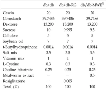
Table 1. Ingredient composition of the experimental diets sup- plemented to mice (%) db/db db/db-RG db/db-MWE1) Casein
Cornstarch Dextrose Sucrose Cellulose Soybean oil t-Butylhydroquinone Salt mix
Vitamin mix L-Cystine Choline bitartrate Mealworm extract Rosiglitazone Total (%)
20 39.7486
13.200 10
5 7 0.0014
3.5 1 0.3 0.25
-
- 100
20 39.7486
13.200 9.995
5 7 0.0014
3.5 1 0.3 0.25
- 0.005
100
20 39.7486
13.200 9.5
5 7 0.0014
3.5 1 0.3 0.25
0.5
- 100
1)db/db (diabetes mellitus control), C57BL/KsJ-db/db mice re- ceiving AIN-93G diet; db/db-RG, C57BL/KsJ-db/db mice re- ceiving AIN-93G diet supplemented with rosiglitazone (0.005 g/100 g diet); db/db-MWE, C57BL/KsJ-db/db mice receiving AIN-93G diet supplemented with mealworm extract (0.5 g/
100 g diet). MWE: mealworm extract.
prandial blood glucose level, and weight gain [13], which are of great concern in the treatment of diabetes. Thus, to reduce these side effects and effectively treat diabetes, the therapeutic use of natural products for treating diabetes has gained immense attention worldwide [33].
For centuries, people have consumed insects as food. The insects have abundant protein, fat, vitamins, and minerals [27], and have been used to treat various diseases [6]. As a representative edible insect, mealworm has been eaten as a traditional food in several countries. Mealworm is the larv- al form of the mealworm beetle, Tenebrio molitor, and it in- habits in stored grain [29]. Similar to other insects, meal- worm is rich in nutrients, and in particular, contains a large amount of unsaturated fatty acids such as linoleic acid (C18:2) and oleic acid (C18:1) [26]. Thus, when consumed as a dietary supplement, mealworm prevents gout and hy- peruricemia [6]. In addition, the beetle species belonging T.
molitor have been used in traditional medicine to curb hep-
atofibrosis and diabetes [32]; however, no study has inves- tigated the use of a mealworm to improve insulin sensitivity in type 2 diabetic mice. Therefore, we designed this study to examine the efficacy of the mealworm extract (MWE) on insulin sensitivity and hyperglycemia in C57BL/Ksj-db/db type 2 diabetic mice.
Materials and Methods
Preparation of materials
Mealworm was purchased from Yongin (Yongin, Gyeong- gi-do, Korea). It was washed with distilled water, and then dried and ground into a powder (Shinhan Science & Tech- nology Co., Kyunggi, Korea). For the extract, mealworm powder was appropriately dissolved in 80% ethanol and fil- tered using Whatman No. 1 filter paper at room temperature overnight. This process was repeated thrice. Later, the meal- worm extract was concentrated in a rotary evaporator and freeze-dried into a powder (BUCHI Co., Flawil, Switzerland).
The mealworm extract powder was preserved in a deep freezer (Samwon Freezing Engineering Co., Busan, Korea).
Animals and feeding diets
Male C57BL/KsJ-db/db mice were purchased from JOONG AH BIO (Suwon-si, Gyeonggi-do, Korea) and db/db mice were fed a pelletized commercial chow diet for 2 weeks after arrival. Before the experiment, the mice were randomly divided into three groups, n=8 per group. The db/db-control
mice group was supplemented with a standard semi-syn- thetic diet (AIN-93G), whereas the mice in the other two groups were fed a standard AIN-39G diet supplemented with either Rosiglitazone (RG, 0.005%, w/w, Sigma, St. Louis, MO, USA) or MWE (0.5%, w/w; Table 1). All mice were placed separately in a cage with controlled light (12 hr light/12 hr dark) and temperature conditions. Thereafter, the mice were given free access to food and water. At the end of the 6-week experiment, mice were anesthetized with ether after a 12 hr fasting period, and blood samples were col- lected from the inferior vena cava to determine the levels of plasma biomarker. All animal experiments were per- formed in accordance with guidelines of the Pusan National University for the care and use of laboratory animals (PNU-2018-1826).
Blood glucose and glycosylated hemoglobin levels
Every week, the glucose concentration in the venous
blood from the mouse-tail vein was measured after a 12 hr
fasting period using a glucometer (Roche Diagnostics
GmbH, Mannheim, Germany). Anticoagulated whole-blood
samples were hemolyzed and the concentration of glycosy-
lated hemoglobin (HbA1c) was measured by using im-
munoturbidimetry.
Plasma insulin level
All blood samples from the inferior vena cava were col- lected into heparin-coated tubes. After centrifugation at 1,000× g for 15 min at 4℃, the plasma was cautiously re- moved from the sample. The levels of plasma insulin were measured via a radioimmunoassay with an enzyme-linked immunosorbent assay ELISA kit (Linco Research, Inc., Bill- erica, MA, USA).
Homeostatic index of insulin resistance
Homeostatic index of insulin resistance (HOMA-IR) was measured as an alternative method to determine insulin sensitivity. HOMA-IR was calculated using the homeostasis model with the following equation (Eq. (1)):
HOMA-IR = {Fasting glucose (mmol/L) × fasting insulin (IU/Ml)}/22.51 (1)
Intraperitoneal glucose tolerance tests
At 5 weeks of MWE treatment, the intraperitoneal glucose tolerance test (IPGTT) was performed on all db/db mice af- ter a 12 hr overnight fast. The mice were injected intra- peritoneally with glucose (0.5 g/kg BW), and later their blood glucose levels were measured at 0, 30, 60, and 120 min by collecting blood from their tails. The IPITT was per- formed at 6 weeks of MWE treatment. After 12 hr overnight fasting, an insulin solution (2 U/kg of BW) was injected in- traperitoneally in the db/db-mice, and the blood sample was collected at 0, 30, 60, and 120 min to check the glucose levels using a blood glucose meter (Roche Diagnistics GmbH, Germany).
Plasma membrane fraction of skeletal muscle Muscle tissue was placed in a buffer (5 mM sodium azide, 0.25 M sucrose, 0.1 Mm phenylmethylsulfonyl fluoride, 10 Mm NaHCO
3, pH 7.0) at 4℃. All reagents were purchased from sigma (Sigma, St. Louis, MO, USA). Subfractionation of muscle membrane was performed as mentioned in a study by Baron et al. [2], using procedures modified by Klip et al. [16]. Dissected skeletal muscle was homogenized and centrifuged at 1,000× g for 10 min, and the supernatant was then collected and stored. The resulting pellet was re- suspended in the buffer and rehomogenized in a glass ho- mogenization tube. The supernatant was collected, com- bined with the first supernatant, and the combination was centrifuged at 9,000× g for 10 min. The resulting supernatant was then centrifuged at 190,000 g for 60 min. The mem-
branes were subsequently applied to a discontinuous su- crose gradient comprising 25, 30, and 35% sucrose (wt/vol) solutions and were centrifuged at 190,000× g for 16 hr.
Membranes were collected from the top layer of each sucrose gradient, resuspended in the buffer, pelleted by centrifuga- tion at 190,000× g for 60 min, and resuspended in the buffer.
Western blot
Western blot analysis was performed on skeletal muscle tissue extract. Skeletal muscle tissues were homogenized in ice-cold lysis buffer, centrifuged at 14,000 rpm, 4℃, for 15 min, and the supernatant was collected. Protein concen- trations in the supernatants were measured using a protein assay kit (Bio-Rad, Herculed, CA, USA). Next, 20 μg protein samples were separated on 12% resolving Tris-HCL gels and transferred to nitrocellulose membranes. The membranes were blocked with 5% skim milk in Tris-buffered saline, 0.1% Tween-20 for 1 hr at room temperature. The blocked membranes were incubated with respective antibodies over- night at 4℃. Antibodies against IRS-1, PI3K, phospho- Akt
Ser473, Akt, and GLUT4 were purchased from Abcam (Cambridge, UK). Antibodies against phospho-IRS-1
Tyr612were purchased from Thermo Fisher Scientific (Rockford, IL, USA). The membranes were then washed and probed with a secondary antibody for 1 hr at room temperature. Each antigen–antibody complex was visualized using enhanced chemiluminescence western blotting detection reagents and detected via chemiluminescence with LAS-1000 plus (Fujifilm, Tokyo, Japan). Band densities were determined by an image analyzer (Multi Gauge V3.1; Fujifilm) and normal- ized to β-actin for the total protein content.
Statistical analyses
The data are presented as means ± SD. Statistical analyses were performed using SAS software (SAS Institute, Inc., Cary, NC, USA). Differences between the groups were eval- uated for significance using one-way analysis of variance (ANOVA) followed by Duncan’s multiple range post-hoc tests.
Results
Body weight, food intake, and water intake During the experiment, body weight, food intake, and wa- ter intake of the db/db-mice were observed every week. Fig.
1 presents the body weight change in mice for 6 weeks.
Table 2. The effects of supplmentation with MWE on food con- sumption and drinking water intake of C57BL/KsJ- db/db mice
db/db db/db-RG db/db-MWE1)
Water intake
(ml/day) 14.48±3.79a 6.21±1.88c 10.84±2.83b Food intake
(g/day) 3.96±0.68NS 3.50±0.74 3.94±0.66
1)db/db (diabetes mellitus control): C57BL/KsJ-db/db mice re- ceiving AIN-93G diet; db/db-RG: C57BL/KsJ-db/db mice re- ceiving AIN-93G diet supplemented with rosiglitazone (0.005 g/100 g diet); db/db-MWE: C57BL/KsJ-db/db mice receiving AIN-93G diet supplemented with mealworm extract (0.5 g/
100 g diet). Values are presented as means ± SD, n=8 per group,
a-cMean values designed by different letters are significantly different between groups (p<0.05). NS: not significant; MWE:
mealworm extract.
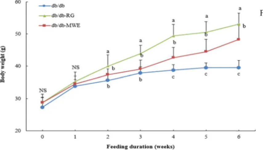
Fig. 1. Weekly changes in body weight in C57BL/KsJ- db/db mice supplemented with mealworm extract. db/db (diabetes mellitus control): C57BL/
KsJ-db/db mice receiving AIN-93G diet; db/
db-RG: C57BL/KsJ-db/db mice receiving AIN- 93G diet supplemented with rosiglitazone (0.005 g/100 g diet); db/db-MWE: C57BL/KsJ- db/db mice receiving AIN-93G diet supple- mented with mealworm extract (0.5 g/100 g di- et). Values are presented as means ± SD, n=8 per group, a-cMean values designed by different letters are significantly different between groups (p<0.05).
Fig. 2. Weekly changes in blood glucose levels in C57BL/
KsJ-db/db mice supplemented with mealworm extract. db/db (diabetes mellitus control): C57BL/
KsJ-db/db mice receiving AIN-93G diet; db/db -RG: C57BL/KsJ-db/db mice receiving AIN-93G diet supplemented with rosiglitazone (0.005 g/
100 g diet); db/db-MWE: C57BL/KsJ-db/db mice receiving AIN-93G diet supplemented with mealworm extract (0.5 g/100 g diet). Values are presented as means ± SD, n=8 per group,
a-cMean values designed by different letters are significantly different between groups (p<0.05).
During the commencement of the experiment, body weights in db/db-control, db/db-RG, and db/db-MWE groups were not significantly different. As the experiment progressed, body weight gradually increased. At the end of the experi- ment, mice in the db/db–RG group displayed significantly higher body weight those in the db/db-control and db/db- MWE groups. Daily food intake and water intake of mice are listed in Table 2. The daily food intake did not differ significantly among the db/db-control, db/db-RG, and db/
db-MWE groups; however, daily water intake was 14.48±
3.79 and 10.84±2.83 in the db/db-control and db/db-MWE groups, respectively, which indicates that the water intake of db/db-control group was significantly higher than that of db/db-MWE group.
Fasting blood glucose
Fig. 2 presents the effect of MWE supplement on fasting blood glucose levels. During the commencement of the ex- periment, blood glucose levels did not significantly differ among the groups; however, fasting blood glucose levels of mice in db/db-control group were elevated throughout the
experiment, presumably indicating the disease progression,
whereas fasting blood glucose levels of mice in db/db-MWE
group were slightly increased. Consequently, at the end of
the experiment, fasting blood glucose was 525.63±57.4 and
A B C
Fig. 3. The effects of supplmentation with MWE on blood glycosylated hemoglobin levels and markers of insulin resistance in C57BL/KsJ-db/db mice. A. Blood glycosylated hemoglobin levels. B. Plasma insulin levels. C. Homeostatic index of insulin resistance in C57BL/KsJ-db/db mice. db/db (diabetes mellitus control): C57BL/KsJ-db/db mice receiving AIN-93G diet;
db/db-RG: C57BL/KsJ-db/db mice receiving AIN-93G diet supplemented with rosiglitazone (0.005 g/100 g diet); db/db- MWE: C57BL/KsJ-db/db mice receiving AIN-93G diet supplemented with mealworm extract (0.5 g/100 g diet). Values are presented as means ± SD, n=8 per group, a-cMean values designed by different letters are siginificantly different between groups (p<0.05). HbA1c: blood glycosylated hemoglobin; HOMA-IR: homeostatic index of insulin resistance.
Fig. 4. The effects of supplementation with MWE on intraperitoneal glucose tolerance tests in C57 BL/KsJ- db/db mice. Blood glucose concentrations were measured at the indicated times. Glucose (0.5 g/kg of body weight) or insulin (2 units/kg of body weight) were injected in db/db-mice at time of zero. db/db (diabetes mellitus control): C57 BL/KsJ-db/db mice receiving AIN-93G diet; db/db-RG:
C57BL/KsJ- db/db mice receiving AIN-93G diet supple- mented with rosiglitazone (0.005 g/100 g diet); db/db- MWE: C57BL/KsJ-db/db mice receiving AIN-93G diet supplemented with mealworm extract (0.5 g/100 g diet).
Values are presented as means ± SD, n=8 per group.
a-cMean values designed by different letters are signifi- cantly different between groups (p<0.05). IPGTT: Intra- peritoneal glucose tolerance tests.
269.50±87.47 in the db/db-control and db/db-MWE groups, respectively, that is, the fasting blood glucose level of mice in the db/db-control group was twice higher than that ob- served in the db/db-MWE group.
HbA1c levels, plasma insulin level, and HOMA-IR As presented in Fig. 3, HbA1c values were 12.99±1.55 and 8.00±3.01 in the db/db-control and db/db-MWE groups, respectively. This indicated that the HbA1c levels, plasma insulin levels, and HOMA-IR values in the db/db-MWE group were significantly lower compared with those in the db/db-control group.
Intraperitoneal glucose and insulin tolerance tests The effect of MWE supplement on glucose tolerance was estimated via IPGTT at 5 weeks of the experiment and the results are presented in Fig. 4. Blood glucose levels of mice in the db/db-control group were higher than those of mice in the db/db-MWE group at all time-points; however, blood glucose levels in the db/db-MWE group peaked at 30 min after glucose injection and recovered to levels close to the basal value at 120 min.
Effect of MWE on the activation of PI3K/Akt pathway To clarify whether MWE supplement promotes the activa- tion of insulin signaling pathway and leads to glucose up- take into cells of skeletal muscles, activation of PI3K/Akt pathway was investigated by observing the phosphorylation levels of IRS-1 and Akt, and by activation of PI3K. As pre- sented in Fig. 5, the levels of IRS-1 phosphorylation and PI3K activation in the db/db-MWE group were significantly
increased compared to the db/db-control group. In the
db/db-MWE groups, levels of IRS-1 phosphorylation were
1.7-fold higher than those in the db/db-control group. The
levels of PI3K activation were 1.8-fold and 1.6-fold higher
in the db/db-RG and db/db-MWE groups, respectively,
than those in the db/db-control group. Moreover, the MWE
supplementation stimulated the phosphorylation of Akt by
A B
Fig. 6. The effect of MWE supplementation on PM-GLUT4 and GLUT4 protein expression in skeletal muscle of C57BL/KsJ-db/db mice. Western blotting was performed, and signal intensities were determined by densitometric analysis using Multi Gauge V3.1 software. A. PM-GLUT4 and GLUT4 protein expression, B. expression levels of PM-GLUT4 and GLUT4. Each value is expressed as mean SD of experiments performed in triplicate. a-cmean values designed by different letters are significantly different between groups (p<0.05). PM-GLUT4: plasma membrane glucose transporter 4.
A
B
Fig. 5. The effect of MWE supplementation on pIRS-1, IRS-1, PI3K, pAkt, and Akt protein expression in skeletal muscle of C57BL/
KsJ-db/db mice. Western blotting was performed, and signal intensities were determined by densitometric analysis using Multi Gauge V3.1 software. A. pIRS-1/IRS-1, PI3K, and pAkt/Akt protein expression, B. expression levels of pIRS-1/IRS-1, PI3K, pAkt/Akt. Each value is expressed as mean SD of experiments performed in triplicate. a-cMean values designed by different letters are significantly different between groups (p<0.05). pIRS-1: phosphorylated insulin receptor substrate-1; PI3K:
phosphatidylinositol-3-kinase; pAkt: phosphorylated Akt.
1.7-fold compared with that in the db/db-control group.
These observations suggested that MWE plays a pivotal role in the activation of PI3K/Akt pathway.
Effect of MWE on PM-GLUT4 expression
GLUT4 translocation from the cytoplasm to plasma mem-
brane is crucial in the absorption of glucose into the cell
[22]. Thus, the effect of MWE supplementation on GLUT4
expression in plasma membrane was investigated. Fig. 6
presents the levels of PM-GLUT4 expression in the db/db-
MWE group compared with those in the db/db-control
group. In db/db-MWE group, the expression of PM-GLUT4
was significantly increased, and the levels were 1.8-fold higher than in the db/db-control group. This result indicates that MWE supplementation may stimulate the GLUT4 trans- location to plasma membrane and, as a consequence, lead to increased glucose uptake into the cell.
Discussion
Diabetes mellitus (DM) is a common metabolic disorder characterized by hyperglycemia, which is caused by im- paired insulin secretion and resistance. During insulin resist- ance, the insulin does not function appropriately due to a decrease in the insulin sensitivity of tissues, thereby causing absorption of blood glucose into the cells and thus leading to hyperglycemia. Thus, improving insulin sensitivity to re- lieve hyperglycemia is essential in the treatment of diabetes [14, 31]. Effective antidiabetic drugs have been developed to treat type 2 diabetes. Thiazolinedione (TZD) class com- prises antidiabetic drugs that are widely used to improve insulin sensitivity and blood glucose control in diabetic pa- tients [5]; however, these drugs may cause side effects such as headache, hypoglycemia, edema, and hypertension [18].
In particular, rosiglitazone, a member of the TZD class, in- creased the adipocyte differentiation, thereby leading to in- creased fat mass and appetite, and decreased physical activity. Due to these side effects of antidiabetic drugs, sev- eral studies have been conducted to improve the insulin sen- sitivity by using natural products with few side effects [17].
In natural products, insects have been used to treat vari- ous diseases. In particular, the mealworms have traditionally been considered as healthy food because they have abundant protein, lipids, vitamins, and minerals [27]. Mealworm is lar- va of T. molitor belonging to the beetle species that have been used in traditional antidiabetic medicines [32]. Nevertheless, no study is available on the use of mealworm as an anti- diabetic in improving insulin sensitivity. Hence, this study aimed to research whether supplementation of the meal- worm extract (MWE) to C57BL/KsJ-db/db type2 diabetic mice improves insulin sensitivity and reduces hyperglyce- mia. RG, an oral anti-diabetic agent, was used to compare the efficacy of MWE.
The daily food intake did not significantly differ among the db/db-control, db/db-RG, and db/db-MWE groups;
however, the water intake was significantly higher in the db/db-control group than that in the db/db-MWE group.
Polydipsia is one of the major symptoms of diabetes mellitus
[24]. A prominent increase of water intake in the db/db-con- trol group reflects polydipsia. Conversely, the supplement of mealworm alleviated the symptoms of diabetes, proving that it can reduce water intake. The body weight of mice in the db/db-MWE group steadily increased for 6 weeks;
however, the increase in the body weight of mice in the db/db-MWE group was significantly lower than that of mice in the db/db-RG group. Such body weight gain in the db/db-RG group is associated with increased adipocyte dif- ferentiation by activation of PPAR-γ as a side effect of rosi- glitazone [17]. Fortunately, the supplementation of MWE did not reveal any side effect such as weight gain.
The progression of type 2 diabetes is related to diabetic complications. As hyperglycemia significantly affects the progression of type 2 diabetes, lowering the blood glucose levels can prevent diabetic complications [5, 19]. The fasting blood glucose levels were significantly lower in the db/db- MWE group than those in the db/db-control group from 2 weeks after the experiment. This phenomenon may be due to the omega-3 fatty acids or chitin and chitosan present in the mealworm. Omega-3 fatty acids have been extensively studied in relation to diabetes, and it is confirmed that ome- ga-3 fatty acids are effective in reducing fasting blood glu- cose levels [10]. Recently, chitin and chitosan have also been extracted from mealworm and have been studied exten- sively in relation to diabetes [35]. Enzymatic digestion of chi- tosan produces low molecular weight chitosan oligo- saccharides, and these have been proven effective in low- ering the fasting blood glucose levels [12, 15]. HbA1c levels are indicative of the average blood glucose levels over the preceding 2-3 months and decreased HbA1c level can reduce the risk of developing macrovascular complications of type 2 diabetes [19]. In the db/db-MWE group, HbA1c levels were significantly decreased compared with those in the db/db-control group. These results suggest that long-term intake of MWE may reduce the blood glucose and further prevent diabetic complications.
Plasma insulin levels in the db/db-MWE and db/db-RG groups were significantly lower than in the db/db-control group. In the early stages of type 2 diabetes, hyperinsuline- mia is observed to overcome insulin resistance in the periph- eral tissues and maintain normal blood glucose levels [7].
In this study, the db/db-control group had hyperinsuline-
mia, whereas the db/db-MWE and db/db-RG groups did
not. To assess the insulin resistance, HOMA-IR was calcu-
lated using fasting glucose and insulin concentrations.
HOMA-IR is homeostatic index of insulin resistance, which is decreased with increasing insulin sensitivity. In this study, HOMA-IR was significantly lower in mice of db/db-MWE group than those of the db/db-control group. These ob- servations imply that MWE supplementation might contrib- ute in improving insulin resistance.
Intraperitoneal glucose tolerance test was performed to assess the insulin sensitivity and utilization of excessive blood glucose [1, 21]. This study demonstrated that blood glucose levels were restored to levels close to the basal value after 120 min of glucose injection in db/db-MWE and db/db-RG groups compared with the db/db-control group.
According to this result, supplementation of MWE increases the insulin sensitivity and effectively controls the blood glu- cose concentrations.
The increase in insulin sensitivity promotes glucose up- take into cells via insulin signaling pathway and thereby re- duces hyperglycemia. To examine the levels of gene ex- pression associated with insulin sensitivity in the insulin sig- naling pathway, western blotting was conducted. In the in- sulin signaling pathway, when insulin binds to the insulin receptor, IRS-1
Tyr612is phosphorylated. The phosphorylated IRS-1
Tyr612binds to the domain of PI3K and activates PI3K, which in turn phosphorylates Akt
Ser473. As a result, GLUT4 is transferred to the plasma membrane to absorb blood glu- cose into the cell [3, 11, 34]. In this study, the supplementa- tion of mealworm extracts significantly increased the levels of gene expression in insulin signaling pathway. MWE sig- nificantly increased phosphorylation of IRS-1
Tyr612and Akt
Ser473, and activation of PI3K in skeletal muscles. In addi- tion, the levels of GLUT4 expression were significantly in- creased in the plasma membrane of skeletal muscles after MWE supplementation.
Omega-3 fatty acids have been found to be associated with the upregulation of genes involved in insulin receptor signaling-1(IRS-1) and GLUT4 [8]. The fatty acids present in the mealworm comprise 77.74% unsaturated fatty acids.
Among these, omega-3 fatty acids account for 46.1% of the unsaturated fatty acids [26]. Recently, published reports re- vealed that α-linolenic acid (C18:3, n−3), one of the omega-3 fatty acids, restored the decrease in palmitic acid-induced glucose uptake, and glucose uptake occurred via mecha- nisms of the insulin signaling pathway including Akt activa- tion in skeletal muscles [23]. In addition to omega-3 fatty acids, chitin and chitosan are involved in lowering blood glucose via insulin signaling pathway [9]. Both high- and
low-molecular weight chitosan could effectively phosphor- ylate Akt and increase the translocation of GLUT4 in skeletal muscles [20]. The isolation of the chitin from the exuvium and whole body of mealworm, the rates were 18.1% and 4.92%, respectively. The relative average yield of chitosan from whole body was 3.65%[30]. According to these results, omega-3 fatty acids or chitin and chitosan in MWE may con- tribute, at least in part, to stimulate insulin signaling path- way in the db/db mice.
In conclusion, MWE supplementation effectively de- creased hyperglycemia in type 2 diabetic db/db-mice. This was because MWE enhanced insulin sensitivity via activa- tion of PI3K/Akt pathway, increased GLUT4 translocation in plasma membrane, and facilitated glucose uptake in the skeletal muscle cells. Therefore, this study suggests that MWE can possess a potential for improving insulin sensi- tivity and alleviating hyperglycemia via insulin signaling pathway in skeletal muscles of db/db-mice.
Acknowledgment
This work was supported by the Health Fellowship Foun- dation.
References
1. Akash, M. S. H., Rehman, K., Sun, H. and Chen, S. 2013.
Interleukin-1 receptor antagonist improves normoglycemia and insulin sensitivity in diabetic Goto-Kakizaki-rats. Eur.
J. Pharmacol. 701, 87-95.
2. Baron, A. D., Zhu, J. S., Zhu, J. H., Weldon, H., Maianu, L. and Garvey, W. T. 1995. Glucosamine induces insulin re- sistance in vivo by affecting GLUT4 translocation in skeletal muscle: Implications for glucose toxicity. J. Clin. Invest. 96, 2792-2801.
3. Baus, D., Heermeier, K., De, H. M., Metz-Weidmann, C., Gassenhuber, J, Dittrich, W., Welte, S. and Tennagels, N.
2008. Identification of a novel AS160 splice variant that reg- ulates GLUT4 translocation and glucose-uptake in rat mus- cle cells. Cell Signal. 20, 2237-2246.
4. Chatterjee, S., Khunti, K. and Davies, M. J. 2017. Type 2 diabetes. Lancet 389, 2239-2251.
5. Durbin, R. J. 2004. Thiazolidinedione therapy in the pre- vention/delay of type 2 diabetes in patients with impaired glucose tolerance and insulin resistance. Diabetes Obes.
Metab. 6, 280-285.
6. Duttaa, P., Dey, T., Dihingia, A., Manna, P. and Kalita, J.
2017. Antioxidant and glucose metabolizing potential of edi- ble insect, Brachytrupes orientalis via modulating Nrf2/
AMPK/GLUT4 signaling pathway. Biomed. Pharmacother. 95, 556-563.
7. Goldstein, B. J. 2002. Insulin resistance as the core defect in type 2 diabetes mellitus. Am. J. Cardiol. 90, 3-10.
8. González-Périz, A., Horrillo, R., Ferre, N., Gronert, K., Dong, B., Moran-Salvador, E., Titos, E. Martinez-Clemente, M., Lopez-Parra, M., Arroyo, V. and Claria, J. 2009. Obesity- induced insulin resistance and hepatic steatosis are alle- viated by omega-3 fatty acids: a role for resolvins and protectins. FASEB J. 23, 1946-1957.
9. Ha, B. G., Park, J. E. and Shon, Y. H. 2016. Stimulatory effect of balanced deep-sea water containing chitosan oligo- saccharides on glucose uptake in C2C12 myotubes. Mar.
Biotechnol. (NY). 18, 475-484.
10. Hartweg, J., Farmer, A. J., Holman, R. R. and Neil, A. 2009.
Potential impact of omega-3 treatment on cardiovascular disease in type 2 diabetes. Curr. Opin. Lipidol. 20, 30-38.
11. Hu, X., Wang, S., Xu, J., Wang, D. B., Chen, Y. and Yang, G. Z. 2014. Triterpenoid saponins from Stauntonia chinensis ameliorate insulin resistance via the AMP-activated protein kinase and IR/IRS-1/PI3K/AKT pathways in insulin-re- sistant HepG2 cells. Int. J. Mol. Sci. 15, 10446-10458.
12. Jo, S. H., Ha, K. S, Moon, K. S., Kim, J. G., Oh, C. G., Kim, Y. C., Apostolidis, E. and Kwon, Y. I. 2013. Molecular weight dependent glucose lowering effect of low molecular weight chitosan oligosaccharide (GO2KA1) on postprandial blood glucose level in SD rats model. Int. J. Mol. Sci. 14, 14214-14224.
13. Joshi S. R., Ramachandran, A., Chadha, M., Chattergee, S., Rathod, R. and Kalra, S. 2014. Acarbose plus metformin fixed-dose combination in the management of type 2 diabetes. Expert Opin. Pharmacother. 15, 1611-1620.
14. Kahn, S. E. 2003. The relative contributions of insulin resist- ance and beta-cell dysfunction to the pathophysiology of type 2 diabetes. Diabetologia 46, 3-19.
15. Kim, J. G., Ha, K. S., Kim, S. C., Kim, Y. C., Apostolidis, E. and Kwon, Y. I. 2014. Effect of long-term supplementation of low molecular weight chitosan oligosaccharide (GO2KA1) on fasting blood glucose and HbA1c in db/db mice model and elucidation of mechanism of action. BMC Complement.
Altern. Med. 14, 272.
16. Klip, A., Ramlal, T., Young, D. A. and Holloszy, J. O. 1987.
Insulin-induced translocation of glucose transporters in rat hindlimb muscles. FEBS Lett. 224, 224-230.
17. Lebovitz, H. E., Dole, J. F., Patwardhan, R., Rappaport, E.
B. and Freed, M. I. 2001. Rosiglitazone monotherapy is effec- tive in patients with type 2 diabetes. Rosiglitazone Clinical Trials Study Group. J. Clin. Endocrinol. Metab. 86, 280-288.
18. Lee, S. H., Min, K. H., Han, J. S., Lee, D. H., Park, D. B., Jung, W. K., Park, P. J., Jeon, B. T., Kim, S. K. and Jeon, Y. J. 2012. Effects of brown alga, Ecklonia cava on glucose and lipid metabolism in C57BL/KsJ-db/db mice, a model of type 2 diabetes mellitus. Food Chem. Toxicol. 50, 575-582.
19. Lind, M., Odén, A., Fahlén, M. and Eliasson, B. 2009. The true value of HbA1c as a predictor of diabetic complications:
Simulations of HbA1c variables. PLoS One 4, e4412.
20. Liu, S. H., Chang, Y. H and Chiang, M. T. 2010. Chitosan reduces gluconeogenesis and increase glucose uptake in skeletal muscle in Streptozotocin-induced diabetic rats. J.
Agric. Food Chem. 12, 5795-5800.
21. Mori, N., Kurata, M., Yamazaki, H., Hosokawa, H., Nadamoto, T., Inoue, K. and Fushiki, T. 2013. Intragastric administration of allyl isothiocyanate reduces hyper- glycemia in intraperitoneal glucose tolerance test (IPGTT) by enhancing blood glucose consumption in mice. J. Nutr.
Sci. Vitaminol. 59, 56-63.
22. Navale, A. M. and Paranjape, A. N. 2016. Glucose trans- porters: physiological and pathological roles. Biophys. Rev.
8, 5-9.
23. Park, S. Y., Kim, M. H., Ahn, J. H., Lee, S. J., Lee, J. H., Eum, W. S., Choi, S. Y. and Kwon, H. Y. 2014. The stimulatory effect of essential fatty acids on glucose uptake involves both Akt and AMPK activation in C2C12 skeletal muscle cells. Kor. J. Physiol. Pharmacol. 18, 255-261.
24. Praythiesh Bruce, M. S. and Vasantha Mallika, M. C. 2019.
Prevalence of complications of diabetes among patients with diabetes mellitus attending a tertiary care centre in Tamil Nadu. Int. J. Community Med. Public Health 6, 1452-1456.
25. Raji, A., Seely, E. W., Bekin, S. A., Williams, G. and Simonson, D. 2003. Rosiglitazone improves insulin sensitivity and low- ers blood pressure in hypertensive patients. Diabetes Care 26, 172-178.
26. Ravzanaadii, N., Kim, S. H., Choi, W. H., Hong, S. J. and Kim, N. J. 2012. Nutritional Value of Mealworm, Tenebrio molitor as Food Source. Int. J. Indust. Entomol. 25, 93-38.
27. Rumpold, B. A. and Schlüter, O. K. 2013. Nutritional compo- sition and safety aspects of edible insects. Mol. Nutr. Food Res. 57, 802-823.
28. Saltiel, A. R. and Kahn, C. R. 2001. Insulin signaling and the regulation of glucose and lipid metabolism. Nature 414, 799-806.
29. Seo, M., Goo, T. W., Chung, M. Y., Baek, M., Hwang, J.
S., Kim, M. A. and Yun, E. Y. 2017. Tenebrio molitor larvae inhibit adipogenesis through AMPK and MAPKs signaling in 3T3-L1 adipocytes and obesity in high-fat diet-induced obese mice. Int. J. Mol. Sci. 18, 158.
30. Song, Y. S., Kim, M. W., Moon, C., Seo, D. J., Han, Y. S., Jo, Y. H., Noh, M. Y., Noh, M. Y., Park, Y. K., Kim, S. A., Kim, Y. W. and Jung, W. J. 2018. Extraction of chitin and chitosan from larval exuvium and whole body of edible mealworm, Tenebrio molitor. Entomol. Res. 48. 227-233.
31. Stuart A. R., Gulve E. A. and Wang M. 2004. Chemistry and biochemistry of type 2 diabetes. Chem. Rev. 104, 1255-1282.
32. Suh, H. J., Kim, S. R., Lee, K. S., Park, S. and Kang, S. C.
2010. Antioxidant activity of various solvent extracts from Allomyrina dichotoma (Arthropoda: Insecta) larvae. J. Photo- chem. Photobiol. B-Biol. 99, 67-73.
33. Takikawa, M., Inoue, S., Horio, F. and Tsuda, T. 2010.
Dietary anthocyanin-rich bilberry extract ameliorates hyper- glycemia and insulin sensitivity via activation of AMP-acti- vated protein kinase in diabetic mice. J. Nutr. 140, 527-533.
34. Velcheti, V. and Govindan, R. 2006. Insulin-like growth fac- tor and lung cancer. J. Thorac. Oncol. 1, 607-610.
35. Won, R. 2017. Insect-based chitin research and its potential application to insect industry in Korea. J. Chitin Chitosan.
22, 215-220.
초록:C57BL/Ksj-db/db 제 2형 당뇨모델을 이용한 갈색거저리 유충(밀웜) 추출물의 인슐린 감수성 및 혈당개선효과
김선영․박재은․한지숙*
(부산대학교 식품영양학과)