• 검색 결과가 없습니다.

Structural and Functional Roles of AIMP2 and TRAF2 in TNF-α Signaling

N/A
N/A
Protected

Academic year: 2021

Share "Structural and Functional Roles of AIMP2 and TRAF2 in TNF-α Signaling"

Copied!
7
0
0

로드 중.... (전체 텍스트 보기)

전체 글

(1)

Structural and Functional Roles of AIMP2 and TRAF2 in TNF-α Signaling

Hyeon Jin Kim

1

, Mi Suk Jeong

2

and Se Bok Jang

1

*

1

Department of Molecular Biology, College of Natural Sciences, Pusan National University, Jangjeon-dong, Geumjeong-gu, Busan 46241, Korea

2

Korea Nanobiotechnology Center, Pusan National University, Jangjeon-dong, Geumjeong-gu, Busan 46241, Korea Received January 13, 2020 /Revised January 27, 2020 /Accepted January 28, 2020

Aminoacyl tRNA synthetase complex interacting multifunctional protein 2 (AIMP2) is a scaffolding protein required for the assembly of multi-tRNA synthetase, and it can exert pro-apoptotic activity in response to DNA damage. In the presence of DNA damage, AIMP2 binds to mouse double minute 2 homolog (MDM2) to protect p53 from MDM2 attack. TGF-β signaling results in the nuclear trans- location of AIMP2, whereby AIMP2 interacts with FUSE-binding protein, and, thus, suppresses c-myc.

TNF receptor-associated factor 2 (TRAF2) is an important mediator between TNF-receptors 1 and 2 which are involved in the signaling of c-Jun N-terminal kinase (JNK), nuclear factor κB (NF-κB), and p38 mitogen-activated protein kinases (MAPKs). TRAF2 is required for the activations of JNK and NF- κB via TNF-α and the mediation of anti-apoptosis signaling. AIMP2 can also enhance pro-apoptosis in the TNF-α signaling. During this signaling, AIMP2 assists the association of E3 ubiquitin ligase, the cellular inhibitor of apoptosis protein 1 (c-IAP1) which is well known and responsible for the degrada- tion of TRAF2. The formation of a complex among AIMP2, TRAF2, and c-IAP1 results in protea- some-mediated TRAF2 degradation. AIMP2 can induce apoptosis via downregulation of TRAF2 to in- teract directly in TNF-α signaling. This review provides new insight into the molecular mechanism responsible for AIMP2 and TRAF2 complex formation and treatments for TNFα-associated diseases.

Key words : AIMP2, apoptosis, c-IAP1, TNF-α, TRAF2

*Corresponding author

*Tel : +82-51-510-2523 , Fax : +82-51-581-2544

*E-mail : [email protected]

This is an Open-Access article distributed under the terms of the Creative Commons Attribution Non-Commercial License (http://creativecommons.org/licenses/by-nc/3.0) which permits unrestricted non-commercial use, distribution, and reproduction in any medium, provided the original work is properly cited.

Journal of Life Science 2020 Vol. 30. No. 1. 106~112 DOI : https://doi.org/10.5352/JLS.2020.30.1.106

TNF receptor signaling

Tumor necrosis factor (TNF)-α induces other inflamma- tory mediators, and thus, regulates inflammatory responses.

TNF-α is involved in inflammation, immunity, tumor pro- gression, cellular homeostasis, transformation, and survival [1, 26, 45]. TNF-α is a proinflammatory cytokine that regu- lates a multitude of biological mechanisms, which are acti- vated by its interaction with TNF-receptor 1 (TNFR1; also called p55/p60) and TNF-receptor 2 (TNFR2; also called p75/p80) [40]. TNFR1 is expressed on almost all cells in man, whereas TNFR2 expression is more limited. More specifi- cally, TNFR2 is expressed on subtypes of neurons, oligoden- drocytes, CD4

+

and CD8

+

T cells, microglia, astrocytes in brain and on endothelial cells, mesenchymal cells, thymo- cytes, and cardiac myocytes. Both receptors can induce the signaling of canonical NF-κB (nuclear factor κB), c-Jun N-ter-

minal kinase (JNK), and p38 mitogen-activated protein kin- ases (MAPKs) [2, 5, 29]. TNFR1 can also initiate apoptotic and necroptotic pathways [41].

Unlike TNFR1, TNFR2 lacks an intracellular death do- main, but it can direct the binding of TRAF1 and TRAF2 in the cytoplasm [14]. This binding leads to the recruitment of E3 ubiquitin ligase and cellular inhibitor of apoptosis pro- tein (c-IAP) 1 and 2 and causes the ubiquitin-dependent deg- radation of TRAF2 [16, 36]. Moreover, the decrease in TRAF2 expression, which has been involved in potentization of TNFR1 can induce apoptosis by TNFR2 [11, 16, 44, 46].

Therefore, TRAF2 is a critical mediator between TNFR1 and TNFR2 and has multiple functions in the TNF stimulation [42]. TNF signals, including apototic and antiaircraft, are ac- tivated and stimulated by the same cytokine. However, TNF signals for cell apoptosis, proliferation and survival have yet to be clarified why cells undergo apoptosis or survival.

TRAF2 regulates cell survival or death on TNFR signaling

TNFR1 is activated by transmembrane TNF and soluble

TNF. Upon TNF binding to TNFR, the cytoplasmic domain

of TNFR1, which contains a death domain, binds TRADD

- Review -

(2)

(TNFR1-associated death domain protein) and recruits to TRAF2, RIPK1 (receptor-interacting serine/threonine-pro- tein kinase 1), c-IAP1 and c-IAP2, and LUBAC (linear ubiq- uitin chain assembly complex) [33]. c-IAP1 and c-IAP2 add Lys63-linked polyubiquitin chains to RIPK1 and LUBAC adds Met1-linked linear ubiquitin chains to RIPK1 [12].

TRAF2 does not act directly as an E3 ubiquitin ligase for RIPK1, and it is possible that TRAF2 plays a role in the re- cruitments of c-IAP1 and c-IAP2 [25, 31]. Ubiquitylated RIPK1 recruits TAK1 (TGF-β-activated kinase 1), TAB2 (TGF-Beta Activated Kinase 1 (MAP3K7) Binding Protein 2), and TAB3, and these recruitments lead to the activations of p38 and JUN N-terminal kinase (JNK) pathways [18]. In ad- dition, TAK/TAB complex activates IKK complex (IκB (inhibitor of κB) kinase complex), which is composed of NEMO (NF-κB essential modulator also called IKKγ), IKKα (IKK subunit α), and IKKβ, which is recruited by ubiquity- lated RIPK1. IKK complex leads to the phosphorylation of IκB, Lys48-linked ubiquitination, and proteasome-mediated degradation. Subsequently, NF-κB translocates to the nu- cleus and activates the transcriptions of genes involved in cell survival and proliferation [12, 15, 17].

TNFR1 induces apoptosis through the formation of DISC (death-inducing signaling complex), which is composed of FAS-associated death domain protein (FADD) and caspase 8, in cytoplasm. Ubiquitylated RIPK1 is deubiquitylated by CYLD (cylindromatosis), which eliminates Lys63 and Met1 –linked polyubiquitin chains from RIPK1; subsequently, deubiquitylated RIPK1 dissociates from membrane-bound TNFR1 signaling [21, 22]. When FLCE-like inhibitory protein (FLIP

L

) levels are low, FADD recruits pro-caspase 8 to form a homodimer, and autocatalytic activations of the pro-do- mains of activated caspase 8 release to cytoplasm and apop- tosis via the activation of pro-caspase 3 [3, 43].

TRAF2 can negatively regulate TNF-induced apoptosis by indirectly modulating RIPK1, and in the absence of TRAF2, non-ubiquitylated RIPK1 triggers pro-apoptotic signaling.

Degradation or depletion of c-IAP1 or c-IAP2 reduces or blocks RIPK1 ubiquitylation. When FLIP

L

levels are low

,

non-ubiquitylated RIPK1 dissociates from membrane-bound TNFR1 signaling and interacts with pro-caspase 8 homo- dimer to initiate an apoptotic cascade. On the other hand, when FLIP

L

levels are high, pro-caspase 8 and FLIP

L

form a heterodimer, which prevents apoptosis and RIPK3-de- pendent necrosis [30].

When caspase 8 or FADD is absent, non-ubiquitylated

RIPK1 forms a complex with RIPK3 and MLKL (pseudoki- nase mixed lineage kinase domain-like) that called a ne- crosome. RIPK3 phosphorylates this complex and induces necroptosis [4]. Petersen et al. found that the binding of TRAF2 to MLKL suppresses the interaction between MLKL and RIPK3 [34].

TNFR2-mediated signaling is not understood anywhere near as well as TNFR1-mediated signaling. Binding of TNF to TNFR2 leads to the recruitment of TRAF2 to the TNFR2 intracellular domain and causes the subsequently recruit- ments of TRAF1, c-IAP1, and c-IAP2 [35]. In addition to TNFR2-mediated NF-κB activation, signaling via TNFR2 can elicit various signaling including p38 MAPK (mitogen-acti- vated protein kinase), JNK (c-jun N-terminal kinase), and PI3K-PKB/Akt (phosphoinositide-3-kinase-protein kinase B/sAkt) [6, 27, 37](Fig. 1A).

Biological function of AIMP2

Aminoacyl-tRNA synthetases (ARSs) are enzymes that li- gate amino acids to their cognate tRNAs before protein synthesis. In humans, eight ARSs form a multi-tRNA synthe- tase complex (MSC) and three non-enzymatic ARS-interact- ing multifunctional proteins (AIMP1-3) [7]. The association and dissociation of this complex are widely involved in cel- lular signaling pathways and human diseases. Of the ARS- interacting multifunctional proteins, AIMP2 plays a scaffold- ing role in the assembly of multi-tRNA synthetase complex and determines cell fate via its antiproliferative and proa- poptotic activities.

When TGF-β is stimulated, AIMP2 is translocated to nu- clei to stimulate the ubiquitination and degradation of FUSE-binding protein (FBP), which is a transcriptional acti- vator of c-myc, and this leads to the transcriptional sup- pression of c-myc. AIMP2-deficient mice have a higher level of FBP during embryonic development, and this leads to the overexpression of c-myc and to the overproliferation of lung epithelial cells [20]. AIMP2 can also promote cell death by directly binding p53. Upon DNA damage, AIMP2 dissociates from MSC and is translocated to nuclei to interact with p53.

AIMP2 prevents the MDM2-dependent ubiquitination and

degradation of p53. Furthermore, AIMP2 depletion increases

resistance to apoptosis-inducing DNA damage, and the in-

troduction of AIMP2 into AIMP2 depleted cells recovers

apoptosis sensibility [13]. AIMP2 is also associated with

Parkinson's disease. Parkin promotes the formation of ubiq-

(3)

A

B

C

Fig. 1. Schematic representations of TNF-α signaling diverse role of AIMP2 and binding among AIMP2, TRAF2, and c-IAP1. (A) TRAF2 mediates crosstalk between the TNFR1 and TNFR2 to induce apoptosis, inflammation, or survival. After TNF stimulation, apoptosis is induced by caspase 8 to FADD binding, and inflammation and survival are promoted through TRAF2 via the JNK cas- cade, the MEKK cascade, and via NF-κB. (B) Diverse roles of AIMP2 in TNF-α, TGF-β and DNA damage are shown. Upon stimuli, AIMP2 released from the MSC binds FBP, p53, and TRAF2 and induces cell death and growth arrest. (C) Schematic representation of binding among AIMP2, TRAF2, and c-IAP1. After formation of the trimeric complex, TRAF2 undergoes ubiquitylation and degradation by c-IAP1, which leads to TNF-α -induced cell death.

uitylated AIMP2 and is induced by AIMP2 accumulation, which may result in neurological disorders (Fig. 1B) [10].

Proapoptotic interactions of AIMP2, TRAF2 and c-IAP1

AIMP2 can enhance pro-apoptosis in the TNF-α signaling and reduce TNF-α-dependent NF-κB activity. During pro-apoptotic signaling, AIMP2 promotes apoptosis by downregulation of TRAF2 in the TNF-α signal pathway. A yeast two-hybrid assay showed that AIMP2 binds to TRAF2, and an investigation involving the deletion of AIMP2 re- gions, showed the aa 84-205 region interacts with TRAF2.

When HEK293 cells were transfected with AIMP2 the level of ubiquitylated-TRAF2 was substantially increased [9].

AIMP2 also downregulates TNF-α-dependent NF-κB activity and I κB levels were found to be remarkably diminished within 30 minutes of TNF-α treatment in AIMP2-deficient cells [9]. However, AIMP2 does not possess E1, E2, or E3 enzyme activities, and therefore, AIMP2 may recruit to E3 ubiquitin ligase c-IAP1, which is known to cause the ubiq- uitylation of TRAF2 [23, 39]. Furthermore, AIMP2 could fa- cilitate c-IAP1 to TRAF2 binding and cause ubiquitin-de- pendent TRAF2 degradation. These results suggest AIMP2, TRAF2, and c-IAP1 form a trimeric complex by direct inter- action, and that this interaction enables AIMP2 to sensitize cells to TNF-α-induced apoptosis via TRAF2 degradation [9]

(Fig. 1C).

The structures of AIMP2, TRAF2, and c-IAP1

AIMP2 is a 35 kDa protein that contains a C-terminal GST domain. In MSC, the GST domains of AIMP3, EPRS (gluta- minyl-prolyl-tRNA synthetase), and MRS (methionyl-tRNA synthetase) interact with each other. The roles of these GST domains remain poorly understood [8], though it has been established that the aa 84-225 region of AIMP2 interacts with TRAF2 [9].

TRAF2 is a 55 kDa protein with an N-terminal RING mo-

tif to mediate E3 ubiquitin ligase activity and five zinc finger

motifs. These domains are required for the TRAF2-mediated

activations of JNK and NF-κB. The TRAF2-N domain is flexi-

ble and forms a triple-helix parallel coiled-coil structure,

whereas the TRAF2-C domain forms a mushroom-shaped

trimer having a β-sandwich domain [28]. The 352-501 resi-

(4)

A

B

AIMP2 TRAF2 c-IAP1

Fig. 2. Structural analyses of AIMP2, TRAF2, and c-IAP1 (A) Domain analyses of AIMP2, TRAF2, and c-IAP1. Schematic representa- tions of full-length AIMP2, TRAF2, and c-IAP1 are shown. AIMP2 consists of a GST domain, whereas TRAF2 has a ring motif, five zinc finger motifs, and a TRAF domain. c-IAP1 consists of BIR domains, a ubiquitin binding domain, a CARD domain, and a RING domain that confers ubiquitin E3 ligase activity. (B) Structures of AIMP2, TRAF2, and c-IAP1 are shown.

Three-dimensional ribbon structures of AIMP2, TRAF2, and c-IAP1 are shown. AIMP2 (residues 106-320; PDB ID: 5Y6L), TRAF2 (residues 267-329; PDB ID: 3M0D), and c-IAP1 (residues 33-116; PDB ID: 3M1D).

dues of TRAF2 forms an eight-stranded antiparallel β-sand- wich, in which strands β1, β8, β5, and β6 are in one sheet and β2, β3, β4, and β7 are in the others. This TRAF2 domain is required for self-association and interactions with re- ceptors [32].

c-IAP1 protein consists of three BIR (baculoviral IAP re- peat) domains followed by a UBA (ubiquitin-associated) do- main, a CARD (caspase recruitment domain), and a RING domain. The BIR domains c-IAP1 contain conserved histi- dine and cysteine residues that coordinate with zinc II. BIR1 residues (34-129) of c-IAP1 interact with TRAF2 residues (267-349)[25, 38]. The CARD domain of c-IAP1 forms a six-helix bundle and the ring domain of c-IAP1 functions as an E3 ligase and transfer of ubiquitin to substrate proteins [24](Fig. 2).

Conclusion

TRAF2 plays an important mediatory role in JNK and NF- κB signaling via cross-talk between itself and TNFR1. TRAF2 is required for the activations of JNK and NF-κB via TNF-α, and mediates anti-apoptotic signaling [19]. Furthermore, TRAF2-mediated signals play significant roles in the regu- lations of cell proliferation, stress response, and cell survival.

AIMP2 controls cell fate in multiple directions. For instance, in the presence of DNA damage, AIMP2 dissociates from multi-tRNA synthetase and translocates to the nucleus where it promotes apoptosis via p53. AIMP2 also suppresses cell proliferation by ubiquitinating FBP, and thus, down- regulating c-myc in the TGF-β signaling.

AIMP2 can also enhance pro-apoptotic activity by down-

regulating TRAF2 expression in the TNF-α signaling, during

(5)

which AIMP2 facilitates the association between E3 ubiq- uitin ligase, c-IAP1 (cellular inhibitor of apoptosis protein 1), and TRAF2. Furthermore, the binding among AIMP2 and TRAF2 and c-IAP1 promotes apoptosis by downregulation of TRAF2 in the TNF-α signaling [9]. This review can pro- vide new insights of the molecular mechanism responsible for AIMP2 and TRAF2 complex formation and treatments for TNF-α-associated diseases.

Acknowledgement

This work was supported by a 2-Year Research Grant from Pusan National University (2019-2020).

The Conflict of Interest Statement

The authors declare that they have no conflicts of interest with the contents of this article.

References

1. Baud, V. and Karin, M. 2001. Signal transduction by tumor necrosis factor and its relatives. Trends Cell Biol. 11, 372-377.

2. Borghi, A., Verstrepen, L. and Beyaert, R. 2016. TRAF2 mul- titasking in TNF receptor-induced signaling to NF-κB, MAP kinases and cell death. Biochem. Pharmacol. 116, 1-10.

3. Bradley, J. R. 2008. TNF-α mediates inflammatory disease.

J. Pathol. 214, 149-160.

4. Brenner, D., Blaser, H. and Mak, T. W. 2015. Regulation of tumour necrosis factor signalling: live or let die. Nat. Rev.

Immunol. 15, 362-374.

5. Cabal-Hierro, L. and Lazo, P. S. 2012. Signal transduction by tumor necrosis factor receptors. Cell Signal. 24, 1297-1305.

6. Cabal-Hierro, L., Rodríguez, M., Artime, N., Iglesias, J., Ugarte, L., Prado, M. A. and Lazo, P. S. 2014. TRAF-medi- ated modulation of NF-kB and JNK activation by TNFR2.

Cell Signal. 26, 2658-2666.

7. Cho, H. Y., Lee, H. J., Choi, Y. S., Kim, D. K., Jin, K. S., Kim, S. and Kang, B. S. 2019. Symmetric assembly of a deca- meric subcomplex in human multi-tRNA synthetase com- plex via interactions between glutathione transferase-ho- mology domains and aspartyl-tRNA synthetase. J. Mol. Biol.

431, 4475-4496.

8. Cho, H. Y., Maeng, S. J., Cho, H. J., Choi, Y. S., Chung, J. M., Lee, S., Kim, H. K., Kim, J. H., Eom, C. Y., Kim, Y.

G., Guo, M., Jung, H. S., Kang, B. S. and Kim, S. 2015.

Assembly of multi-tRNA synthetase complex via hetero- tetrameric glutathione transferase-homology domains. J. Biol.

Chem. 290, 29313-29328.

9. Choi, J. W., Kim, D. G., Park, M. C., Um, J. Y., Han, J. M., Park, S. G., Choi, E. C. and Kim, S. 2009. AIMP2 promotes TNFα-dependent apoptosis via ubiquitin-mediated degra-

dation of TRAF2. J. Cell Sci. 122, 2710-2715.

10. Corti, O., Hampe, C., Koutnikova, H., Darios, F., Jacquier, S., Prigent, A., Robinson, J. C., Pradier, L., Ruberg, M., Mirande, M., Hirsch, E., Rooney, T., Fournier, A. and Brice, A. 2003. The p38 subunit of the aminoacyl-tRNA synthetase complex is a Parkin substrate: linking protein biosynthesis and neurodegeneration. Hum. Mol. Genet. 12, 1427-1437.

11. Duckett, C. S. and Thompson, C. B. 1997. CD30-dependent degradation of TRAF2: implications for negative regulation of TRAF signaling and the control of cell survival. Genes Dev. 11, 2810-2821.

12. Haas, T. L., Emmerich, C. H., Gerlach, B., Schmukle, A. C., Cordier, S. M., Rieser, E., Feltham, R., Vince, J., Warnken, U., Wenger, T., Koschny, R., Komander, D., Silke, J. and Walczak, H. 2009. Recruitment of the linear ubiquitin chain assembly complex stabilizes the TNF-R1 signaling complex and is required for TNF-mediated gene induction. Mol. Cell 36, 831- 844.

13. Han, J. M., Park, B. J., Park, S. G., Oh, Y. S., Choi, S. J., Lee, S. W., Hwang, S. K., Chang, S. H., Cho, M. H. and Kim, S. 2008. AIMP2/p38, the scaffold for the multi-tRNA synthetase complex, responds to genotoxic stresses via p53.

Proc. Natl. Acad. Sci. USA. 105, 11206-11211.

14. Inoue, J. I., Ishida, T., Tsukamoto, N., Kobayashi, N., Naito, A., Azuma, S. and Yamamoto, T. 2000. Tumor necrosis fac- tor receptor-associated factor (TRAF) family: adapter pro- teins that mediate cytokine signaling. Exp. Cell Res. 254, 14- 24.

15. Israël, A. 2010. The IKK complex, a central regulator of NF-κ B activation. Cold Spring Harb. Perspect. Biol. 2, a000158.

16. Chan, F. K. and Lenardo, M. J. 2000. A crucial role for p80 TNFR2 in amplifying p60 TNFR1 apoptosis signals in T lymphocytes. Eur. J. Immunol. 30, 652-660.

17. Kalliolias, G. D. and Ivashkiv, L. B. 2016. TNF biology, pathogenic mechanisms and emerging therapeutic strategies.

Nat. Rev. Rheumatol. 12, 49-62.

18. Kanayama, A., Seth, R. B., Sun, L., Ea, C. K., Hong, M., Shaito, A., Chiu, Y. H., Deng, L. and Chen, Z. J. 2004. TAB2 and TAB3 activate the NF-κB pathway through binding to polyubiquitin chains. Mol. Cell 15, 535-548.

19. Kim, J. Y., Morgan, M., Kim, D. G., Lee, J. Y., Bai, L., Lin, Y., Liu, Z. G. and Kim, Y. S. 2011. TNFα induced non- canonical NF-κB activation is attenuated by RIP1 through stabilization of TRAF2. J. Cell Sci. 124, 647-656.

20. Kim, M. J., Park, B. J., Kang, Y. S., Kim, H. J., Park, J. H., Kang, J. W., Lee, S. W., Han, J. M., Lee, H. W. and Kim, S. 2003. Downregulation of FUSE-binding protein and c-myc by tRNA synthetase cofactor p38 is required for lung cell differentiation. Nat. Genet. 34, 330-336.

21. Komander, D. and Rape, M. 2012. The ubiquitin code. Ann.

Revi. Biochem. 81, 203-229.

22. Kovalenko, A., Chable-Bessia, C., Cantarella, G., Israël, A., Wallach, D. and Courtois, G. 2003. The tumour suppressor CYLD negatively regulates NF-κB signalling by deubiquiti- nation. Nature 424, 801-805.

23. Li, X., Yang, Y. and Ashwell, J. D. 2002. TNF-RII and c-IAP1

(6)

mediate ubiquitination and degradation of TRAF2. Nature 416, 345-347.

24. Mace, P., Shirley, S. and Day, C. 2010. Assembling the build- ing blocks: structure and function of inhibitor of apoptosis proteins. Cell Death Differ. 17, 46-53.

25. Mace, P. D., Smits, C., Vaux, D. L., Silke, J. and Day, C. L.

2010. Asymmetric recruitment of cIAPs by TRAF2. J. Mol.

Biol. 400, 8-15.

26. MacEwan, D. J. 2002. TNF ligands and receptors–a matter of life and death. Br. J. Pharmacol. 135, 855-875.

27. Marchetti, L., Klein, M., Schlett, K., Pfizenmaier, K. and Eisel, U. L. 2004. Tumor necrosis factor (TNF)-mediated neuro- protection against glutamate-induced excitotoxicity is en- hanced by N-Methyl-D-aspartate receptor activation Essen- tial role of a TNF receptor 2-mediated phosphatidylinositol 3-kinase-dependent NF-κB pathway. J. Biol. Chem. 279, 32869- 32881.

28. McWhirter, S. M., Pullen, S. S., Holton, J. M., Crute, J. J., Kehry, M. R. and Alber, T. 1999. Crystallographic analysis of CD40 recognition and signaling by human TRAF2. Proc.

Natl. Acad. Sci. USA. 96, 8408-8413.

29. Micheau, O. and Tschopp, J. 2003. Induction of TNF re- ceptor I-mediated apoptosis via two sequential signaling complexes. Cell 114, 181-190.

30. Oberst, A. and Green, D. R. 2011. It cuts both ways: reconcil- ing the dual roles of caspase 8 in cell death and survival.

Nat. Rev. Mol. Cell Biol. 12, 757-763.

31. Park, S. M., Yoon, J. B. and Lee, T. H. 2004. Receptor inter- acting protein is ubiquitinated by cellular inhibitor of apop- tosis proteins (c-IAP1 and c-IAP2) in vitro. FEBS Lett. 566, 151-156.

32. Park, Y. C., Burkitt, V., Villa, A. R., Tong, L. and Wu, H. 1999.

Structural basis for self-association and receptor recognition of human TRAF2. Nature 398, 533-538.

33. Park, Y. C., Ye, H., Hsia, C., Segal, D., Rich, R. L., Liou, H.

C., Myszka, D. G. and Wu, H. 2000. A novel mechanism of TRAF signaling revealed by structural and functional analyses of the TRADD-TRAF2 interaction. Cell 101, 777-787.

34. Petersen, S. L., Chen, T. T., Lawrence, D. A., Marsters, S.

A., Gonzalvez, F. and Ashkenazi, A. 2015. TRAF2 is a bio- logically important necroptosis suppressor. Cell Death Differ.

22, 1846-1857.

35. Rodríguez, M., Cabal-Hierro, L., Carcedo, M. T., Iglesias,

J. M., Artime, N., Darnay, B. G. and Lazo, P. S. 2011. NF-κB signal triggering and termination by tumor necrosis factor receptor 2. J. Biol. Chem. 286, 22814-22824.

36. Rothe, M., Pan, M. G., Henzel, W. J., Ayres, T. M. and Goeddel, D. V. 1995. The TNFR2-TRAF signaling complex contains two novel proteins related to baculoviral inhibitor of apoptosis proteins. Cell 83, 1243-1252.

37. Ruspi, G., Schmidt, E. M., McCann, F., Feldmann, M., Williams, R. O., Stoop, A. A. and Dean, J. L. 2014. TNFR2 increases the sensitivity of ligand-induced activation of the p38 MAPK and NF-κB pathways and signals TRAF2 protein degradation in macrophages. Cell Signal. 26, 683-690.

38. Samuel, T., Welsh, K., Lober, T., Togo, S. H., Zapata, J. M.

and Reed, J. C. 2006. Distinct BIR domains of cIAP1 mediate binding to and ubiquitination of tumor necrosis factor re- ceptor-associated factor 2 and second mitochondrial activa- tor of caspases. J. Biol. Chem. 281, 1080-1090.

39. Silke, J. and Brink, R. 2010. Regulation of TNFRSF and in- nate immune signalling complexes by TRAFs and cIAPs.

Cell Death Differ. 17, 35-45.

40. Tartaglia, L. A. and Goeddel, D. V. 1992. Two TNF receptors.

Immunol. Today 13, 151-153.

41. Vandenabeele, P., Declercq, W., Beyaert, R. and Fiers, W.

1995. Two tumour necrosis factor receptors: structure and function. Trends Cell Biol. 5, 392-399.

42. Wajant, H. and Scheurich, P. 2001. Tumor necrosis factor receptor-associated factor (TRAF) 2 and its role in TNF signaling. Int. J. Biochem. Cell Biol. 33, 19-32.

43. Wang, L., Du, F. and Wang, X. 2008. TNF-α induces two distinct caspase-8 activation pathways. Cell 133, 693-703.

44. Wu, C. J., Conze, D. B., Li, X., Ying, S. X., Hanover, J. A.

and Ashwell, J. D. 2005. TNF-α induced c-IAP1/TRAF2 complex translocation to a Ubc6-containing compartment and TRAF2 ubiquitination. EMBO J. 24, 1886-1898.

45. Wu, Y. and Zhou, B. P. 2010. TNF-α/NF-κB/Snail pathway in cancer cell migration and invasion. Br. J. Cancer 102, 639- 644.

46. Yeh, W. C., Shahinian, A., Speiser, D., Kraunus, J., Billia, F., Wakeham, A., de la Pompa, J. L., Ferrick, D., Hum, B., Iscove, N., Ohashi, P., Rothe, M., Goeddel, D. V. and Mak, T. W.

1997. Early lethality, functional NF-κB activation, and in-

creased sensitivity to TNF-induced cell death in

TRAF2-deficient mice. Immunity 7, 715-725.

(7)

초록:TNF-α 신호에서 AIMP2와 TRAF2의 구조적 및 기능적 역할

김현진

1

․정미숙

2

․장세복

1

*

(

1

부산대학교 자연과학대학 분자생물학과,

2

부산대학교 한국나노바이오테크놀러지센터)

아미노아실-트랜스퍼 리보핵산 합성효소-상호작용 다기능 단백질 2(AIMP2)는 여러 tRNA 합성효소들과의 결 합체를 이루게 하는 기능을 하며, DNA 손상에 대한 반응으로 세포사멸 활성을 나타낼 수 있다. DNA에 손상이 발생하면 AIMP2는 MDM2 공격으로부터 p53을 보호하기 위해 MDM2에 결합한다. TGF-β 신호에서 AIMP2는 세포 핵으로 들어가 FUSE 결합 단백질(FBP)과 결합하여 c-myc을 억제한다. TNF 수용체 관련 인자 2(TRAF2)는 c-Jun N-말단 키나아제(JNK), NF-κB 및 p38 미토겐 활성화 단백질 키나아제(MAPKs)의 신호에서 실행되는 두 수용체, TNF 수용체 1과 2 사이의 중요한 중재자이다. TARF2는 TNF-α 신호에서 JNK와 NF-κB의 활성화에 필요 하며, 세포사멸 신호를 막는 중재자 역할을 수행한다. 또한 TNF-α 신호에서 AIMP2는 세포사멸을 향상시킨다.

이 신호에서, AIMP2는 TRAF2를 분해하는 것으로 잘 알려진 E3 유비키틴 효소인 c-IAP1과의 결합을 향상시킨다.

AIMP2, TRAF2 및 c-IAP1을 포함한 복합체의 형성은 proteasome을 매개로 하여 TRAF2의 분해를 초래한다. 이러

한 연구 결과는 AIMP2가 TNF-α 신호에서 직접적인 상호작용을 통해 TRAF2를 하향 조절시켜 세포사멸을 유도할

수 있음을 시사한다.

수치

Fig.  1.  Schematic  representations  of  TNF-α  signaling  diverse  role  of  AIMP2  and  binding  among  AIMP2,  TRAF2,  and  c-IAP1
Fig.  2.  Structural  analyses  of  AIMP2,  TRAF2,  and  c-IAP1  (A)  Domain  analyses  of  AIMP2,  TRAF2,  and  c-IAP1

참조

관련 문서

Micro- and nano-sized pores were formed on the surface of the alloy using PEO and anodization methods, and the pore shape change according to the Zr

In order to analyze thermal stability and capacity loss according to the temperature of the battery cell by applying different C-rate discharge and

Second, exploratory factor analysis and Cronbach's α were conducted to analyze the validity and reliability of the questionnaire regarding participation

Glutamate excitotoxicity induced by excessive activation of NMDA receptor causes various damage to cells, which leads to cell death.. In previous studies, increased ROS

Consider the motion of a particle of mass m which is constrained to move on the surface of a cone of half-angle α and which is subject to a gravitational force g. Let the

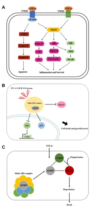
Taken together, these results suggested that latex containing the ficin inhibited the cell growth and induced apoptosis by caspase and Bcl-2 family signaling pathway in

Laser-induced doping of amorphous copper diselenide (α-CuSe 2 ) thin film with gallium (Ga) and indium (In) was performed to control/improve their electrical

Degradation of PE and PP into fuel oil by using solid acid and non-acid catalyst (Sakata et.. of liquid products from thermal and catalytic degradation of (a) HDPE at 430 °C