1. 서 론
먹는물 등에 존재하는 미생물을 분석할 때 대상 미생물이 살아 있는지 죽어 있는지의 여부를 판단하는 것은 매우 중요 하다. 일반적으로 미생물의 deoxyribonucleic acids (DNA)는 세포가 죽은 뒤에도 수일에서 3주 정도 보존되어 남아 있을 수 있다고 한다(Josephson et al., 1993, Masters et al., 1994, Nocker et al., 2006). 따라서 DNA 를 분석하는 polymerase chain reaction(PCR) 등의 유전
자 분석법을 이용할 경우 물에 존재하는 살아 있는 미생물의 DNA 뿐만 아니라 죽어 있는 미생물의 DNA까지도 증폭하 여 검출하게 되므로 실제 살아있는 미생물의 수가 과대평가 될 수도 있다는 단점이 있다(Nocker et al., 2006). 미생물 이 살아 있는지 또는 손상되었거나 죽어 있는지를 판단하는 중요한 기준은 바로 세포막의 활성이 남아있는지의 여부이 다. 살아있는 미생물의 세포막은 그 활성을 유지하고 있기 때문에 ethidium bromide(EtBr) 등의 DNA 염색 시약이 세포의 내부로 침투하지 못하게 하는 기작을 유지하고 있다.
수환경에서 살아 있는 대장균의 검출을 위한 ethidium monoazide-중합효소연쇄반응법
Ethidium monoazide-PCR for the detection of viable Escherichia coli in aquatic environments
이규철
1*․김현정
1․이병기
1․권순복
1․김기돈
1․이상태
1․이찬희
2Gyucheol Lee1*․Hyunjeong Kim1․Byunggi Lee1․Soonbok Kwon1 Gidon Kim1․Sangtae Lee1․ Chanhee Lee2
1 한국수자원공사 K-water 연구원 수돗물분석연구센터, 2 충북대학교 생명과학부 미생물학과 (2009년 2월 3일 접수; 2009년 3월 18일 수정; 2009년 3월 25일 채택)
Abstract
It is very important to differentiate of DNA derived from live or dead bacteria within mixed microbial communities in aquatic environments. Ethidium monoazide (EMA) is a DNA intercalating agent and the treatment of EMA with strong visible light cleaves the genomic DNA of bacteria. In dead bacterial cells, EMA intercalates into the genomic DNA, induces the cleavage of DNA, and inhibits the PCR amplification. In this study, we developed the EMA-PCR and EMA real-time PCR to detect the DNA derived from viable Escherichia coli(E.coli) in mixed cultures of live and deadE.coli. The treatment of EMA, 50 ㎍/mL, and 650 W visible halogen light exposure for 2 minutes cleaved the genomic DNA derived from heat killed E.colibut did not those of live E.coli. EMA-PCR could detect the DNA from live E.coli in mixed culture samples of live and dead E.coliat various ratio and there was no DNA amplification in only dead E.colicultures. Similar results were observed in EMA real-time PCR. Further studies are needed to develop various EMA-PCR methods to detect viable waterborne pathogens such as Helicobacter pylori, Giardia lamblia, and so on.
Key words : ethidium monoazide, polymerase chain reaction, real-time polymerase chain reaction
주 제 어 : EMA, 중합효소연쇄반응법, 실시간 중합효소연쇄반응법
*Corresponding author Tel:+82-42-629-20349, Fax:+82-42-629-2079, E-mail: [email protected](Lee, G.)
그러나 손상되었거나 죽어 있는 미생물은 세포막이 손상되 어 있기 때문에 DNA 염색시약이 세포내부로 침투하는 것을 막아내지 못한다. 이러한 원리는 형광을 갖는 DNA 염색시 약과 유세포분류기(flowcytometry)를 이용하여 살아있는 세포와 죽어있는 세포를 구분하는 연구에 많이 사용되고 있 다(Nebe-von -Caron et al., 1998, 2000).
최근에는 ethidium monoazide(EMA)라는 DNA 염색시 약을 이용한 방법이 살아있는 세균을 검출하기 위한 현미경 방법이나 유세포분류법(flowcytometry)의 대체 방법으로 서 연구되고 있다(Nogva et al., 2003, Rudi et al., 2005a,b, Wang and Levin, 2006). EMA는 DNA 염색 시 약의 일종으로 halogen 같은 매우 밝은 빛에 의해 DNA와 공유결합을 하여 nitrene을 형성하는 것으로 알려져 있다 (Coffman et al., 1982, DeTraglia et al., 1978, Hixon et al., 1975). 이러한 빛에 의해 유도된 EMA와 DNA의 화학적 결합은 genomic DNA의 깨짐(cleavage)을 유도하 므로 PCR에 의해 DNA가 증폭되는 것을 억제한다 (Seojima et al., 2007, Chang et al., 2009). 이러한 원리 로 EMA를 이용하면 살아있는 세균과 죽어있는 세균을 분리 할 수 있는 PCR법의 개발이 가능하다. 또한 DNA와 반응하 지 않은 EMA는 물에 존재하는 분자들과 반응하여 자연스럽 게 불활성화 되는 것으로 알려져 있어(DeTraglia et al., 1978), DNA를 추출하기 위해 세포막을 인위적으로 제거해 도 이로부터 유리된 DNA가 잔류 EMA와 반응하여 PCR 증폭이 저해될 가능성은 거의 없다. 최근 이러한 원리를 이 용한 다양한 미생물에 대한 EMA-PCR법이 여러 연구자들 에 의해 개발되어 보고되고 있다(Flenka et al., 2007, Pisz et al., 2007, Cawthorn and Witthuhn, 2008, Inoue et al., 2008). 본 연구에서는 한국 수계에 존재하는 다양한 세균들에 대한 EMA-PCR법을 개발하기에 앞서 그 응용 가능성을 알아보고자 16S rDNA에 대한 PCR법을 이용하 여 대장균을 대상으로 EMA의 처리 조건과 빛의 처리 조건, 그리고 EMA-PCR 조건을 최적화시켜 살아있는 대장균과 죽어있는 대장균이 섞여있는 조건에서 살아있는 대장균만 을 선별하여 검출할 수 있는 EMA-PCR법을 정립하고자 하였다.
2. 재료 및 방법
2.1. 세균 및 배양
본 실험을 위한 세균으로 대장균(Escherichia coli(E.
coli), Bioball, BTF, Pittsburgh, PA, USA)을 사용하였다.
먼저 대장균 콜로니를 취하여 Luria Bertani(LB) 배지(BD DifcoTM, Franklin Lakes, NJ, USA)에 접종한 후 12~14
시간 동안 37℃에서 진탕배양하였으며, 실험을 시작하기 4 시간 전에 대장균 배양액을 다시 신선한 LB배지에 접종하여 OD600가 0.5~0.8 사이가 되도록 배양하여 실험에 사용하였 다. 인위적으로 세균을 죽이기 위한 조건으로 72℃에서 10 분간 열처리하였다(Nocker et al., 2006, Nokcer and Camper, 2006).
2.2. EMA의 처리
EMA(Invitrogen, Carlsbad, CA, USA)의 적절한 처 리농도를 결정하기 위해 살아있는 대장균과 죽어있는 대 장균 배양액 500 uL(OD600=0.5~0.8)에 각각 0, 1, 10, 25, 50, 100 ㎍/mL의 농도로 EMA를 처리하고 세균용 G-spinTM Genomic DNA Extraction Kit(iNtRON BIOTECHNOLOGY, Seongnam, Korea)를 사용하여 제 조사에서 제안한 방법에 따라 genomic DNA를 추출한 후 1.5% agarose gel에 전기영동하여 적절한 EMA의 농도를 결정하였다. 광반응(photoactivation)을 시키기 위한 광원 (light source)으로 650 W급 할로겐램프(PHILIPS Broadway, Suresnes, France)를 사용하였으며, 대장균이 담긴 튜브를 얼음위에 놓고 20 cm 거리에서 2분간 빛을 조사하였다.
2.3. 16S rDNA PCR과 real-time PCR
대장균을 검출하기 위한 PCR과 real-time PCR을 위해 16S rDNA 부위를 target으로 하였으며, primer는 DG74 5'-AGG AGG TGA TCC AAC CGA A-3'와 RW01 5'-ACG TGG AGG AAG AAG GTG GGG AT-3'를 사용 하였다(Greisen et al., 1994). PCR 반응조건은 10×
buffer 5 μ L, 25 mM MgCl2 5 μL, 10 mM dNTP 1 μL, forward와 reverse primer 20 pmole/μL 각 1 μL, DNA Taq polymerase 0.5 μL(Roche, Mannheim, Germany), DNA template 1 μL, 그리고 nuclease free water (Ambion, Austin, TX, USA) 총 volume이 50 μL가 되도록 넣어주었으며, 95℃에서 3분간 반응시키고, denaturation 반 응을 위해 95℃ 25초, annealing 반응 60℃ 25초, extension 반응 72℃ 30초 반응을 40 cycles 반복하고, 최종 extension 반응을 위해 72℃에서 2분간 반응시켰다.
1.5% agarose gel에서 전기영동하였으며, EtBr로 염색하 고 GEL Logic 200 imaging system(Kodak Molecular Imaging Systems, Rochester, NY, USA)을 이용하여 PCR 산물을 분석하였다. Real-time PCR의 경우 SYBR GREEN PCR Master Mix(Applied Biosystems, Foster City, CA, USA)와 7500 Real Time PCR System (Applied Biosystems)을 사용하였다. 반응 조건은 SYBR
GREEN PCR Master Mix(2×) 25 μL, 각 primers(7 pmole/μL) 1 μL씩, template DNA 1 μL, 그리고 nuclease free water(Ambion) 22 μL를 넣고, 95℃에서 3분간 반응시키고, denaturation 반응을 위해 95℃ 25초, annealing 반응 60℃ 25초, extension 반응 72℃ 30초 반 응을 40 cycles 반복하였다.
3. 결 과
3.1. 대장균의 열처리 사멸 조건 최적화
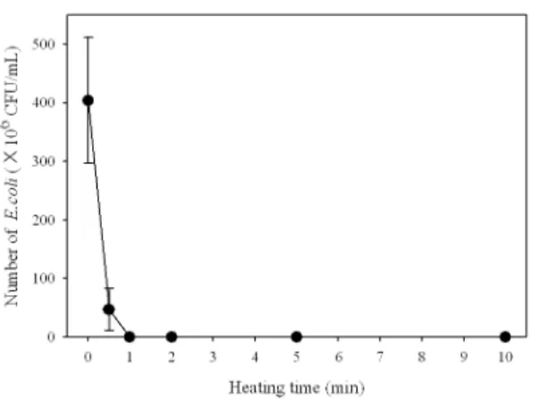
본 연구의 목적은 살아있는 세균과 죽어있는 세균이 함께 존재할 경우 살아있는 세균만을 PCR법으로 검출해 내는 방 법을 정립하는 것이다. 먼저 죽어있는 세균을 얻기 위한 열 처리 조건을 최적화 시킬 필요가 있었다. 일반적으로 70%
ethanol을 10~20분간 처리함으로 세균을 죽이거나, 72℃
에서 5~15분간 열처리를 함으로서 세균을 죽이는 방법들이 알려져 있다. 본 연구에서는 72℃에서 열처리를 하는 방법 을 선택하였다. 72℃에서 시간대별(0, 0.5, 1, 2, 5, 그리고 10분)로 열처리를 가한 후 세균을 LB 고체평판배지에서 배
양하여 단일 집락수를 계수하여 세균이 모두 사멸되는 조건 을 선택하였다. Fig. 1에서 보는바와 같이 대장균은 72℃에 서 최소 1분 이상 열처리 할 경우 모두 사멸됨을 알 수 있었 다. 위 결과를 토대로 본 연구에서 대장균을 확실하게 죽이 는 조건으로 72℃에서 10분간 열처리를 하는 것으로 결정하 였다.
3.2. EMA 및 빛 처리 조건 최적화
살아있는 세균만을 죽어있는 세균과 분리하여 검출할 수 있도록 적절한 EMA 처리 농도를 구하고자 살아있는 세균과 죽어있는 세균에 각각 EMA를 농도별로(0, 1, 10, 25, 50, 그리고 100 ㎍/mL) 처리한 후 650 W급 할로겐램프를 사용 하여 20 cm 거리에서 2분간 빛을 조사하였다. 이로부터 genomic DNA를 추출한 후 전기영동하여 DNA의 추출 여 부를 확인하였다. 2회 반복 실험한 결과 Fig. 2에서 보는바 와 같이 살아있는 세균의 경우 모든 농도에서 genomic DNA가 제대로 추출되었다. 죽어있는 세균의 경우 EMA를 0, 1, 그리고 10 μg/mL를 처리한 경우에서는 genomic DNA가 관찰되었고, 25 ㎍/mL를 처리한 경우 한번은 DNA
Samples Ⅰ Ⅱ Ⅲ Ⅳ Ⅴ Ⅵ
Live*1 0 10 100 250 500 1000
Dead*2 1000 990 900 750 500 0
Live*1: Escherichia coli cells were cultured in LB broth at OD600=0.8 for about four hours; Dead*2: Live E.coli were heat-killed for 10 minutes at 72℃.
Table 1. Mixed culture samples of live and dead Escherichia coli cells.
(μL)
Fig. 1. Number of Escherichia coli cells dependent on heat-killed time.
Escherichia coli cells were cultured in LB broth at OD600=0.5 for about four hours and then heat-killed at 7 2℃ for various time.
Fig. 2. Extraction of genomic DNA from live and dead Escherichia coli cultures treated with various concentration of EMA.
(a) and (b) were results of duplicate experiments.
가 추출되었음을 관찰하였고(Fig. 2a), 한번은 DNA가 추출 되지 않았음을 관찰할 수 있었다(Fig. 2b). 그러나 EMA를 50 μg/mL와 100 μg/mL를 처리한 경우에는 genomic DNA가 관찰되지 않음을 알 수 있었다(Fig. 2a와 2b). 이상 의 결과로 미루어 본 연구에서는 살아있는 세균과 죽어있는 세균을 구별하기 위한 EMA 처리 농도를 50 μg/mL로 결정 하였다.
3.3. EMA-PCR에 의한 살아있는 대장균의 검출 앞서 실험에서 EMA 처리 조건을 최적화 시켰다. 실제 물에는 살아있는 세균과 죽어있는 세균이 함께 존재한다.
이런 경우 EMA를 처리하여 PCR을 수행하게 되면 살아있는 세균에서만 PCR 산물이 증폭되어 검출된다. 실제 환경에서 살아있는 세균과 죽어있는 세균이 혼재되어 있는 것과 유사 한 조건을 만들기 위해 배양한 대장균(OD600=0.8)을 일부 는 72℃에서 10분간 열처리하여 죽인 후 각각 적절한 비율 로 살아있는 세균과 죽어있는 세균을 혼합하였다(Table 1).
살아있는 세균과 죽어있는 세균이 제대로 혼합되었는지 그 리고 살아있는 세균수를 알아보기 위해 LB 고체평판배지에 접종하여 배양한 후 세균수를 알아보았다. 그 결과 살아있는 세균수의 비율에 따라 적절히 살아있는 세균수가 증가하고 있음을 알 수 있었다(Fig. 3).
살아있는 대장균과 죽어있는 대장균의 혼합이 적절히 이 루어졌음을 확인하였으므로 genomic DNA 역시 혼합비에 따라 제대로 추출되고 있는지를 확인하기 위해 EMA를 처리 하고 빛을 조사한 후 genomic DNA를 추출하여 전기영동으 로 확인하였다. 그 결과 EMA를 처리하지 않은 경우 살아있 는 대장균과 죽어있는 대장균의 혼합 비율에 관계없이 모든 시료에서 genomic DNA를 관찰할 수 있었으며, EMA를 처
리한 경우에서는 살아있는 세균이 10 μL 첨가된 Ⅱ번 시료 까지 genomic DNA가 제대로 추출되고 있음을 관찰하였고, 살아있는 대장균이 전혀 첨가되지 않은 Ⅰ번 시료의 경우 genomic DNA가 전혀 관찰되지 않았다(Fig. 4a). 이상의 결과로부터 살아있는 대장균과 죽어있는 대장균이 혼합되 어 있는 조건에서도 EMA 처리에 의해 죽어있는 대장균의 경우 genomic DNA가 모두 깨져 전혀 추출되지 않고, 살아 있는 대장균으로부터만 DNA가 제대로 추출되고 있음을 알 수 있었다.
최종적으로 EMA-PCR이 살아있는 대장균으로부터 추 출된 DNA를 template로 하여 제대로 PCR 산물을 증폭시 킬 수 있는지 알아보기 위해 이상의 시료들(Ⅰ~Ⅵ)로부터 분리한 DNA를 이용하여 16S rDNA에 대한 PCR을 수행했 다. 그 결과 Fig. 4b에서 보는바와 같이 EMA를 처리하지 않은 경우 모든 시료에서 DNA가 증폭되어 EMA를 처리하 지 않은 일반 PCR 방법으로는 살아있는 대장균과 죽어있는 대장균을 구분하지 못한다는 것을 알 수 있었다. 그러나 EMA를 처리한 경우 살아있는 대장균이 포함된 Ⅱ~Ⅵ번 시 료에서는 DNA가 증폭되는 것을 알 수 있었으며, 살아있는 대장균의 양이 증가함에 따라 PCR 산물의 양도 함께 증가하 는 것을 알 수 있었다(Fig. 4b).
3.4. EMA-real-time PCR에 의한 살아있는 대장균 검출
앞서 결과에서 살아있는 대장균의 양에 따라 EMA-PCR 산물의 양이 증가하고 있음을 알 수 있었으나, 일반적으로 PCR로는 살아있는 대장균의 정량분석이 불가능하고 real- time PCR법을 사용할 경우 정량이 가능하다. 따라서 본 연 구에서도 시료(Ⅰ~Ⅵ)에 EMA를 처리한 후 일반 PCR에서 Fig. 3. Number of Escherichia coli cells mixed with live and dead
Escherichia coli at various ratio (referred at Table. 1).
Fig. 4. Gel electrophoresis of genomic DNA and EMA-PCR with the genomic DNA extracted from mixed culture samples of live and dead Escherichia coli treated with EMA or not.
(a), gel electrophoresis of genomic DNA extracted from mixed cultures of live and dead Escherichia coli treated with EMA or not;
(b), EMA-PCR amplification of the genomic DNA
사용한 16S rDNA에 대한 동일한 primers(DG71과 RW01)을 이용하여 SYBR green dye를 이용한 real-time PCR을 수행하였다. 그 결과 살아있는 대장균의 양이 증가함 에 따라 Ct value가 감소하는 것, 즉 살아있는 대장균의 양이 많을수록 PCR 산물이 보다 빠르게 증폭되고 있는 것을 확인 할 수 있었다(Fig. 5). 그러나 살아있는 대장균이 전혀 들어 있지 않은 Ⅰ번 시료에서도 형광이 검출되어 마치 DNA가 증폭되고 있는 것처럼 보였으나 확인실험 결과 이는 primers의 dimer 형성에 따른 background 형광으로 확인 되었다(data not shown).
4. 고 찰
한국의 먹는물 수질기준 및 검사 등에 관한 규칙(2008)에 따르면 일반세균, 대장균, 총대장균군, 분원성대장균군, 분 원성 연쇄상구균, 녹농균, 살모넬라, 쉬겔라, 아황산환원혐 기성포자형성균, 여시니아 균을 먹는물에서 검사해야 한다 (환경부, 2008). 대부분의 세균들이 배양법에 기초하고 있 어 분석시간이 최소 2~3일 이상 걸린다는 단점이 있다. 또 한 미국 환경보호청(U.S. Environmental Protection Agency)에서 지정한 오염후보군 목록(Contamination Candidate Lists)에 있는 Aeromonas hydrophila, Helicobacter pylori, Mycobacterium avium complex, Campylobacter jejuni, E. coli(O157), Legionella pneumophila, Salmonella enterica, Shigella sonnei, 그 리고 Vibrio cholerae의 경우 역시 배양을 통해 분석할 경우 시간이 오래 걸리거나 선택배지의 선정이 까다로운 등 대상 미생물을 정확하고 신속하게 분석하기 어렵다는 단점이 있 다. 물론 이러한 단점들을 해결하기 위해 유전자 분석법에
기초한 PCR 방법 등에 관한 연구가 진행되고 있으나 (Mazari-Hiriart et al., 2001, Kong et al., 2002, Yanez et al., 2005, Chomvarin et al., 2007), 앞서 언급한 대로 살아있는 세균만을 정확하게 검출할 수 없기 때문에 실제 현장 시료를 대상으로 한 분석법으로 직접 사용하기에 부적 합할 수도 있다. 그러나 EMA를 이용한 PCR법의 경우 위 문제들을 해결할 수 있다. 앞서 언급한 대로 살아있는 대장 균의 경우 세포막의 활성이 유지되고 있으므로 EMA를 처리 하여도 EMA가 세포내로 침투하지 못하나 죽어있는 세균의 경우 세포막의 활성이 유지되고 있지 못하므로 EMA가 세포 내로 침투하게 되어 DNA의 깨짐(cleavage)을 유도하므로 genomic DNA를 추출하여 전기영동할 경우 genomic DNA 가 관찰되지 않는다(Seojima et al., 2007, Chang et al., 2009). 이러한 원리로 죽어있는 세균의 DNA를 추출하여 PCR을 수행하게 되면 PCR 산물이 증폭되지 않고, 반대로 살아있는 세균에서만 PCR 산물이 증폭되어 PCR법으로 살 아있는 세균만을 검출할 수 있게 되는 것이다. 따라서 본 연구에서는 대장균을 대상으로 EMA-PCR법을 정립하여 물에서 살아있는 대장균만을 선별하여 검출할 수 있는지의 여부를 알아보고자 하였다. 본 연구 결과 대장균의 경우 EMA를 50 ㎍/mL로 처리하고 650W급 할로겐 램프를 이용 하여 20 cm 거리에서 2분간 조사한 후 genomic DNA를 추출하여 PCR을 수행할 경우 살아있는 대장균의 DNA만을 선별하여 검출할 수 있음을 확인하였다. 따라서 향후 검출하 고자 하는 대상 미생물이 있을 경우 PCR법만 정립하게 되면 PCR 전 단계에 EMA를 처리하고 광반응을 시킴으로서 살아 있는 대상 세균만을 선별적으로 검출할 수 있는 분자생물학 적 방법의 개발이 가능하게 될 것이다. 아울러 살아있는 세 균만을 검출할 수 있으므로 이러한 방법들을 수질 분석에 직접 응용할 수 있을것으로 기대된다. 아쉽게도 바이러스의 경우는 숙주세포 밖의 환경에서는 활성이 유지되는 세포막 을 갖지 못하기 때문에, EMA-PCR법을 이용한 검출법을 응용할 수 없을 것으로 생각되나, 주요한 수인성 병원체 중 하나인 원생동물의 경우 EMA-PCR을 이용한 분석이 가능 할 것으로 판단된다. 특히 원생동물 중 Cryptosporidium과 Giardia의 경우 heat shock을 이용한 hsp70 유전자에 대한 RT-PCR법이 정립되어 활성이 있는 것만을 검출할 수 있는 방법이 개발되어 있으므로(Nam et al., 2007) 이 방법의 전 단계에 EMA만 처리한다면 보다 정확하게 살아있는 원생동 물의 검출이 가능한 방법을 개발할 수 있을 것으로 사료된다.
향후 정량분석을 위해 본 연구에서 시도되었던 real- time PCR법의 단점을 보완하기 위해 SYBR green dye를 이용하지 않고 specific probe를 이용한다면, primer dimers에 의해 발생되었던 문제점들을 해결할 수 있을 것으 Fig. 5. EMA-real-time PCR with genomic DNA extracted
from mixed cultures of live and dead Escherichia coli treated with EMA.
로 기대된다. 아울러 최적화 시킨 EMA-PCR법과 EMA-real-time PCR법을 실제 현장 시료 분석에도 적용 이 가능한지 등의 추가 연구가 필요할 것으로 판단된다.
5. 결 론
본 연구에서는 살아있는 대장균과 죽어있는 대장균이 혼 재되어 존재하는 시료에서 배양법이 아닌 PCR 방법을 이용 하여 살아있는 대장균만을 보다 빠르게 검출할 수 있는 EMA-PCR법을 개발하였다. 이 방법은 향후 EMA-real -time PCR법을 보완할 경우 정량분석까지도 가능할 것으 로 기대되며, 다양한 수인성 병원성 세균과 원생동물의 분석 법 개발에 매우 중요한 자료가 될 것으로 생각된다.
감사의 글
본 연구는 한국수자원공사 K-water 연구원 수돗물분석연 구센터에서 수행되었음(과제번호: KIWE-WARC-07-05)
참고문헌
1. 환경부 (2008) 먹는 물 수질기준 및 검사 등에 관한 규칙. 환경 부령 제276호.
2. Cawthorn, D.M. and Witthuhn, R.C. (2008) Selective PCR detection of viable Enterobacter sakazakii cells utilizing propidium monoazide or ethidium bromide monoazide, J. Appl. Microbiol., 105(4):1178-1185.
3. Chang, B., Sugiyama, K., Taguri, T., Amemura-Maekawa, J., Kura, F. and Watanabe, H. (2009) Specific detection of viable Legionella cells by combined use of photoactivated ethidium monoazide and PCR/real-time PCR, Appl. Environ. Microbiol., 75(1):147-153.
4. Chomvarin, C., Namwat, W., Wongwajana, S., Alam, M., Thaew-Nonngiew, K., Sinchaturus, A. and Engchanil, C.
(2007) Application of duplex-PCR in rapid and reliable detection of toxigenic Vibrio cholerae in water samples in Thailand, J. Gen. Appl. Microbiol., 53(4):229-237.
5. Coffman, G.L., Gaubatz, J.W., Yielding, K.L. and Yielding, L,W. (1982) Demonstration of specific high affinity binding sites in plasmid DNA by photoaffinity labeling with ethidium analog, J. Biol. Chem., 257(22):
13205-13207.
6. DeTraglia, M.C., Brand, J.S. and Tometski, A.M. (1978) Characterization of azidobenzamidines as photoaffinity
labeling for trypsin, J. Biol. Chem., 253(6):1846-1852.
7. Flekna, G., Stefanic, P., Wagner, M., Smulders, F.J., Mozina, S.S. and Hein, I. (2007) Insufficient differentiation of live and dead Campylobacter jejuni and Listeria monocytogenes cells by ethidium monoazide (EMA) compromises EMA/real-time PCR, Res.
Microbiol., 158(5):405-412.
8. Greisen, K., Loeffelholz, M., Purohit, A. and Leong, D.
(1994) PCR primers and probes for the 16S rRNA gene of most species of pathogenic bacteria, including bacteria found in cerebrospinal fluid, J. Clin. Microbiol., 32(2):335-351.
9. Hixon, S.C., White, W.E. and Yielding, K,L. (1975) Selective covalent binding of an ethidium analog to mitochondrial DNA with production of petite mutants in yeast by photoaffinity labeling, J. Mol. Biol., 92(2):319-329.
10. Inoue, D., Tsutsui, H., Yamazaki, Y., Sei, K., Soda, S., Fujita, M. and Ike, M. (2008) Application of real-time polymerase chain reaction (PCR) coupled with ethidium monoazide treatment for selective quantification of viable bacteria in aquatic environment, Water Sci.
Technol., 58(5):1107-1112.
11. Josephson, K.L., Gerba, C.P. and Pepper, I.L. (1993) Polymerase chain reaction detection of nonviable bacterial pathogens, Appl. Environ. Microbiol., 59(10):3513-3515.
12. Kong, R.Y., Lee, S.K., Law, T.W., Law, S.H. and Wu, R.S.
(2002) Rapid detection of six types of bacterial pathogens in marine waters by multiplex PCR, Water Res., 36(11):2802-2812.
13. Masters, C.I., Shallcross, J.A. and Mackey, B.M. (1994) Effect of stress treatments on the detection of Listeria monocytogenes and enterotoxigenic Escherichia coli by the polymerase chain reaction. J. Appl. Bacteriol., 77(1):73-79.
14. Mazari-Hiriart, M., Lόpez-Vidal, Y., Castillo-Rojas, G., Ponce de Leόn, S. and Cravioto, A. (2001) Helicobacter pylori and other enteric bacteria in freshwater environments in Mexico City, Arch. Med. Res., 32(5):458-467.
15. Nam, S.H., Lee, J.I., Lee, S.T. and Lee, G.C. (2007) Detection of viable Cryptosporidium parvum by real-time RT-PCR, J. Kor. Soc. Environ. Anal.,
10(3):134-140.
16. Nebe-von-Caron, G., Stephens, P. and Badley, R.A.
(1998) Assessment of bacterial viability status by flow cytometry and single cell sorting, J. Appl. Microbiol., 84(6):988-998.
17. Nebe-von-Caron, G., Stephens, P.J., Hewitt, C.J., Powell, J.R. and Badley, R.A. (2000) Analysis of bacterial function by multi-colour fluorescence flow cytometry and single cell sorting, J. Microbiol. Methods, 42(1):97-114.
18. Nocker, A. and Camper, A.K. (2006) Selective removal of DNA from dead cells of mixed bacterial communities by use of ethidium monoazide, Appl. Environ. Microbiol., 72(3):1997-2004.
19. Nocker, A., Cheung, C.Y. and Camper, A.K. (2006) Comparison of propidium monoazide with ethidium monoazide for differentiation of live vs. dead bacteria by selective removal of DNA from dead cells. J. Microbiol.
Methods, 67(2):310-320.
20. Nogva, H.K., Drømtorp, S.M., Nissen, H. and Rudi, K.
(2003) Ethidium monoazide for DNA-based differentiation of viable and dead bacteria by 5'-nuclease PCR, Biotechniques, 34(4):804-808, 810, 812-813.
21. Pisz, J.M., Lawrence, J.R., Schafer, A.N. and Siciliano,
S.D. (2007) Differentiation of genes extracted from non-viable versus viable micro-organisms in environmental samples using ethidium monoazide bromide, J. Microbiol. Methods, 71(3):312-318.
22. Rudi, K., Moen, B., Drømtorp, S.M. and Holck, A.L.
(2005a) Use of ethidium monoazide and PCR in combination for quantification of viable and dead cells in complex samples, Appl. Environ. Microbiol., 71(2):1018-1024.
23. Rudi, K., Naterstad, K., Drømtorp, S.M. and Holo, H.
(2005b) Detection of viable and dead Listeria monocytogenes on gouda-like cheeses by real-time PCR, Lett. Appl. Microbiol., 40(4):301-306.
24. Soejima, T., Iida, K., Qin, T., Taniai, H., Seki, M. and Yoshida, S. (2008) Method to detect only live bacteria during PCR amplification, J. Clin. Microbiol., 46(7):2305-2313.
25. Wang, S. and Levin, R.E. (2006) Discrimination of viable Vibrio vulnificus cells from dead cells in real-time PCR, J. Microbiol. Methods, 64(1):1-8.
26. Yanez, M.A., Carrasco-Serrano, C., Barbera, V.M. and Catalan, V. (2005) Quantitative detection of Legionella pneumophila in water samples by immunomagnetic purification and real-time PCR amplification of the dotA gene, Appl. Environ. Microbiol., 71(7):3433-3441.