한국인의 측두하악관절에서 Dental cone-beam CT를 이용한 관절융기의 경사도에 대한
방사선학적 평가
원광대학교 치과대학, 치과보철학교실 오상천․한지석
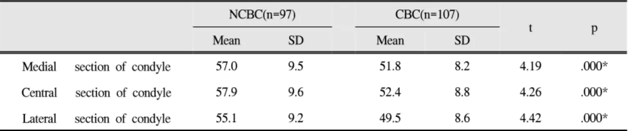
하악의 개구운동 시, 과두-관절원판 복합체는 관절융기를 따라서 활주운동을 하게 되므로 관절융기의 형태는 측두하악관절의 기능적인 움직임을 이해하는데 매우 중요한 부분이다. 본 연구의 목적은 치과용 cone-beam CT를 이용하여 관절융기의 후방경사를 계측하고, 관절융기의 경사도에 대한 과두의 병적 변화의 영향을 평가하며, 성 별과 연령에 따른 관절융기의 차이를 비교 하는 것이다. 이를 위해 원광대학교 산본치과병원에 내원한 102명(남 자:43명, 여자:49명, 평균나이: 37.7세)의 204개 측두하악관절의 cone-beam CT 영상을 평가하였으며, 모든 영상은 측두하악관절 분석모드로 전환하여 양측 관절의 연속된 시상 단면 이미지와 관상 단면 이미지를 관찰하였다. 신 뢰성있는 데이터를 얻기 위해 숙련된 3명의 치과의사가 동일한 이미지 파일을 보며 판독 작업을 시행하였고, 3명 중 2명의 판독 결과가 동일할 경우만 최종 판독 결과로 기록하였다. 정상과두(NCBC)와 골변화를 동반한 과두 (CBC)의 관절융기 경사도는 내측이 57.0°(NCBC)과 51.8°(CBC), 중심이 57.9°(NCBC)과 52.4°(CBC), 그리고 외측이 55.1°(NCBC)과 49.5°(CBC)를 나타냈고, 골변화를 보이는 과두가 정상과두보다 낮은 관절융기 경사도를 보였다.
이러한 차이는 성별이나 연령에 따른 통계적 유의성은 없었다.
주요어: 한국인의 측두하악관절, Cone-beam CT, 관절융기 경사도, 방사선학적 평가
(
구강회복응용과학지 2013:29(2):163~173)
서 론
하악과두, 관절와, 관절융기, 관절원판, 부속인 대로 이루어져 있는 측두하악관절에서 관절융기 는 관절와의 전방부에 위치한 볼록한 골융기로 정의된다.
1하악이 개구운동을 할 때 일반적으로
교신저자:
오상천
원광대학교 치과대학 보철학교실
경기도 군포시 산본동 1142, 435-040, 대한민국
Fax: +82 31 390 2777, Tel: +82 31 390 2899, E-mail: [email protected]
원고접수일: 2013년 3월 23일, 원고수정일: 2013년 5월 15일, 원고채택일: 2013년 6월 25일
하악과두는 접번운동에 이어 활주운동을 하고 관절원판은 하악과두와 기능적인 복합체를 이루 어 관절융기의 후방 경사면을 따라 활주운동을 하게 되므로 관절융기의 후방 경사도는 이런 운 동에 관여하는 중요한 요인 중의 하나이다.
2관절융기의 가파른 경사는 관절원판 변위와
관련된 측두하악관절 내장증을 유발하는 요인으 로 간주되어 관절융기의 경사도와 관절원판 변 위 사이의 관련성을 규명하기 위한 많은 연구들 이 진행되어 왔다. Atkinson과 Bates
3는 측두하악 관절장애 환자에서 관절융기의 후방 경사도를 측정하여 하악과두 및 관절원판의 변위는 관절 융기의 급격한 후방 경사와 밀접한 관련성이 있 다고 보고하였고, Hall 등
4은 관절잡음이, Kerstens 등
5은 관절원판 전방변위가 있는 측두하 악관절과 정상 측두하악관절의 관절융기 경사도 를 파노라마 방사선 사진 상에서 비교한 결과, 관절잡음 및 관절원판 변위가 있는 측두하악관 절이 더 큰 관절융기의 후방 경사도를 나타내는 것으로 보고하였다. 이와는 반대로 Panmekiate 등
6과 Pullinger 등
7그리고 Galante 등
8은 정상인 과 관절원판이 전방으로 변위된 환자에서 관절 융기의 후방 경사도를 비교한 결과, 관절융기의 가파른 후방 경사와 관절원판 전방 변위 사이에 는 관련성이 없다고 하였으며, Ren 등
9도 관절원 판이 변위된 환자에서 오히려 관절융기 경사도 가 더 작은 것으로 보고하면서 관절융기의 경사 도는 관절원판의 변위보다는 하악과두의 형태변 화를 의미하는 골변화와 더 깊은 관련이 있는 것 으로 보고하였다. 이처럼 측두하악관절에서 관 절융기의 경사도와 관절원판의 변위와의 관련성 에 대해서는 많은 연구가 있었음에도 아직까지 도 그 관련성이 명확히 규명되지 않고 있으며, 특히 하악과두의 골변화와 관절융기의 경사도 사이의 관련성에 대해서는 더 많은 연구가 필요 한 상태이다.
일반적으로 측두하악관절의 형태 연구를 위하 여 경두개방사선사진이나 파노라마방사선사진 을 많이 이용해 왔다. 그러나 측두하악관절은 해 부학적 형태가 매우 다양하고 복잡한 양상을 띄 므로 일반 방사선사진 상에서는 측두하악관절에 대한 명확한 관찰이 어렵고 방사선의 조사각도에 따라 형태가 다양하게 나타나는 한계가 있었으 며,
10이를 보완하기 위해 좀더 정확하고 다양한
영상 정보를 얻을 수 있는 일반단층사진, 전산화 단층사진, 측두하악관절조영사진, 측두하악관절 조영 단층사진, 자기공명영상 등 다양한 방법들 이 소개되어 왔다.
11특히 최근에는 높은 공간 분 해능과 다면영상재구성시 상의 왜곡이 없는 cone -beam형 전산화단층영상(Cone-Beam Computed Tomography, CBCT)이 소개되어 측두하악관절과 같은 해부학적으로 복잡한 구조의 형태를 평가하 고 계측하는데 많이 활용되고 있다.
12-14따라서 본 연구의 목적은 정확한 치과용 CBCT를 이용하여 한국인의 관절융기의 후방 경 사도를 계측하고, 하악과두의 골변화와 관절융 기의 후방 경사도의 관계를 평가하기 위함이다.
연구 재료 및 방법 1. 연구 재료
2007년 7월부터 2008년 9월까지 원광대학교 산본치과병원에 내원한 환자 중 i-CAT Cone- Beam 3-D Dental Imaging System(Imaging Sciences International, USA)을 이용하여 진단받은 환자들의 전산화단층 영상을 대상으로 측두하악 관절 분석 모드로 전환하여 양쪽 측두하악관절 의 영상을 분석하였다. 환자들의 나이는 평균 37.7세(13세~90세) 였으며, 43명의 남자와 59명의 여자를 포함하여 총 102명의 환자, 204개의 측두 하악관절을 관찰하였다. 측두하악관절은 하악과 두의 골변화 유무를 관찰하여 정상 하악과두 (n=97)와 골변화가 일어난 하악과두(n=107)로 구 분하여 관절융기의 후방 경사도를 측정하였다.
2. 연구 방법
1) 측두하악관절 분석 준비
촬영된 전산화단층 영상을 이용하여 하악과두
의 골형태와 관절융기의 후방 경사도를 측정하
기 위한 준비를 하였다. 관절융기가 Frankfort
Fig. 1. CT image adjusted to FH plane.
horizontal(FH) plane 또는 교합평면이나 구개평면 (palatal plane) 같은 수평기준평면과 이루는 각도 로 정의되는
15관절융기의 경사도 계측을 위해 본 연구에서는 FH plane을 기준으로 삼았다. 측 두하악관절 3차원 분석을 위하여 Invivo 5 (Anatomage, USA)프로그램을 이용하였으며, 영 상을 분석하기에 앞서 촬영된 모든 이미지가 FH plane에 수평이 되도록 하기 위해 전산화단층 영 상을 골분석 모드로 전환하여 양측의 외이공 최 상방점(porion, Po)과 우측의 안와 최하방점 (orbitalle, Or)에 점을 찍고 이 점들을 연결한 FH plane을 설정한 후 영상을 FH plane에 수평이 되 도록 재구성하였다(Fig. 1). 그 후에 측두하악관
Fig. 2. The serial sagittal images of the TMJs.
절 분석모드로 전환하여 양측 관절의 연속된 시 상 단면 이미지와 관상 단면 이미지를 관찰하였 다(Fig. 2).
2) 하악과두의 병적 형태변화 관찰
하악과두의 골 이상 형태에 대한 판단 기준은 Uemura 등
16과 Koyama 등
17의 분류를 참고하였 고, 하악과두의 flattening, erosion, concavity, osteophyte가 있을 경우 골의 형태 변화가 일어난 것으로 간주하였다(Fig. 3). 하악과두의 골변화 상태를 관찰하기 위하여 숙련된 3명의 치과의사 가 동일한 이미지 파일을 보며 판독 작업을 시행 하였고, 3명 중 2명의 판독 결과가 동일한 경우 최종 판독 결과로 기록하였다.
Fig. 3. Classification of condylar bone change.
(a) A normal condyle, displaying typical morphology. (b) Flattening, flattened contour on the functional surface of the condyle. (c) Erosion, localized area of decreased bone density on the superior area of the condylar surface.
(d) Concavity, deformed contour with a
concavity on the condylar surface. (e)
Osteophyte, bony protrusion formed on
the condylar surface, usually on the
anterior part.
3) 관절융기의 경사도 계측
Ren 등
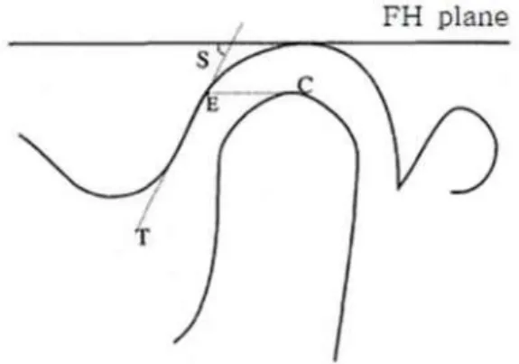
9의 연구방법을 참고하여 각각의 측두 하악관절은 하악과두의 외측, 중앙, 내측으로 세 등분하여 측정하였으며(Fig. 4), 관절융기의 후방 경사도는 Lawther
18의 방법을 이용하여 계측하였 다(Fig. 5). 먼저 관절와의 천장(the roof of glenoid fossa)에 FH plane과 나란한 기준선을 그리고 이 와 평행한 선을 하악과두의 최상방점(C)에서 관 절융기의 후방 경사면 쪽으로 그려서 만나는 한 점을 기준점(E)으로 삼고 여기를 통과하면서 관 절융기의 경사면에 접하는 선(T)을 그려 이 선 (T)과 FH plane이 이루는 각도, 즉 관절융기의 경 사도 값을 측정하였다.
4) 통계분석
측정된 관절융기 경사도의 통계적 유의성을 검증하기 위하여 t-test와 one-way ANOVA를 시 행하였다. 사후 검증은 Scheffe test를 이용하였으 며, P<0.05 유의수준으로 신뢰도를 평가하였다.
통계처리에는 SPSS WIN 12.0(SPSS Inc., USA)를 이용하였다.
Fig. 4. Three tomographic sections perpendi- cular to long axis of the condyle were chosen to represent lateral, central, and medial parts of the TMJ.
결 과
1. 하악과두의 골변화가 관절융기의 경사도에 미치는 영향
정상 하악과두의 경우 관절융기의 경사도는 하 악과두의 내측에서 57.0°, 중앙에서 57.9°, 외측에 서 55.1°로 나타났다. 하악과두에서 골변화가 일 어난 경우에는 내측에서 51.8°, 중앙에서 52.4°, 외 측에서 49.5°로 나타나 하악과두의 골변화가 일 어난 경우 관절융기의 경사도가 더 편평한 것으 로 나타났으며, 내측(t=4.192, p<0.05), 중앙 (t=4.257, p<0.05), 외측(t=4.426, p<0.05) 모두에서 통계적으로 유의한 차이를 보였다 (Table Ⅰ, Fig.
6).
Fig. 5. Measurement of steepness of articular
eminence on serial tomograms: C, top
of the condyle; E, intersection of a line
parallel to the Frankfurt horizontal
plane through point C with the
posterior slope of the articular
eminence; T, line through point E and
tangential to the posterior slope of the
articular eminence; S, steepness of
articular eminence.
NCBC(n=97) CBC(n=107)
t p
Mean SD Mean SD
Medial section of condyle 57.0 9.5 51.8 8.2 4.19 .000*
Central section of condyle 57.9 9.6 52.4 8.8 4.26 .000*
Lateral section of condyle 55.1 9.2 49.5 8.6 4.42 .000*
*Statistically significant (p<0.05)
NCBC, no condylar bone change; CBC, condylar bone change
Table Ⅰ. Effect of condylar bone change on the steepness of articular eminence (degrees) at medial, central, and lateral section of TMJ
Fig. 6. Steepness of eminence at lateral, central, and mesial section of joint.
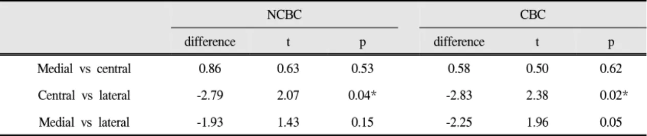
2. 측두하악관절에서 위치에 따른 관절융기의 경사도
측두하악관절에서의 위치에 따른 관절융기의 경사도를 비교 했을 때 정상인의 경우는 중앙부 위에서 가장 경사도가 크고 내측, 외측의 순으로 작게 나타났으며, 하악과두의 골변화가 있는 경 우에도 역시 중앙, 내측, 외측의 순으로 경사도 가 작아지는 것으로 나타났다. 중앙 부위와 외측 부위의 경사도에서는 통계적으로 유의한 차이가 있었다(p<0.05) (Table Ⅱ, Fig 7).
Fig. 7. Comparison of steepness of articular eminence between sections of TMJ.
NCBC, no condylar bone change; CBC, condylar bone change.
3. 성별과 나이에 따른 관절융기의 경사도 하악과두의 형태가 정상인 경우 내측, 중앙, 외측 모두에서 남성의 경우 여성보다 관절융기 의 경사도가 좀더 컸고 하악과두의 골변화가 있 는 경우에도 역시 여성에 비해 남성에서 관절융 기의 경사도가 더 크게 나타났으나 통계적으로 유의한 차이는 나타나지 않았다(Table Ⅲ, p>0.05). 또한 나이에 따른 관절융기의 경사도를 비교했을 때도 내측, 중앙, 외측 모두에서 통계 적으로 유의한 차이는 나타나지 않았다(Table
Ⅳ, p>0.05).
NCBC CBC
difference t p difference t p
Medial vs central 0.86 0.63 0.53 0.58 0.50 0.62
Central vs lateral -2.79 2.07 0.04* -2.83 2.38 0.02*
Medial vs lateral -1.93 1.43 0.15 -2.25 1.96 0.05
*Statistically significant (p<0.05)
NCBC, no condylar bone change; CBC, condylar bone change
Table Ⅱ. Comparison of steepness of articular eminence between sections of TMJ
NCBC CBC
Men Women t p Men Women t p
Medial 57.3 56.7 0.35 0.73 52.5 51.4 0.63 0.53
Central 58.8 56.9 0.95 0.35 52.7 52.2 0.33 0.74
Lateral 56.9 53.3 1.92 0.06 49.7 49.5 0.10 0.92
*Statistically significant (p<0.05)
NCBC, no condylar bone change; CBC, condylar bone change
Table Ⅲ. Comparison of steepness of articular eminence at medial, central, lateral sections of TMJ between men and women
< 20 21~40 41~60 > 60 F P
Medial 56.4 58.4 56.4 52.6 1.03 0.38
Central 57.3 58.6 57.5 56.0 0.23 0.87
Lateral 53.7 55.7 55.3 52.9 0.36 0.79
*Statistically significant (p<0.05)
Table Ⅳ. Comparison of steepness of articular eminence at medial, central, lateral sections of TMJ among ages
고 찰
하악 운동은 크게 전치의 전방유도와 같은 전 방조절 요소와 측두하악관절 같은 후방 조절요소
에 의해서 조절된다. 특히 후방 조절요소는 관절
융기의 경사도에 의해 영향을 많이 받는데 하악
이 기능운동을 할 때 전내하방적 이동에 따른 하
악과두의 경로, 즉 과두유도각(condylar guidance
angle)을 결정하므로 하악운동에서 매우 중요한 요소가 된다. 이러한 관절융기의 경사도, 즉 과로 (condylar path)는 건강한 사람에서는 변하지 않는 고정된 요소로 간주되지만 특정 상황, 즉 외상이 나 수술 또는 지속적인 병변에 의해서도 변화되 는 것으로 보고된다.
1따라서 이러한 관절융기의 경사도에 대한 많 은 연구들이 진행되었고, 이를 측정하기 위한 다 양한 방법들이 이용되었다. Kerstens 등
5은 파노 라마 방사선사진을 이용하였는데 적용이 용이한 반면에 관절융기의 가장 외측 부분밖에 관찰할 수 없는 한계가 있었고, Panmekiate 등
6과 Ren 등
9은 관절조영술을 이용하면 관절융기뿐만이 아니 라 관절원판의 변위와 골의 상태 까지도 동시에 관찰할 수 있는 장점을 강조하였으며, Major 등
19과 Galante 등
8은 관절원판의 위치를 정확히 관찰 할 수 있는 자기공명영상를 제시하면서 골의 형 태를 정확하게 관찰하기 어려운 자기공명영상의 단점을 극복하기 위해 단층촬영법을 함께 이용 하기도 하였다. 본 연구에서는 CBCT를 이용하 여 측두하악관절을 관찰하였는데 CBCT는 일반 단층방사선사진이나 파노라마방사선 사진에 비 해 훨씬 정확도가 높고, 해상도도 일반 의과용 helical computed tomography(Helical CT)에 비해 더 높은 것으로 알려져 있으며. 한 번의 촬영으 로 얻은 영상 데이터를 다면 재구성 영상프로그 램을 이용하여 여러 평면의 단면상을 모두 관찰 할 수는 장점을 보이는 것으로 보고된다.
20본 연 구에서도 타 연구에서와는 달리 이러한 프로그 램을 이용하여 한 과두에서 3개의 단면을 얻어 좀더 세분화된 데이터를 얻을 수 있었다.
측두하악관절의 해부학적 형태와 관련된 연구 로 Ingervall
21은 관절융기의 높이가 하악과두의 운동 경로에 미치는 연구에서 관절융기의 높이 가 높을수록 하악과두의 운동경로가 더욱 수직 적이었다고 보고하였고, Lawther
18는 연령에 따 른 관절와의 고경과 관절융기의 후방 경사도 변 화를 관찰한 결과, 관절융기의 후방경사도는 연 령에 의해 크게 영향을 받지 않는다고 하였으며,
Ricketts
22는 5세에서 22세 사이에 관절와의 고경 이 5.5mm에서 7.0mm로 증가됨을 보고한바 있다.
또한 Kurita 등
23과 de Leeuw 등
24은 측두하악관절 내장증과 하악과두 크기의 관련성을 조사한 연 구에서 측두하악관절 내장증과 골관절염 (osteoarthritis)이 있는 환자에서 하악과두의 내외 측 장경이 감소된다고 보고하기도 하였다.
이런 골관절염(osteoarthritis)은 하악과두에 대 한 외상이나 과도한 부하로 인해 하악과두의 관 절면 연골에 퇴행성 변화가 유발되고, 이차적으 로 연골하 골의 형태적 변화가 일어나는 것으로 알려져 있는데,
25Akerman 등
26에 의하면 골경화 나 골침식은 측두하악관절에 발생된 퇴행성 질 환의 전형적인 방사선학적 소견인 반면에 편평 화나 골극(osteophyte) 등은 점진적인 골개형 (bone remodeling) 과정으로 설명하였다. 이러한 측두하악관절의 골관절염은 주변 구조물에도 영 향을 미치는데 Tsuruta 등
27은 하악과두의 골변화 가 있을 경우 측두하악관절 부위의 증가된 부하 를 견딜 수 있도록 관절와의 천장에 보상성 골형 성이 일어나 두께가 증가한다고 하였고, Yamada 등
28은 하악과두의 골변화가 있는 경우에 과두유 도(condylar path)가 더 편평하다고 하였다. 또한 Estomaguio 등
29은 하악과두의 골변화는 하악의 형태뿐만 아니라 관절와와 두개저(cranial base)의 형태에도 영향을 미치는데 이러한 변화는 증가 된 하중에 대한 적응성 변화라고 하였다. 이처럼 하악과두의 골관절염과 관련된 측두하악관절의 변형된 형태는 관절융기를 비롯한 측두하악관절 의 해부학적 형태에 많은 영향을 미치는 중요한 요소이다.
본 연구에서 관절융기의 경사도를 측정했을
때 하악과두의 형태가 정상인 경우보다 하악과
두의 골변화가 있을 경우에 관절융기의 내측, 중
앙, 외측 모두에서 경사도가 더욱 편평한 것으로
나타났다. 이는 관절융기의 형태에 대한 이전의
연구 결과들과 비슷한 결과로 Ren 등
9은 하악과
두의 골변화가 있을 경우 관절융기의 후방 경사
도가 현저히 감소되었다는 내용과 일치함을 보
였다. Angel
30과 Granados
31도 골관절염을 보이는 측두하악관절은 정상적인 측두하악관절에 비하 여 관절융기의 형태가 편평하다고 하였고, 특히 Yamada 등
32은 정상 하악과두와 골변화가 일어 난 하악과두에서 관절융기의 경사도를 비교하 고, 이를 다시 osteophyte 그룹과 erosion 그룹으로 나누어 비교 하였는데 정상 하악과두와 erosion 그룹 사이에는 큰 차이가 없었던 것에 반해 osteophyte 그룹에서는 관절융기의 경사도가 뚜 렷하게 감소하였다고 하였다. 측두하악관절에서 골관절염의 진행과정이 정상 하악과두에서 erosion을 거쳐 osteophyte에 이르게 된다는 점을 고려하면
33하악과두의 골변화가 심해질수록 관 절융기의 경사도가 감소된다고 할 수 있다. 본 연구를 포함한 이러한 결과들을 종합해 보았을 때 골관절염에 의한 하악과두의 점진적인 개형 이 발생하면 관절융기의 경사면에서도 적응성 골변화가 일어나, 관절융기의 경사면이 더욱 편 평해지면서 단위 면적당 가해지는 부하의 양이 감소하는 효과가 나타나도록 하는 생역학적 변 화가 아닐까 사료되었다.
측두하악관절에서 내외측 위치에 따른 관절융 기의 경사도를 측정했을 때 정상 하악과두의 경 우와 골변화가 일어난 경우 모두 내측과 중앙에 비해 외측에서 경사도가 가장 작은 것으로 나타 났다. 이는 dry skull을 이용하여 관절의 중앙부 분이 외측 부분에 비하여 더욱 경사도가 크다고 보고한 이전의 연구와 비슷한 결과였다.
34또한 측두하악관절 내장증이 있는 환자들의 경우 골 의 개형 또는 골관절염성 변화가 대부분 관절의 외측에서 많이 발생한다고 알려져 있는데
35, 이 와 같은 이유로 하악과두의 골변화가 있는 경우 관절융기의 외측에서 경사도가 더 편평하게 나 타난 것으로 사료되었다. 또한 기존 연구에서 관 절융기의 경사도를 Ren 등
9은 평균 64.4°로, Isberg 등
36은 평균 68.7°로 보고하였는데 본 연구 에서는 하악과두의 형태가 정상인 경우 하악과 두의 중앙 부위에서 평균 57.9°로 측정되어 서양 인에 비해 관절융기의 경사면이 다소 편평한 것
으로 나타났는데 이는 환자의 선택이나 인종간 의 다양성에 기인한 것으로도 추정되어 추후에 이 부분에 대한 연구가 필요하리라 사료되었다.
결 론
본 연구는 한국인의 cone-beam CT 측두하악관 절 영상을 대상으로 하악과두의 골변화가 관절 융기의 경사도에 미치는 영향과 측두하악관절에 서 위치에 따른 관절융기의 경사도, 그리고 성별 과 나이에 따른 관절융기의 후방 경사도를 평가 해 본 결과 다음과 같은 결론을 얻었다.
1. 정상 하악과두의 관절융기 경사도는 하악과두 의 내측이 57.0°, 중앙이 57.9°, 외측이 55.1°를 나타냈고, 하악과두에서 골변화가 일어난 경 우에서 관절융기의 경사도가 더 편평한 것으 로 나타났다.
2. 측두하악관절에서 내외적 위치에 따른 관절융 기의 경사도를 비교 했을 때 정상인과 하악과 두의 골변화가 있는 모든 경우에서 중앙과 내 측에 비해 외측의 경사도가 더 작은 것으로 나 타났다.
3. 하악과두의 형태가 정상인 경우나 하악과두의 골변화가 있는 경우, 모두에서 남성과 여성의 관절융기 경사도의 유의한 차이는 보이지 않 았으며, 나이에 따른 통계학적 유의한 차이도 나타나지 않았다.
연구비 지원 및 사의
본 연구는 2011년도 원광대학교 교내연구비 지원에 의해 이루어졌음.
REFERENCES
1. Okeson JP. Fundamental of occlusion and temporo- mandibular disorder. 1st ed. St. Louis. Mosby Inc . 1985:9-25
2. Westesson PL, Kurita K, Eriksson L, Katzberg RW.
Cryosectional observations of functional anatomy of the temporomandibular joint. Oral Surg Oral Med Oral Pathol . 1989;68:247-251
3. Atkinson WB, Bates RE, Jr. The effects of the angle of the articular eminence on anterior disk displace- ment. J Prosthet Dent . 1983;49:554-555
4. Hall MB, Gibbs CG, Sclar AG. Association between the prominence of the articular eminence and displaced tmj disks. J Craniomandib Pract . 1985;3:
237-239.
5. Kerstens HC, Tuinzing DB, Golding RP, Van der Kwast WA. Inclination of the temporomandibular joint eminence and anterior disc displacement. Int J Oral Maxillofac Surg . 1989;18:228-232
6. Panmekiate S, Petersson A, Akerman S. Angulation and prominence of the posterior slope of the eminence of the temporomandibular joint in relation to disc position. Dentomaxillofac Radiol . 1991;20:
205-208
7. Pullinger AG, Bibb CA, Ding X, Baldioceda F.
Contour mapping of the tmj temporal component and the relationship to articular soft tissue thickness and disk displacement. Oral Surg Oral Med Oral Pathol . 1993;76:636-646
8. Galante G, Paesani D, Tallents RH, Hatala MA, Katzberg RW, Murphy W. Angle of the articular eminence in patients with temporomandibular joint dysfunction and asymptomatic volunteers. Oral Surg Oral Med Oral Pathol Oral Radiol Endod . 1995;80:
242-249
9. Ren YF, Isberg A, Westesson PL. Steepness of the articular eminence in the temporomandibular joint.
Tomographic comparison between asymptomatic volunteers with normal disk position and patients with disk displacement. Oral Surg Oral Med Oral Pathol Oral Radiol Endod . 1995;80:258-266 10. Katzberg RW, Dolwick MF, Helms CA, Bales DJ.
Arthrographic evaluation of the temporomandibular joint. J Oral Surg . 1979;37:793-799
11. Eckerdal O, Lundberg M. Temporomandibular joint relations as revealed by conventional radiographic techniques. A comparison with the morphology and tomographic images. Dentomaxillofac Radiol . 1979;8:
65-70
12. Hilgers ML, Scarfe WC, Scheetz JP, Farman AG.
Accuracy of linear temporomandibular joint measure- ments with cone beam computed tomography and digital cephalometric radiography. Am J Orthod Dentofacial Orthop . 2005;128:803-811
13. Honda K AY, Kashima M, Takano Y, Sawada K, Ejima K. Evaluation of the usefulness of the limited cone-beam ct (3dx) in the assessment of the thickness of the roof of the glenoid fossa of the temporo- mandibular joint. Dentomaxillofac Radiol . 2004;33:
391-395
14. Tsiklakis KK, Syriopoulos K, Stamatakis HC.
Radiographic examination of the temporomandibular joint using cone beam computed tomography.
Dentomaxillofac Radiol . 2004;33:196-201
15. Katsavrias EG. Changes in articular eminence inclination during the craniofacial growth period.
Angle orthod . 2002;72:258-264
16. Uemura S, Nakamura M, Iwasaki H, Fuchihata H. A roentgenological study on temporomandibular joint disorders, morphological changes of tmj arthrosis.
Dent Radiol . 1979;19:224-237
17. Koyama J, Nishiyama H, Hayashi T. Follow-up study of condylar bony changes using helical computed tomography in patients with temporomandibular disorder. Dentomaxillofac Radiol . 2007;36:472-477 18. Lawther W. Roentgenographic study of the temporo-
mandibular joint using a special head positioner.
Angle Orthod . 1956;26:22-33
19. Ingervall B. Relation between height of the articular tubercle of the temporomandibular joint clicking.
Angle Orthod . 1974;41:15-24
20. Ricketts R. Variation of the temporomandibular joint as revealed by cephalometric laminagraphy. Am J Orthod . 1950;36:877-896
21. Kurita H, Ohtsuka A, Kobayashi H, Kurashina K.
Alteration of the horizontal mandibular condyle size associated with temporomandibular joint internal derangement in adult females. Dentomaxillofac Radiol . 2002;31:373-378
22. de Leeuw R, Boering G, van der Kuijl B, Stegenga
B. Hard and soft tissue imaging of the temporoman-
dibular joint 30 years after diagnosis of osteoarthrosis and internal derangement. J Oral Maxillofac Surg . 1996;54:1270-1280; discussion 1280-1271
23. Major PW, Kinniburgh RD, Nebbe B, Prasad NG, Glover KE. Tomographic assessment of temporo- mandibular joint osseous articular surface contour and spatial relationships associated with disc displacement and disc length. Am J Orthod Dentofacial Orthop . 2002;121:152-161
24. Cohnen M, Kemper J, Mobes O, Pawelzik J, Modder U. Radiation dose in dental radiology. Eur Radiol . 2002;12:634-637
25. Wiberg B, Wanman A. Signs of osteoarthrosis of the temporomandibular joints in young patients: A clinical and radiographic study. Oral Surg Oral Med Oral Pathol Oral Radiol Endod . 1998;86:158-164 26. Akerman S, Kopp S, Rohlin M. Macroscopic and
microscopic appearance of radiologic findings in temporomandibular joints from elderly individuals.
An autopsy study. Int J Oral Maxillofac Surg . 1988;
17:58-63
27. Tsuruta A, Yamada K, Hanada K, Hosogai A, Tanaka R, Koyama J, Hayashi T. Thickness of the roof of the glenoid fossa and condylar bone change: A ct study. Dentomaxillofac Radiol . 2003;32:217-221 28. Yamada K, Tsuruta A, Hosogai A, Kohno S, Hayashi
T, Hanada K. Condylar bone change and sagittal incisal and condylar paths during mandibular protrusive excursion. Cranio . 2005;23:179-187
29. Estomaguio GA, Yamada K, Ochi K, Hayashi T, Hanada K. Craniofacial morphology and inclination of the posterior slope of the articular eminence in female patients with and without condylar bone change. Cranio . 2005;23:257-263
30. Angel JL. Factors in temporomandibular joint form.
Am J Anat . 1948;83:223-246
31. Granados JI. The influence of the loss of teeth and attrition on the articular eminence. J Prosthet Dent . 1979;42:78-85
32. Yamada K, Tsuruta A, Hanada K, Hayashi T.
Morphology of the articular eminence in temporo- mandibular joints and condylar bone change. J Oral Rehabil . 2004;31:438-444
33. Hatcher D, McEvoy S, Mah R, Faulkner M.
Distribution of local general stresses in the stomato- gnathic system. In:McNeil C, ed. Science and Practice of Occlusion. Chicago: Quintessence . 1997:259 34. Ichikawa W, Laskin DM. Anatomic study of the
angulation of the lateral and midpoint inclined planes of the articular eminence. Cranio . 1989;7:22-26 35. Bean L, Omnell K-A, Oberg T. Comparison between
radiologic observations and macroscopic tissue changes in temporomandibular joints. Dentoma- xillofac Radiol . 1977;6:90-106
36. Isberg A, Westesson PL. Steepness of articular
eminence and movement of the condyle and disk in
asymptomatic temporomandibular joints. Oral Surg
Oral Med Oral Pathol Oral Radiol Endod .
1998;86:152-157
Radiographic Evaluation of Stiffness of Articular Eminence in the Temporomandibular Joint(TMJ) of Korean Using Dental
cone-beam CT
Sang-chun Oh, Ji-seok Han
Department of Prosthodontics, School of Dentistry, Wonkwang University
When the mandible performs opening movement, the condyle-disk complex conducts sliding movement along the articular eminence. Thus, anatomic configuration of articular eminence is very important to normal movement of TMJ. The purpose of this study was to measure the posterior slope of the articular eminence and evaluate the effect of a pathologic bone change in the condylar head on the stiffness of articular eminence, and compare the differences of the articular eminence slope by gender and age using dental cone-beam CT. As using i-CAT Cone-Beam Computed Tomography, the CT images of 204 TMJs of 102 patients(43 men and 59 women, mean age: 37.7 years) who were diagnosed at Wonkwang University Sanbon Dental Hospital were evaluated. All images were converted into a TMJ analysis mode to observe the continuous sagittal section images and coronal section images of the joints. To observe and assess bone changes in the condyle, three dentists measured the stiffness of the articular eminence on the same images, and when two of the three dentists agreed on their reading, these results were adopted and recorded. The articular eminence slope, considering the condylar anatomic configuration, was measured in three regions, namely, lateral part, central part, and medial part of the condyle. In the cases of a normal condyle(NCBC) and a condyle(CBC) with bone change, the articular eminence slopes were 57.0°(NCBC) and 51.8°(CBC) at the medial part, 57.9°(NCBC) and 52.4°(CBC) at the central part, and 55.1°(NCBC) and 49.5°(CBC) at the lateral part of the condyle. And the articular eminence slope of the condyle with bone change demonstrated less steepness than that of normal condyle (p<0.05). The articular eminence slope showed mediolaterally that it was the steepest at the central, followed by at the medial, and at the lateral (p<0.05). There were no significant differences by the gender and the age (p.0.05).
Key words: Temporomandibular Joint of Korean, Dental Cone-beam CT, Stiffness of Articular Eminence, Radiographic Evaluation
Correspondence to : Sang-Chun Oh
Dept. of Prosthodontics, College of Dentistry, Wonkwang University 435-040, 1142 Sanbon-Dong, Gunpo, Gyeonggi-Do, Korea
Fax: +82 31 390 2777, Tel: +82 31 390 2899, E-mail: [email protected]
Received: March 23, 2013, Last Revision: May 15, 2013, Accepted: June 25, 2013