ANIMAL
Effect of acute heat stress on feed intake and plasma concentrations of tumor necrosis factor-α and free amino acids in growing pigs
Byeonghyeon Kim1, Minji Kim1, Hye Ran Kim1, Sang Yun Ji1, Ki Hyun Kim2, Ju Lan Chun2, Youl-Chang Baek1, Yookyung Lee1, Jin Young Jeong1, Sung Dae Lee1,*
1Animal Nutrition & Physiology Team, National Institute of Animal Science, Rural Development Administration, Wanju 55365, Korea
2Animal Welfare Team, National Institute of Animal Science, Rural Development Administration, Wanju 55365, Korea
*Corresponding Author: [email protected]
Abstract
Exposure to heat stress (HS) has negative effects on pig production and health. Plasma concentrations of amino acids (AAs) can be used as indicators of HS. Therefore, this study was conducted to evaluate the effect of acute HS on feed intake, water drink, and plasma AAs in pigs. A total of 6 growing pigs (n = 6, 3 boars and 3 gilts) were raised in thermal neutral (TN;
25℃) conditions for the 5-d adaptation period as a control. After the adaptation, pigs were exposed to HS at 33℃ (HS33) for 24 h. All the pigs were fed the same diet formulated to meet or exceed predicted requirements during the whole experimental period. Blood samples were collected after the adaptation and heat treatment to verify the AAs. Measurements were the average daily feed intake (ADFI), average daily water intake (ADWI), water loss, tumor necrosis factor (TNF)-α, nitrate, total nitric oxide, and AAs in the plasma samples. Data were analyzed using the PROC GLM of SAS. HS33 had a lower ADFI (p < 0.05) and a tendency for an increased ADWI and water loss compared to TN. The level of TNF-α was significantly decreased (p <
0.05) in HS33. HS33 had a lower concentration of histidine and sarcosine compared to TN;
however, there were no differences in the levels of nitrates and total nitric oxide. In conclusion, the results of this study show the differential effect of HS on the plasma concentration of AAs.
We expect that the changed AAs could be potential indicators of HS.
Keywords: histidine, indicators, sarcosine, water intake, water loss OPEN ACCESS
Accepted: July 30, 2020 Revised: July 23, 2020 Received: May 30, 2020
Citation: Kim B, Kim M, Kim HR, Ji SY, Kim KH, Chun JL, Baek YC, Lee Y, Jeong JY, Lee SD. 2020. Effect of acute heat stress on feed intake and plasma concentrations of tumor necrosis factor-α and free amino acids in growing pigs. Korean Journal of Agricultural Science 47:597-604. https://doi.org/10.7744/
kjoas.20200047
증가하는 여름철 기온으로 인한 높아진 체온을 조절하기 위해 땀샘을 통해 땀을 배출하거나 호흡을 통해 체내 의 열을 밖으로 내보낸다. 하지만 돼지는 땀샘의 기능이 약하고 몸의 크기에 비해 폐가 상대적으로 작아 열에 취 약하다(Baumgard and Rhoads, 2013; Ross et al., 2015). 또한, 심부의 열을 피부로 이동시켜 체온을 조절해야 하지만, 돼지의 두꺼운 피하지방층이 심부의 열을 피부로 전달하는 것을 방해한다(Ross et al., 2015; Mayorga et al., 2019).
그리고 수컷 돼지는 암컷에 비해 피하지방층이 두껍고 근육과 지방의 비율이 달라 온도에 대한 반응성이 다르다 (Armero et al., 1999; Jaturasitha et al., 2008; Muhlisin et al., 2014).
고온 스트레스로 인해 혈액이 심부에서 피부 표면으로 이동하게 되면 장관으로 이동하는 혈류량이 줄어들어 장 세포에 산소 공급이 줄어들게 된다(Pearce et al., 2013b). 이로 인해, 산화적 스트레스와 장 세포 손상이 발생하고 삼 투압을 증가시켜 장으로 들어온 독소가 쉽게 체내로 들어오게 된다(Lambert, 2009; Pearce et al., 2012). 뿐만 아니라, 고온 스트레스로 인한 사료 섭취량 감소와 혈류를 통해 장 세포로 공급되는 영양소 공급의 감소는 에너지, 단백질, 그리고 지방 관련 대사 과정과 이와 관련된 대사물질의 변화를 일으킨다(Pearce et al., 2013a; Pearce et al., 2014). 예를 들어, 섭취량 감소로 인한 glucose의 제한된 공급을 보완하고 장 세포를 보호하기 위해 장관 내 glucose transporter의 양을 증가시키거나, 근육 내 단백질의 손상을 일으키고 지방의 축적을 유발한다(Pearce et al., 2012; Lu et al., 2018).
고온 스트레스로 인한 대사 과정의 변화와 이에 따른 생산성 감소를 관리하기 위해 혈액이나 타액에서의 대사 물질의 변화에 대한 연구가 진행되었다(Escribano et al., 2013; Dou et al., 2017). 또한, 가축에서 고온 스트레스로 발 생하는 아미노산의 이화작용과 혈액 내 아미노산의 변화에 대한 연구가 진행되었으며(Morales et al., 2016a; Guo et al., 2018; Lu et al., 2018), 이러한 대사물질의 변화를 통해 돼지를 고온 스트레스로부터 관리하기 위한 생체 지표 개 발이 가능할 것으로 보인다. 따라서, 고온 스트레스와 혈액 내 유리아미노산의 변화에 대한 상관성을 구명하고, 증 가하는 여름철 폭염 일수 따른 양돈 관리를 위한 잠재적인 생체 지표를 제시하고자 본 연구를 진행하였다.
Materials and Methods
본 실험은 국립축산과학원 동물사육시설에서 실시되었으며, 동물실험윤리위원회(IACUC) 운영 규정을 준수하 고 승인을 받았다(No. 2017-245).
공시 동물 및 시험구 배치
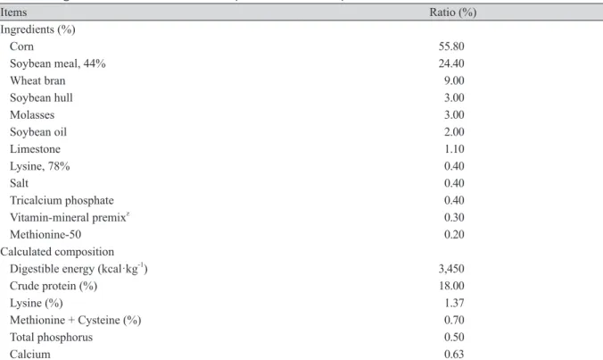
시험 동물은 체중 56.1 kg 전후 듀록종 암, 수 각각 3마리를 공시하였다. 시험 동물은 항온·항습이 유지되는 챔버 에서 대사틀에 5일간 사육 온도 25℃, 상대습도 60%의 조건(thermal neutral, TN)으로 적응기간을 두었다. 적응 기간 이후 사육 온도 33℃, 상대습도 60%의 조건(heat stress at 33℃, HS33)으로 24시간 후 혈액을 채취하였다. 시험기간 동안 급여 사료는 옥수수·대두박 위주로 한국돼지사양표준(NIAS, 2017) 영양소 요구량 이상 배합한 사료를 1일 2 회로 2 kg씩 급여하였고, 물은 자유 채식하였다. 시험 사료의 원료 구성과 화학적 조성은 Table 1과 같다.
조사 항목 및 조사 방법
돼지의 일당 사료 섭취량, 일일 음수량, 물 허실량을 조사하였고, 혈액 시료는 10:00에 경정맥에서 채취하여 헤파 린 처리 후 1시간 이내에 원심분리기로 4℃에서 15분간 원심분리(1,660 × g) 후 상층액을 액체 질소로 급속 동결하 여 분석 전까지 - 80℃에서 냉동 보관하였다.
일당 사료 섭취량(average daily feed intake, ADFI)은 전체 사료섭취량에서 시료채취시점에 남은 사료 량을 제외하 여 계산하였다. 일일 음수량은 유량계(Daesung, Seoul, Korea)를 니플에 설치하여 매일 투입량을 측정하였고, 워터컵 에서 배출되는 물의 양을 회수하여 측정하여 계산하였다.
혈장에서 tumor necrosis factor (TNF)-α 분석은 ELISA 분석 kit (R&D system, Minneapolis, USA)를 이용하여 분석 하였다. 그리고 nitrate (μmol·L-1)와 total nitric oxide (μmol·L-1) 분석은 parameter 분석 kit (R&D systems, Minneapolis,
USA)를 이용하여 분석하였다. 혈장에서 유리아미노산 분석은 이전 연구에서 제시된 방법으로 전처리 하였다 (Reverter et al., 1997). 그 후, 아미노산 전용 분석기(Hitachi L8900, Tokyo, Japan)를 이용하여 혈장 내 유리아미노산 을 정량 분석하였다.
Table 1. Ingredients and chemical composition of the experimental diets.
Items Ratio (%)
Ingredients (%)
Corn 55.80
Soybean meal, 44% 24.40
Wheat bran 9.00
Soybean hull 3.00
Molasses 3.00
Soybean oil 2.00
Limestone 1.10
Lysine, 78% 0.40
Salt 0.40
Tricalcium phosphate 0.40
Vitamin-mineral premixz 0.30
Methionine-50 0.20
Calculated composition
Digestible energy (kcal·kg-1) 3,450
Crude protein (%) 18.00
Lysine (%) 1.37
Methionine + Cysteine (%) 0.70
Total phosphorus 0.50
Calcium 0.63
z Supplied per kg of diet: Vit A 5,000,000 IU; Vit D3 1,000,000 IU; Vit E 1,000 mg; Vit B1 150 mg; Vit B2 300 mg; Vit B12 1,500 mg; niacin amide 1,500 mg; DL-calcium pantothenate 1,000 mg; folic acid 200 mg; Vit H 10 mg; choline chloride 2,000 mg;
Mn 3,800 mg; Zn 1,500 mg; Fe 4000 mg; Cu 500 mg; I 250 mg; Co 100 mg; Mg 200 mg.
Results and Discussion
돼지가 고온 스트레스를 받지 않고 정상적인 몸 상태를 유지하기 위해서는 열적 중성(thermal neutral, TN) 온도 가 필요하며, 이 온도의 구간은 열적 중성권(TN zone)이다. 일반적으로, 상대적으로 체중이 많이 나가는 육성·비 육돈의 TN zone이 이유자돈보다 낮고, 고온에 대한 반응성 또한 이유자돈에 비해 육성·비육돈이 더 크다(Cottrell
et al., 2015). 본 연구에서 사용된 육성돈의 TN온도는 25℃이하이다(Quiniou et al., 2001). 하지만, HS33 처리구에 서 사료섭취량이 TN보다 유의적으로(p < 0.05; Table 2) 감소하였고, 음수량과 물 허실량이 증가하는 경향(p < 0.10;
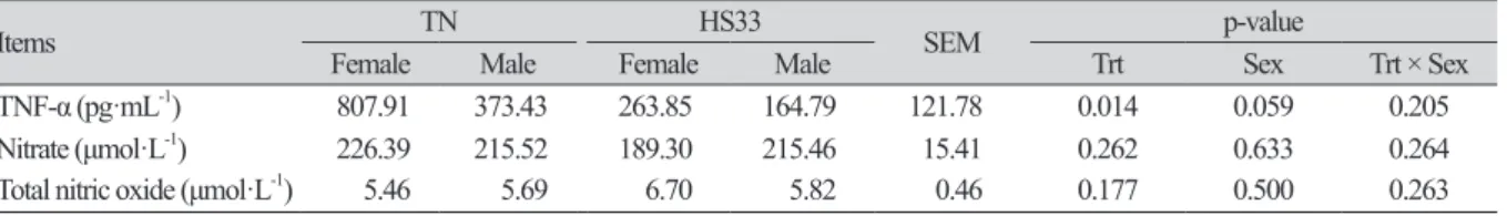
Table 2)으로 보아 돼지들은 고온 스트레스를 받은 것으로 보인다. 사료섭취량이 감소한 이유는 고온 스트레스로 부터 몸을 보호하기 위해 체내 대사열 생산을 최소화하기 위한 것으로 보이며, 이전 연구에 의하면 고온 스트레스 처리 후 24시간 동안 사료효율 감소로 인한 체내 이화작용과 탈수증상을 일으킨 것으로 보고되었다(Pearce et al., 2013a; Park et al., 2016b; Lu et al., 2018). 뿐만 아니라, 고온 스트레스를 받은 돼지들은 몸을 보호하기 위해 친염증성 사이토카인인 TNF-α의 발현량을 조절한 것으로 보인다. HS33은 TNF-α를 TN과 비교하여 유의적(p < 0.05; Table 3)으로 감소시켰으며, 이는 TNF-α로 인해 발생하는 apoptosis로부터 몸을 보호하기 위한 방어기작으로 보인다(Qu and Ajuwon, 2018). 하지만, 수컷의 TNF-α 수치가 암컷에 비해 낮은 경향(p < 0.10; Table 3)으로 보아 고온 스트레스 로부터 몸을 보호하기위한 수컷의 방어기작이 암컷보다 더 강했을 것으로 보인다. 또한, 암컷보다 상대적으로 두 꺼운 수컷의 피하지방층과 암컷의 성호르몬에 의한 영향이 성별에 의한 결과의 차이를 보여준 것으로 판단된다 (Armero et al., 1999; Wang et al., 2007).
Table 2. Effect of heat stress on feed intake, water intake, and water loss in growing pigs under thermal neutral (TN) or heat stress (HS) conditions.
Items TN HS33 SEM p-value
Female Male Female Male Trt Sex Trt × Sex
ADFI (kg·d-1) 2.50 2.83 1.41 1.69 0.33 0.011 0.394 0.936
ADWI (L·d-1) 7.76 12.90 20.75 22.66 5.23 0.079 0.545 0.780
Water loss (L·d-1) 10.68 14.10 26.75 58.00 12.75 0.062 0.241 0.337
HS33, heat stress at 33℃; SEM, standard error of the mean; Trt, treatment; Trt × Sex, treatment × sex interaction; ADFI, average daily feed intake; ADWI, average daily water intake.
Table 3. Effect of heat stress on plasma concentrations of tumor necrosis factor (TNF)-α, nitrate, and total nitric oxide in growing pigs under thermal neutral (TN) or heat stress (HS) conditions.
Items TN HS33 SEM p-value
Female Male Female Male Trt Sex Trt × Sex
TNF-α (pg·mL-1) 807.91 373.43 263.85 164.79 121.78 0.014 0.059 0.205
Nitrate (μmol·L-1) 226.39 215.52 189.30 215.46 15.41 0.262 0.633 0.264
Total nitric oxide (μmol·L-1) 5.46 5.69 6.70 5.82 0.46 0.177 0.500 0.263
HS33, heat stress at 33℃; SEM, standard error of the mean; Trt, treatment; Trt × Sex, treatment × sex interaction.
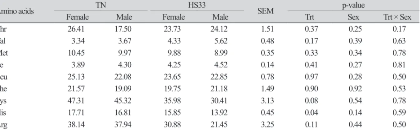
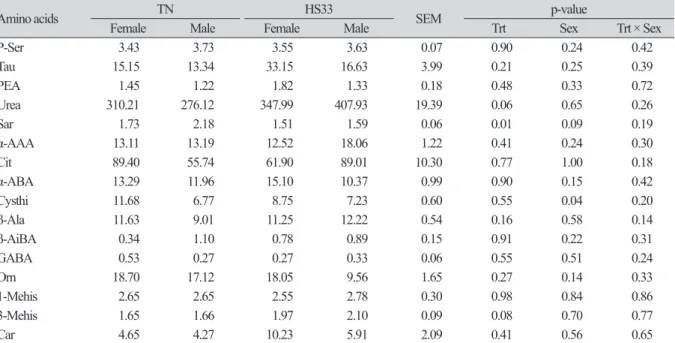
고온 스트레스는 혈장 내 필수 아미노산인 histidine (His)과 비단백질 아미노산인 sarcosine (Sar)의 결과에 영향을 주었고, 이것은 고온 스트레스에 의한 산화적 스트레스로부터 몸을 보호하기 위한 작용때문이다. 하지만, 혈장 내 비필수 아미노산의 함량은 변화가 없었다(Table 5). His은 장 염증을 완화시켜주는 물질이며, 항산화 기능을 가진 carnosine (Car)합성의 전구체이고, His과 Sar은 고온 스트레스에 의해 발생하는 활성산소를 줄여준다(Boldyrev et al., 2013; Akbarian et al., 2016; Morales et al., 2016b). 본 실험에서 HS33 처리구의 His과 Sar은 TN에 비해 유의적(p < 0.05)으 로 감소하였다(Table 4 and Table 6). 따라서, 고온 스트레스를 받은 돼지들의 혈장에서 항산화 기능을 가진 두 아미노 산(His과 Sar)의 감소는 항산화 스트레스를 유발하는 물질들을 줄이기 위해 사용되었을 것이다(Morales et al., 2016a).
하지만, 이전 연구결과에 의하면 만성 고온 스트레스를 받은 육성돈의 혈청 내 Car과 Sar 모두 감소하였지만, 본 실험 에서 급성 고온 스트레스를 받은 육성돈의 혈장 내 Car의 양은 유의적인 차이가 없었다(Table 6; Morales et al., 2016a).
이는 각 실험에서 사용된 고온 스트레스 조건(만성 vs. 급성)의 차이 때문으로 판단된다(Morales et al., 2016a).
Table 4. Effect of heat stress on plasma concentrations (μg·mL-1) of indispensable amino acids in growing pigs under thermal neutral (TN) or heat stress (HS) conditions.
Amino acids TN HS33 SEM p-value
Female Male Female Male Trt Sex Trt × Sex
Thr 26.41 17.50 23.73 24.12 1.51 0.37 0.25 0.17
Val 3.34 3.67 4.33 5.62 0.48 0.17 0.39 0.63
Met 10.45 9.97 9.88 8.99 0.35 0.33 0.34 0.78
Ile 3.89 4.30 4.25 4.52 0.14 0.41 0.27 0.81
Leu 25.13 22.08 23.65 22.85 0.78 0.97 0.28 0.50
Phe 21.57 19.09 19.75 21.18 1.49 0.90 0.92 0.53
Lys 47.31 45.32 35.98 30.41 3.13 0.08 0.54 0.78
His 17.71 16.81 15.85 13.92 0.45 0.04 0.14 0.59
Arg 38.14 37.94 30.88 21.45 3.25 0.11 0.44 0.50
HS33, heat stress at 33℃; SEM, standard error of the mean; Trt, treatment; Trt × Sex, treatment × sex interaction; Thr, threonine; Val, valine;
Met, methionine; Ile, isoleucine; Leu, leucine; Phe, phenylalanine; Lys, lysine; His, histidine; Arg, arginine.
Table 5. Effect of heat stress on plasma concentrations (μg·mL-1) of dispensable amino acids in growing pigs under thermal neutral (TN) or heat stress (HS) conditions.
Amino acids TN HS33
SEM p-value
Female Male Female Male Trt Sex Trt × Sex
Asp 0.55 0.70 0.68 0.52 0.06 0.76 0.85 0.24
Ser 18.46 17.35 15.99 14.47 0.95 0.22 0.50 0.92
Gly 28.34 25.00 28.75 28.03 1.12 0.39 0.43 0.58
Ala 95.93 118.61 104.29 86.59 5.93 0.27 0.98 0.13
Cys 113.09 97.06 106.11 105.48 2.91 0.70 0.24 0.23
Tyr 50.09 45.61 47.69 46.89 1.56 0.97 0.46 0.57
Pro 16.23 16.57 15.59 24.78 1.63 0.28 0.15 0.22
Conclusion
본 연구는 급성 고온 스트레스 처리에 따른 육성돈의 사료 섭취량, TNF-α, 그리고 혈장 내 아미노산의 비교를 통하여 고온 스트레스가 돼지에게 미치는 영향을 평가하였다. 고온 스트레스는 사료섭취량과 TNF-α를 감소시켰으며, histidine 과 sarcosine의 농도를 감소시켰다. 사료섭취량과 TNF-α의 감소는 사료 섭취에 의한 대사열을 감소시키기 위함이고, TNF-α에 의해 발생하는 apoptosis로부터 몸을 보호하기 위한 방어기작으로 판단된다. 또한, histidine과 sarcosine의 감소 는 고온 스트레스로 인해 발생하는 산화적 스트레스를 줄이기 위해 이용되었기 때문에 줄어든 것으로 판단된다. 따라 서, 본 연구 결과에서 고온 스트레스에 의해 변화하는 아미노산들은 고온 스트레스 지표로써 가능성을 보여준다.
Acknowledgements
본 연구는 농촌진흥청 연구사업(세부과제번호: PJ01277102)의 지원 및 2019년도 농촌진흥청 국립축산과학원 전 문연구원 과정 지원사업에 의해 수행되었습니다.
고온 스트레스는 육성돈의 근육 내 단백질의 손상과 이화작용을 일으켰을 것이며, 이는 혈액 내 urea와 3-methylhistidine (3-Mehis)의 변화와 연관이 있다. 본 실험에서 HS33처리에 의해 urea와 3-Mehis 모두 TN보다 증가하는 경향(p < 0.10; Table 6)을 보였으며, 이러한 결과는 고온 스트레스에 의해 근육의 손상과 분해가 일어난 것을 암시한다(Pearce et al., 2013a;
Morales et al., 2016a). 하지만, 유의적인 차이가 없었기 때문에 본 실험에서 사용된 급성 고온 스트레스는 육성돈의 근육 내 단백질에 미친 영향이 크진 않았음을 의미한다.
Table 6. Effect of heat stress on plasma concentrations (μg·mL-1) of nonprotein amino acids in growing pigs under thermal neutral (TN) or heat stress (HS) conditions.
Amino acids TN HS33 SEM p-value
Female Male Female Male Trt Sex Trt × Sex
P-Ser 3.43 3.73 3.55 3.63 0.07 0.90 0.24 0.42
Tau 15.15 13.34 33.15 16.63 3.99 0.21 0.25 0.39
PEA 1.45 1.22 1.82 1.33 0.18 0.48 0.33 0.72
Urea 310.21 276.12 347.99 407.93 19.39 0.06 0.65 0.26
Sar 1.73 2.18 1.51 1.59 0.06 0.01 0.09 0.19
α-AAA 13.11 13.19 12.52 18.06 1.22 0.41 0.24 0.30
Cit 89.40 55.74 61.90 89.01 10.30 0.77 1.00 0.18
α-ABA 13.29 11.96 15.10 10.37 0.99 0.90 0.15 0.42
Cysthi 11.68 6.77 8.75 7.23 0.60 0.55 0.04 0.20
β-Ala 11.63 9.01 11.25 12.22 0.54 0.16 0.58 0.14
β-AiBA 0.34 1.10 0.78 0.89 0.15 0.91 0.22 0.31
GABA 0.53 0.27 0.27 0.33 0.06 0.55 0.51 0.24
Orn 18.70 17.12 18.05 9.56 1.65 0.27 0.14 0.33
1-Mehis 2.65 2.65 2.55 2.78 0.30 0.98 0.84 0.86
3-Mehis 1.65 1.66 1.97 2.10 0.09 0.08 0.70 0.77
Car 4.65 4.27 10.23 5.91 2.09 0.41 0.56 0.65
HS33, heat stress at 33℃; SEM, standard error of the mean; Trt, treatment; Trt × Sex, treatment × sex interaction; P-Ser, phosphoserine;
Tau, taurine; PEA, phosphoethanolamine; Sar, sarcosine; α-AAA, α-aminoadipic acid; Cit, citrulline; α-ABA, α-aminobutyric acid; Cysthi, Cystathionine; β-Ala, β-alanine; β-AiBA, β-aminoisobutric acid; GABA, γ-aminobutyric acid; Orn, ornithine; 1-Mehis, 1-methylhistidine;
3-Mehis, 3-methylhistidine; Car, carnosine.
Authors Information
Byeonghyeon Kim, https://orcid.org/0000-0003-4651-6857 Minji Kim, https://orcid.org/0000-0003-2106-1921 Hye Ran Kim, https://orcid.org/0000-0003-2207-3668 Sang Yun Ji, https://orcid.org/0000-0001-7235-3655 Ki Hyun Kim, https://orcid.org/0000-0002-9834-2126 Ju Lan Chun, https://orcid.org/0000-0002-4618-586X Youl-Chang Baek, https://orcid.org/0000-0003-4454-5339 Yookyung Lee, https://orcid.org/0000-0002-9896-4152 Jin Young Jeong, https://orcid.org/0000-0002-8670-7036 Sung Dae Lee, Rural https://orcid.org/0000-0002-9167-4099
References
Akbarian A, Michiels J, Degroote J, Majdeddin M, Golian A, De Smet S. 2016. Association between heat stress and oxidative stress in poultry; mitochondrial dysfunction and dietary interventions with phytochemicals. Journal of Animal Science and Biotechnology 7:37.
Armero E, Flores M, Toldra F, Barbosa JA, Olivet J, Pla M, Baselga M. 1999. Effects of pig sire type and sex on carcass traits, meat quality and sensory quality of dry-cured ham. Journal of the Science of Food and Agriculture 79:1147- 1154.
Baumgard LH, Rhoads RP. 2013. Effects of heat stress on postabsorptive metabolism and energetics. Annual Review of Animal Biosciences 1:311-337.
Boldyrev AA, Aldini G, Derave W. 2013. Physiology and pathophysiology of carnosine. Physiological Reviews 93:1803- 1845.
Cottrell JJ, Liu F, Hung AT, DiGiacomo K, Chauhan SS, Leury BJ, Furness JB, Celi P, Dunshea FR. 2015. Nutritional strategies to alleviate heat stress in pigs. Animal Production Science 55:1391-1402.
Dou S, Villa-Vialaneix N, Liaubet L, Billon Y, Giorgi M, Gilbert H, Gourdine JL, Riquet J, Renaudeau D. 2017. 1HNMR- Based metabolomic profiling method to develop plasma biomarkers for sensitivity to chronic heat stress in growing pigs. PLoS One 12:1-18.
Escribano D, Soler L, Gutiérrez AM, Martínez-Subiela S, Cerón JJ. 2013. Measurement of chromogranin A in porcine saliva: Validation of a time-resolved immunofluorometric assay and evaluation of its application as a marker of acute stress. Animal 7:640-647.
Guo J, Gao S, Quan S, Zhang Y, Bu D, Wang J. 2018. Blood amino acids profile responding to heat stress in dairy cows.
Asian-Australasian Journal of Animal Sciences 31:47-53.
Jaturasitha S, Srikanchai T, Chakeredza S, ter Meulen U, Wicke M. 2008. Backfat characteristics of barrows and gilts
Morales A, Cota SEM, Ibarra NO, Arce N, Htoo JK, Cervantes M. 2016a. Effect of heat stress on the serum concentrations of free amino acids and some of their metabolites in growing pigs. Journal of Animal Science 94:2835-2842.
Morales A, Hernández L, Buenabad L, Avelar E, Bernal H, Baumgard LH, Cervantes M. 2016b. Effect of heat stress on the endogenous intestinal loss of amino acids in growing pigs. Journal of Animal Science 94:165-172.
Muhlisin, Panjono, Lee SJ, Lee JK, Lee SK. 2014. Effects of crossbreeding and gender on the carcass traits and meat quality of Korean native black pig and duroc crossbred. Asian-Australasian Journal of Animal Sciences 27:1019- 1025.
NIAS (National Institute of Animal Science). 2017. Korean feeding standard for swine. NIAS, RDA, Wanju, Korea.
Park JE, Heo BY, Young S. 2016a. A study on human damage duo to heat wave by region. Journal of Korean Society of Hazard Mitigation 16:103-109. [in Korean]
Park S, Kim B, Kim Y, Kim S, Jang K, Kim Y, Park J, Song M, Oh S. 2016b. Nutrition and feed approach according to pig physiology. Korean Journal of Agricultural Science 43:750-760. [in Korean]
Pearce SC, Gabler NK, Ross J, Escobar J, Patience JF, Rhoads RP, Baumgard LH. 2013a. The effects of heat stress and plane of nutrition on metabolism in growing pigs. Journal of Animal Science 91:2108-2118.
Pearce SC, Mani V, Boddicker RL, Johnson JS, Weber TE, Ross JW, Baumgard LH, Gabler NK. 2012. Heat stress reduces barrier function and alters intestinal metabolism in growing pigs. Journal of Animal Science 90:257-259.
Pearce SC, Mani V, Weber TE, Rhoads RP, Patience JF, Baumgard LH, Gabler NK. 2013b. Heat stress and reduced plane of nutrition decreases intestinal integrity and function in pigs. Journal of Animal Science 91:5183-5193.
Pearce SC, Sanz-Fernandez MV, Hollis JH, Baumgard LH, Gabler NK. 2014. Short-term exposure to heat stress attenuates appetite and intestinal integrity in growing pigs. Journal of Animal Science 92:5444-5454.
Qu H, Ajuwon KM. 2018. Adipose tissue-specific responses reveal an important role of lipogenesis during heat stress adaptation in pigs. Journal of Animal Science 96:975-989.
Quiniou N, Noblet J, van Milgen J, Dubois S. 2001. Modelling heat production and energy balance in group-housed growing pigs exposed to low or high ambient temperatures. British Journal of Nutrition 85:97-106.
Reverter M, Lundh T, Lindberg JE. 1997. Determination of free amino acids in pig plasma by precolumn derivatization with 6-N-aminoquinolyl-N-hydroxysuccinimidyl carbamate and high-performance liquid chromatography.
Journal of Chromatography B: Biomedical Applications 696:1-8.
Ross JW, Hale BJ, Gabler NK, Rhoads RP, Keating AF, Baumgard LH. 2015. Physiological consequences of heat stress in pigs. Animal Production Science 55:1381-1390.
Wang J, Korczykowski M, Rao H, Fan Y, Pluta J, Gur RC, McEwen BC, Detre JA. 2007. Gender difference in neural response to psychological stress. Social Cognitive and Affective Neuroscience 2:227-239.