다공성 칼슘포스파이트에 대한 파괴분석
박진홍
*· 배지용
*,**· 심재범
***,전인수
†Numerical analysis of fracture mechanisms for porous calcium phosphate
Jinhong Park , Jiyong Bae, Jaebum shin, Insu Jeon
Key Words :
Calcium phosphate(칼슘포스파이트), Micro focus X-ray CT system(마이크로 초점 X- 레이 단층촬영 시스템), 3D reconstruction(3 차원 재구축), finite element
analysis(유한요소 해석)
Abstract
In this study, the fracture strength for fracture mechanism porous calcium phosphate made from sintered with ß- tricalcium phosphate obtained by wet precipitation procedure is analyzed using finite element method and experiment measurement. First, three 3×3×3 ㎜³and 5×5×5 ㎜³ specimens are prepared and tomographic images of one 5×5×5 ㎜³ specimen are obtained by micro focus X-ray CT. The compression tests using the specimens are carried out to measure the elastic modulus and fracture strength to analyze the fracture mechanism of porous calcium phosphate specimen. The tomographic images are reconstructed by 3D reconstruction program. The finite elements are directly built up in the reconstructed specimen. The numerical simulation for the compression tests is performed using the element. The mechanism of calcium phosphate of simulation are obtained by the compression tests using there cylindric specimen of height 19.5 ㎜ and diameter 10 ㎜. From the results, the applicability of porous calcium phosphate is evaluated to care fracture and vacant bone of a patient as the reinforcement material.
1. 서 론
캴슘포스페이트는 인조골의 재료로써 다양한
임플란트와 스카폴더(1-3) 제작뿐만 아니라
임플란트의 코팅(4-6), 골 시멘트(7) 등으로 사용되어 왔다. 파손된 뼈의 대용물질로서
사용할 수 있으며 구강 이식조직 편에 충전재 및 보강, 활성지지대로 활용할 수 있는 생체 적합성을 갖춘 골 대체 및 재생재료이다
2. 실험방법
2.1 시편에 대한 3 차원 재구축
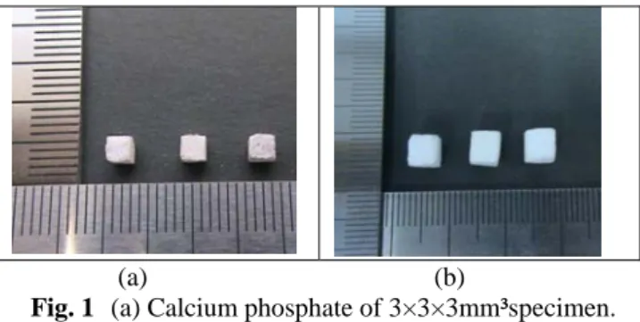
먼저 3×3×3mm³시편과 5×5×5mm³의 작은 다 공성 재료의 시편을 제작 한 후 마이크로 초점 X-선 CT 장비(Micro focus X-ray CT system)를 이용 하여 재료의 내부구조를 단층 촬영한다 (Fig. 1).
†
회원, 전남대학교 기계시스템공학부 E-mail : [email protected]TEL : (062)530-1688 FAX : (062)530-1689
*
전남대학교 기계공학과**
화순전남대학교병원 관절센터***
주)경원메디칼1301 대한기계학회 2008년도 추계학술대회 논문집
(a) (b)
Fig. 1 (a) Calcium phosphate of 3×3×3mm³specimen.
(b) Calcium phosphate of 5×5×5mm³specimen.
촬영한 내부 구조의 Tomographic 사진으로 촬영 하고 촬영한 Tomographic 사진에 대한 전체구조 를 3 차원적으로 재구축한다 (Fig. 2).
(a) (b)
Fig. 2 (a) A tomography image of calcium phosphate.
(b) 3D reconstruction image of calcium phosphate.
2.2 3 차원 유한 요소 모델링
재구축된 다공성 칼슘포스파이트 시편의 3 차원 모델에 PATRAN 을 사용하여 직접 3 차원 유한 요 소들을 형성시킨다 (Fig. 3).
Fig. 3 Finite element model of calcium phosphate.
3. 수치해석
3.1 압축 시험
시편의 압축 변형에 대한 수치해석을 위하여 사 용한 재료의 물성치는 압축 시험기를 사용하여 칼 슘포스파이트 시편 지름 10mm 높이 19.5 mm 의 시편 이용하여 얻는다. 시뮬레이션을 위한 경계조
건은 X,Y 방향은 압축시험기 중앙에 구속되며 Z 축 변위는 5.31%의 변형이 위쪽 압반에 걸린다.
4. 향후 계획
4.1 유한요소해석을 실시한 후 그 계산결과를 실험결과와 비교하여 파손 및 손실된 뼈를 위한 보강재로써 발포 칼슘포스파이트의 활용 가능성을 평가한다.
참고문헌
(1) Xiaoshu Dai, Satya Shivkumar, 2008, "Hybrid analogs for production of porous calcium phosphate scaffolds," Material science and engineering, vol.28 pp336-340
(2) Julian R. jones, Gowsihan Poologasundarampillai, Robert C. Atwood, Dominique Bernard, Peter D. Lee, 2006, "Non-destructive quantitative 3D analysis for the optimization of tissue scaffolds," biomaterials, vol. 28, pp1404~1413
(3) Yan Li, In-Seop Lee, Fu-Zhai Cui, Seong-Ho Choi, 2008, "The biocompatibility of nanostructured calcium phosphate coated on micro-arc oxidized titanium,"
biomaterials, vol. 29, pp2025~2032
(4) Qassilis Karageorgiou, David Kaplan, 2005,
"Porosity of 3D biomaterial scaffolds and psteogenesis"
biomaterials, vol. 26 pp5474~5491
(5) Mangal Roy, b. Vamsi Krishna, Amit Bandyopadhyay, Susmita Bose, 2008, "Laser processing of bioactive tricalcium phosphate coating on titanium for load-bearing implant," Acta biomaterialia vol. 4, pp324~333
(6) C.X Resende, J.Dille, G.M.Platt, I.N.Bastos, G.A.Soares, 2008"characterization of coating produced on titanium surface by a designed solution containing calcium and phosphate ions," materials chemistry and physics, vol. 109, pp429-435
(7) Sabine H. Dickens, Glenn M. Flaim, 2008, "Effect of a bonding agent on in vitro biochemical activities of remineralizing resin-based calcium phosphate cements,"
Dental materials pp1~8
1302