https://doi.org/10.3350/cmh.2020.0126 Clinical and Molecular Hepatology 2020;26:562-576
Original Article
Clinical outcomes of coronavirus disease 2019 in
patients with pre-existing liver diseases: A multicenter study in South Korea
Yu Rim Lee
1,2,3,*, Min Kyu Kang
3,4,*, Jeong Eun Song
3,5, Hyun Jung Kim
3,6, Young Oh Kweon
1,2,3, Won Young Tak
1,2,3, Se Young Jang
1,2,3, Jung Gil Park
3,4, Changhyeong Lee
3,5, Jae Seok Hwang
3,6, Byoung Kuk Jang
3,6, Jeong Ill Suh
3,7, Woo Jin Chung
3,6, Byung Seok Kim
3,5, and Soo Young Park
1,2,31Department of Internal Medicine, School of Medicine, Kyungpook National University, Daegu; 2Department of Internal Medicine, Kyungpook National University Hospital, Daegu; 3Daegu-Gyeongbuk Liver Study Group; 4Department of Internal Medicine, Yeungnam University College of Medicine, Daegu; 5Department of Internal Medicine, Daegu Catholic University School of Medicine, Daegu;
6Department of Internal Medicine, Keimyung University School of Medicine, Daegu; 7Department of Internal Medicine, Dongguk Univer- sity Gyeongju Hospital, Dongguk University College of Medicine, Gyeongju, Korea
Graphical Abstract
Background/Aims: Although coronavirus disease 2019 (COVID-19) has spread rapidly worldwide, the implication of
pre-existing liver disease on the outcome of COVID-19 remains unresolved.
Methods: A total of 1,005 patients who were admitted to five tertiary hospitals in South Korea with laboratory-con-
firmed COVID-19 were included in this study. Clinical outcomes in COVID-19 patients with coexisting liver disease as well as the predictors of disease severity and mortality of COVID-19 were assessed.
Results: Of the 47 patients (4.7%) who had liver-related comorbidities, 14 patients (1.4%) had liver cirrhosis. Liver cirrho-
sis was more common in COVID-19 patients with severe pneumonia than in those with non-severe pneumonia (4.5% vs.
0.9%, P=0.006). Compared to patients without liver cirrhosis, a higher proportion of patients with liver cirrhosis required oxygen therapy; were admitted to the intensive care unit; had septic shock, acute respiratory distress syndrome, or acute kidney injury; and died (P<0.05). The overall survival rate was significantly lower in patients with liver cirrhosis than in those without liver cirrhosis (log-rank test, P=0.003). Along with old age and diabetes, the presence of liver cirrhosis was found to be an independent predictor of severe disease (odds ratio, 4.52; 95% confidence interval [CI], 1.20–17.02;
P=0.026) and death (hazard ratio, 2.86; 95% CI, 1.04–9.30; P=0.042) in COVID-19 patients.
Conclusions: This study suggests liver cirrhosis is a significant risk factor for COVID-19. Stronger personal protection and
more intensive treatment for COVID-19 are recommended in these patients. (Clin Mol Hepatol 2020;26:562-576)
Keywords: Liver diseases; Liver cirrhosis; Prognosis; Mortality; COVID-19Study Highlights
Liver cirrhosis was more common in COVID-19 patients with severe pneumonia than in those with non-severe pneumonia. Compared to patients without liver cirrhosis, patients with liver cirrhosis had a significantly higher proportion of oxygen therapy, admission to ICU, septic shock, acute re- spiratory distress syndrome, acute kidney injury, and death. The presence of liver cirrhosis was significantly associated with disease severity and death in COVID-19 patients.
Corresponding author : Woo Jin Chung
Department of Internal Medicine, Keimyung University School of Medicine, 1035 dalgubeul-daero, Dalseo-gu, Daegu 42601, Korea Tel: +82-53-258-7200, Fax: +82-53-258-6008
E-mail: [email protected]
https://orcid.org/0000-0002-3736-1067 Byung Seok Kim
Department of Internal Medicine, Daegu Catholic University School of Medicine, 33 Duryugongwon-ro 17-gil, Nam-gu, Daegu 42472, Korea Tel: +82-53-650-4090, Fax: +82-53-656-3281
E-mail: [email protected]
https://orcid.org/0000-0002-4318-4570 Soo Young Park
Department of Internal Medicine, Kyungpook National University Hospital, School of Medicine, Kyungpook National University, 130 Dongdeok-ro, Jung-gu, Daegu 41944, Korea
Tel: +82-53-200-5519, Fax: +82-53-426-8773 E-mail: [email protected] https://orcid.org/0000-0002-4944-4396 Abbreviations:
ACLF, acute-on-chronic liver failure; ALT, alanine aminotransferase; ARDS, acute respiratory distress syndrome; BMI, body mass index; CI, confidence interval; CLIF-C, Chronic Liver Failure Consortium; COPD, chronic obstructive pulmonary disease; COVID-19, coronavirus disease 2019; HCC, hepatocellular carcinoma; HR, hazard ratio; HSI, hepatic steatosis index; ICU, intensive care unit;
IQR, interquartile range; MELD, model for end-stage liver disease; NAFLD, non- alcoholic fatty liver disease; OF, organ failure; OR, odds radio; SARS-CoV-2, severe acute respiratory syndrome coronavirus 2; WHO, World Health Organization
Received : Jun. 4, 2020 / Revised : Jul. 31, 2020 / Accepted : Aug. 4, 2020 Editor: Ju Dong Yang, Cedars-Sinai Medical Center, USA
*Yu Rim Lee and Min Kyu Kang have equally contributed to this work.
INTRODUCTION
Since the first case of coronavirus disease 2019 (COVID-19) was identified in Wuhan, China in December 2019, as of June 2, 2020, a total of 6,194,533 laboratory-confirmed COVID-19 cases and 376,320 deaths had been documented worldwide.1 The extensive expansion of COVID-19, caused by severe acute respiratory syn- drome coronavirus 2 (SARS-CoV-2), represents a great concern globally.2,3 Due to the rapid spread of COVID-19 and its high mor- tality rate, it is necessary to identify the risk factors affecting the clinical outcomes of COVID-19. Knowledge about the risk factors of COVID-19 can help in the decision-making process for early management of COVID-19 patients.
Previous studies have shown that COVID-19 patients with co- morbidities had poor clinical outcomes.3-5 Underlying comorbidi- ties including diabetes, hypertension, chronic obstructive pulmo- nary disease (COPD), and cardiovascular disease are known to be risk factors of COVID-19. However, the impact of the presence of liver disease, which is one of the major public health problems worldwide, on the outcome of COVID-19 remains unclear.6 Glob- ally, liver disease accounts for approximately 2 million deaths per year.7 Moreover, the incidence and severity of infection are higher in cirrhosis patients compared to the general population.8,9 Only a few studies have been performed on liver disease and/or liver function as prognostic factors of COVID-19.10,11 In previous stud- ies, data were obtained from retrospective studies with a relative- ly small number of patients, and there were differences in the re- sults among studies.3-5 In addition, those studies did not show the clinical outcomes according to the exact cause of liver disease, such as chronic hepatitis or liver cirrhosis. Therefore, studies on liver-related comorbidities in COVID-19 patients are urgently needed.
In South Korea, the first confirmed case of COVID-19 was a Chi- nese woman who traveled from Wuhan, China on January 20, 2020. Since then, there have been 11,590 confirmed cases of COVID-19 in South Korea, and 273 of them have died as of June 3, 2020.12,13 As a large number of confirmed patients were linked to a religious group and hospitalized psychiatric patients in a closed ward, there was an explosive outbreak in the country with hundreds of newly confirmed cases per day.12,14 With a significant surge in new cases, deaths during home isolation while waiting for hospitalization or pending test results have occurred.14 There- fore, the need to monitor individuals with the risk factors for COVID-19, including old age and/or underlying disease, has emerged.
In this study, we evaluated the implication of existing liver-relat- ed comorbidities on the clinical outcomes of COVID-19 patients.
PATIENTS AND METHODS Patients
From February 17 to April 6, 2020, patients with laborato- ry-confirmed COVID-19 who were admitted to five designated tertiary hospitals in Daegu, South Korea were included in this study. COVID-19 was diagnosed according to the interim guidance of the World Health Organization (WHO), mainly by the detection of the SARS-CoV-2 sequence in the nasopharyngeal and oro- pharyngeal swab.15,16
The study was performed in accordance with the ethical guide- lines of the Helsinki Declaration of 1975, as revised in 2013. The study protocol was approved by the Institutional Review Board of Kyungpook National University Hospital (IRB No. KNUCH- 2020-04-017). Informed consent from study participants was waived by the ethics committee of the designated hospitals.
Data collection and study definition
We obtained medical records such as demographic variables, comorbidities, clinical symptoms, laboratory findings, and radio- logic imaging studies on admission, treatments, complications, and clinical outcomes from electronic records. Laboratory testing included a complete blood count, serum C-reactive protein, serum procalcitonin, liver and renal function test, coagulation testing, serum lactate dehydrogenase, and serum reatine kinase. The data cut-off point for the study was April 30, 2020.
The degree of severity of COVID-19 (severe vs. non-severe) was defined based on the WHO interim guidance for COVID-19.17 Shock and acute respiratory distress syndrome (ARDS) were also determined in accordance with the WHO interim guidance.17 Acute kidney injury was diagnosed by the highest serum creatinine level and urine output.18 Secondary infection was identified if the pa- tients had both clinical symptoms or signs of bacteremia or pneu- monia, and a positive culture of a new pathogen from blood sam- ples or a lower respiratory tract specimen. For patients requiring oxygen administration, the most intense level of oxygen therapy (nasal cannula or venturi mask, high-flow nasal cannula, invasive mechanical ventilation, or invasive mechanical ventilation with extracorporeal membrane oxygenation) was recorded.
Table 1. Baseline characteristics of COVID-19 patients with and without pre-existing liver disease (n=1,005) All (n=1,005)Liver disease (n=47, 4.7%)Without liver disease (n=958, 95.3%)P-value*LC (n=14, 1.4%)No LC (n=991, 98.6%)P-value* Demographic variable Age (years)61 (48–72)63 (54–70)60 (48–72)0.26966 (60–81)60 (48–72)0.051 Age, ≥65 years407/1,005 (40.5)20/47 (42.6)387/958 (40.4)0.7697/14 (50.0)400/991 (40.4)0.466 Male gender361/1,005 (35.9)26/47 (55.3)335/958 (35.0)0.00510/14 (71.4)351/991 (35.4)0.005 Height (cm)162 (155–168)166 (157–170)161 (155–168)0.217166 (158–174)161 (155–168)0.209 Body weight (kg)62 (54–70)64 (56–74)62 (54–70)0.33065 (56–73)62 (54–70)0.463 BMI (kg/m2 )23.7 (21.5–26.0)23.8 (22.6–26.1)23.7 (21.5–26.0)0.52723.0 (21.7–27.4)23.7 (21.5–26.0)0.997 Overweight, BMI ≥25142/435 (32.6)5/20 (25.0)137/415 (33.0)0.4552/8 (25.0)140/427 (32.8)0.642 Obese, BMI ≥3020/435 (4.6)1/20 (5.0)19/415 (4.6)0.9301/8 (12.5)19/427 (4.4)0.316 Smoking history Former or current smokers81/853 (9.5)9/41 (22.0)72/812 (8.9)0.0116/13 (46.2)75/840 (8.9)0.001 Other comorbidity Diabetes mellitus191/1,004 (19.0)13/47 (27.7)178/957 (18.6)0.1225/14 (35.7)186/990 (18.8)0.159 Hypertension311/1,004 (31.0)14/46 (30.4)297/958 (31.0)0.9355/14 (35.7)306/990 (30.9)0.772 Cardiovascular disease38/1,004 (3.8)3/47 (6.4)35/957 (3.7)0.4172/14 (14.3)36/990 (3.6)0.095 Chronic obstructive pulmonary disease14/1,004 (1.4)2/47 (4.3)12/957 (1.3)0.1370/14 (0.0)14/990 (1.4)0.654 Chronic renal disease16/1,003 (1.6)2/47 (4.3)14/956 (1.5)0.1701/14 (7.1)15/989 (1.5)0.203 Symptoms on admission Fever/chill452/995 (45.4)27/47 (57.4)425/948 (44.8)0.0908/14 (57.1)444/981 (45.3)0.375 Cough567/993 (57.1)23/47 (48.9)544/946 (57.5)0.2475/14 (35.7)562/979 (57.4)0.103 Shortness of breath247/995 (24.8)15/47 (31.9)232/948 (24.5)0.2496/14 (42.9)241/981 (24.6)0.125 Gastrointestinal symptoms (vomiting/ diarrhea)237/995 (23.8)11/47 (23.4)226/948 (23.8)0.9452/14 (14.3)235/981 (24.0)0.538 Myalgia325/994 (32.7)9/47 (19.1)316/947 (33.4)0.0431/14 (7.1)324/980 (33.1)0.044 Headache253/994 (25.6)8/47 (17.0)245/947 (25.9)0.1742/14 (14.3)251/980 (25.6)0.537 Time from symptom onset to admission (days)6 (3–9)7 (2–10)6 (3–9)0.7398 (1–8)6 (3–9)0.512 Vital signs at presentation Body temperature (°C)36.9 (36.5–37.3)36.9 (36.4–37.3)36.9 (36.5–37.4)0.51236.6 (36.2–37.3)36.9 (36.5–37.4)0.071 Respiratory rate (breath/min)20 (18–20)20 (18–20)20 (18–20)0.94020 (18–20)20 (18–20)0.658 Oxygen saturation (%)97 (95–98)97 (95–98)97 (95–98)0.83196 (94–98)97 (95–98)0.542
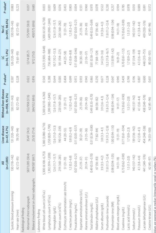
All (n=1,005)Liver disease (n=47, 4.7%)Without liver disease (n=958, 95.3%)P-value*LC (n=14, 1.4%)No LC (n=991, 98.6%)P-value* Systolic blood pressure (mmHg)130 (118–142)131 (120–140)130 (117–143)0.507126 (120–140)130 (118–143)0.883 Heart rate (/min)82 (72–95)78 (70–94)82 (72–95)0.22875 (61–95)82 (72–95)0.223 Radiological finding Abnormal chest radiograph587/1,005 (58.4)35/47 (74.5)552/958 (57.6)0.02212/14 (85.7)575/991 (58.0)0.037 Bilateral involvement on chest radiographs409/587 (69.7)25/35 (71.4)384/552 (69.6)0.8169/12 (75.0)400/575 (69.6)0.685 Laboratory finding White blood cell count (×103 /uL)5,300 (4,108–6,753)5,380 (3,980–6,710)5,300 (4,110–6,760)0.5435,225 (1,905–9,448)5,300 (4,120–6,730)0.833 Lymphocyte count (×103 /uL)1,392 (974–1,838)1,150 (820–1,790)1,399 (983–1,840)0.288736 (484–988)1,409 (990–1,840)0.001 Hemoglobin (g/dL)12.6 (11.7–13.7)12.6 (11.0–13.3)12.6 (11.8–13.7)0.64410.7 (10.2–13.3)12.6 (11.8–13.7)0.609 Platelet count (×109 /L)219 (168–281)190 (126–239)220 (169–283)0.005181 (104–221)220 (168–282)0.016 Erythrocyte sedimentation rate (mm/h)51 (31–70)44 (20–55)51 (31–71)0.40444 (25–58)51 (31–70)0.453 C-reactive protein (mg/L)1.2 (0.2–6.8)0.8 (0.2–6.9)1.2 (0.2–6.8)0.6994.5 (0.8–8.8)1.2 (0.2–6.7)0.803 Procalcitonin (ng/mL)0.07 (0.03–0.21)0.11 (0.05–0.57)0.07 (0.03–0.21)0.9720.11 (0.08–0.46)0.07 (0.03–0.21)0.834 Aspartate aminotransferase (U/L)25 (19–37)30 (21–39)25 (19–36)0.93336 (30–59)25 (19–36)0.551 Alanine aminotransferase (U/L)20 (14–31)23 (15–36)20 (14–30)0.94322 (16–37)20 (14–30)0.960 Total bilirubin (mg/dL)0.49 (0.33–0.70)0.51 (0.34–0.80)0.49 (0.33–0.70)0.8650.81 (0.58–1.21)0.49 (0.33–0.69)0.970 Alkaline phosphatase (U/L)67 (56–83)77 (62–84)66 (56–82)0.25077 (68–124)67 (56–82)0.078 Serum albumin (g/dL)3.9 (3.5–4.2)3.8 (3.3–4.1)3.9 (3.6–4.3)0.6773.4 (2.9–3.6)3.9 (3.6–4.3)0.522 Prothrombin time (seconds)11.8 (11.3–12.4)11.9 (11.2–12.8)11.8 (11.3–12.4)0.22813.2 (11.8–16.7)11.8 (11.2–12.4)0.230 Prothrombin time (INR)0.98 (0.94–1.05)1.00 (0.94–1.10)0.98 (0.94–1.04)0.6471.13 (0.99–1.42)0.98 (0.94–1.04)0.257 Blood urea nitrogen (mg/dL)14 (11–18)15 (11–19)14 (11–17)0.94516 (9–21)14 (11–18)0.934 Creatinine (mg/dL)0.73 (0.62–0.90)0.77 (0.61–0.94)0.73 (0.62–0.90)0.4390.70 (0.61–1.12)0.73 (0.62–0.90)0.785 Lactic acid (mmol/L)1.5 (1.1–2.0)1.7 (0.9–2.3)1.5 (1.1–2.0)0.7491.7 (0.9–2.7)1.5 (1.1–2.0)0.978 Sodium (mmol/L)140 (137–142)139 (136–142)140 (137–142)0.196137 (134–139)140 (137–142)0.010 Potassium (mmol/L)4.1 (3.8–4.4)4.2 (3.8–4.5)4.1 (3.8–4.4)0.8074.3 (3.6–4.6)4.1 (3.8–4.4)0.889 Lactate dehydrogenase (U/L)441 (349–576)454 (348–538)438 (349–578)0.600448 (313–751)439 (350–576)0.891 Creatine kinase (U/L)62 (45–97)74 (53–100)62 (45–96)0.63180 (50–101)62 (45–96)0.572 Values are expressed as median (interquartile range) or numbers (%). COVID-19, coronavirus disease 2019; LC, liver cirrhosis; BMI, body mass index; INR, internatinal normalized ratio. *Calculated by Student’s t test (or the Mann-Whitney U test, if appropriate) and chi-squared test (or Fisher’s exact test, if appropriate).
Table 1. Continued
Table 2. Treatments and clinical outcomes of COVID-19 patients with and without pre-existing liver disease (n=1,005) All (n=1,005)Liver disease (n=47, 4.7%)Without liver disease (n=958, 95.3%)P-value*LC (n=14, 1.4%)No LC (n=991, 98.6%)P-value* Treatments Antiviral therapy584/1,005 (58.1)27/47 (57.4)557/958 (58.1)0.92510/14 (71.4)574/991 (57.9)0.309 Lopinavir/ritonavir563/1,005 (56.0)27/47 (57.4)536/958 (55.9)0.84010/14 (71.4)553/991 (55.8)0.242 Darunavir/cobicistat51/,1005 (5.1)2/47 (4.3)49/958 (5.1)0.7930/14 (0.0)51/991 (5.1)1.000 Hydroxychloroquine537/1,005 (53.4)24/47 (51.1)513/958 (53.5)0.7397/14 (50.0)530/991 (53.5)0.795 Systemic glucocorticoid168/1,005 (16.7)10/47 (21.3)158/958 (16.5)0.3915/14 (35.7)163/991 (16.4)0.068 Immunoglobulin26/1,005 (2.6)3/47 (6.4)23/958 (2.4)0.1172/14 (14.3)24/991 (2.4)0.048 Oxygen support289/1,005 (28.8)15/47 (31.9)274/958 (28.6)0.6248/14 (57.1)281/991 (28.4)0.032 High-flow nasal cannula+invasive mechanical ventilation127/1,005 (12.6)11/47 (23.4)116/958 (12.1)0.0236/14 (42.9)121/991 (12.2)0.005 High-flow nasal cannula 57/1,005 (5.7)7/47 (14.9)50/958 (5.2)0.0143/14 (21.4)54/991 (5.4)0.040 Invasive mechanical ventilation70/1,005 (7.0)4/47 (8.5)66/958 (6.9)0.5623/14 (21.4)67/991 (6.8)0.068 Invasive mechanical ventilation and ECMO18/1,005 (1.8)1/47 (2.1)17/958 (1.8)0.5810/14 (0.0)18/991 (1.8)1.000 Complication ICU admission97/1,005 (9.7)8/47 (17.0)89/958 (9.3)0.1225/14 (35.7)92/991 (9.3)0.007 Septic shock80/1,005 (8.0)5/47 (10.6)75/958 (7.8)0.4144/14 (28.6)76/991 (7.7)0.020 Acute respiratory distress syndrome113/1,005 (11.2)8/47 (17.0)105/958 (11.0)0.1995/14 (35.7)108/991 (10.9)0.014 Acute kidney injury55/1,005 (5.5)3/47 (6.4)52/958 (5.4)0.7393/14 (21.4)52/991 (5.2)0.037 Continuous renal-replacement therapy22/1,005 (2.2)1/47 (2.1)21/958 (2.2)0.9761/14 (7.1)21/991 (2.1)0.268 Secondary infection41/1,005 (4.1)3/47 (6.4)38/958 (4.0)0.4341/14 (7.1)40/991 (4.0)0.444 Clinical outcomes Death77/1,005 (7.7)7/47 (14.9)70/958 (7.3)0.0824/14 (28.6)73/991 (7.4)0.018 Discharge816/1,005 (81.2)35/47 (74.5)781/958 (81.5)0.2277/14 (50.0)809/991 (81.6)0.008 Hospital stay (days)22 (15–31)22 (16–31)22 (15–31)0.81023 (16–32)22 (15–31)0.889 Values are expressed as median (interquartile range) or numbers (%). COVID-19, coronavirus disease 2019; LC, liver cirrhosis; ECMO, extracorporeal membrane oxygenation; ICU, intensive care unit. *Calculated by Student’s t test (or the Mann-Whitney U test, if appropriate) and chi-squared test (or Fisher’s exact test, if appropriate).
Chronic hepatitis B or C infection, liver cirrhosis, and hepatocel- lular carcinoma (HCC) were identified by history taking, serology test, and/or imaging studies such as computed tomography.19 Non-alcoholic fatty liver disease (NAFLD) was defined using he- patic steatosis index [HSI = 8 × (alanine aminotransferase [ALT] / aspartate aminotransferase ratio) + body mass index (BMI) + (+2, if diabetes mellitus; +2, if female)] of more than 36 points.20 For each patient with liver cirrhosis, Child-Pugh class, model for end- stage liver disease (MELD), chronic liver failure organ failure (CLIF- OF) score, and CLIF Consortium acute-on-chronic liver failure (CLIF-C ACLF) score were assessed on admission. ACLF on admis- sion was also assessed and graded according to the EASL-CLIF definition at diagnosis of COVID-19.21 Hepatic flare was defined as ALT level ≥5 × upper limit of normality.
Study outcomes
The primary endpoint was clinical outcomes (including oxygen supplementation, the use of high-flow oxygen therapy and inva- sive mechanical ventilation, admission to intensive care unit [ICU], ARDS, or death) in COVID-19 patients with coexisting liver dis- ease. The secondary endpoint was independent predictors of dis- ease severity and mortality in COVID-19.
Statistical analysis
Data are presented as the median and interquartile range (IQR) or numbers (%) as appropriate. No imputation was conducted for
missing data. Categorical variables were compared using the Chi- square test (or Fisher’s exact test), while Student’s t-test (or Mann-Whitney’s test) was used to compare continuous variables.
The cumulative overall survival rates were estimated using the Ka- plan-Meier method. Factors related to the severity of COVID-19 were identified with univariate and multivariate logistic regression analysis. The multivariate Cox proportional hazards regression test was used to identify the factors associated with mortality.
The odds radio (OR), hazard ratio (HR), and 95% confidence inter- val (CI) were also determined. A P-value<0.05 was considered statistically significant. Statistical analyses were performed using IBM SPSS Statistics for Windows version 25.0 (IBM Corp., Ar- monk, NY, USA).
RESULTS
Baseline characteristics and clinical outcomes of COVID-19
From February 17 to April 6, 2020, a total of 1,005 patients (361 men and 644 women) were included in this study. Baseline characteristics of the patients are summarized in Table 1. The me- dian age was 61 years. The most common symptoms of COVID-19 were cough (n=567, 57.1%), followed by fever and chills (n=452, 45.4%). Eight patients were healthcare-related COVID-19, and all of these patients had no pre-existing liver disease. Radiologic ab- normality was found in 587 patients (58.4%). Oxygen therapy
Figure 1. Kaplan-Meier plots of overall survival according to the presence of liver cirrhosis. Survival rate was signifi- cantly lower in patients with liver cirrhosis than in those without liver cirrhosis (log-rank test, P=0.003).
Patients without liver cirrhosis
Patients without liver cirrhosis
Log-rank test: P=0.003
Number at risk
Patients with liver cirrhosis
Patients with liver cirrhosis
991 584 143 17
14 9 1 0
0 20 40 60
Time (days) Survival probability 1.0
0.8 0.6 0.4
was administered to 289 patients (28.8%); among them, 70 pa- tients (7.0%) underwent invasive mechanical ventilation. Nine- ty-seven patients (9.7%) were admitted to the ICU, and 113 pa- tients (11.2%) had ARDS. Seventy-seven patients (7.7%) died after a median of 11 (6–25) days from COVID-19 diagnosis (Table 2). All patients died of respiratory failure due to progression of COVID-19.
When the patients were divided into severe (n=133, 13.2%) and non-severe (n=872, 86.8%) disease in accordance with WHO in- terim guidance for COVID-19,17 patients with severe pneumonia had significantly older age, higher body mass index, and more fre- quent smoking history (P<0.05) (Supplementary Table 1). The presence of comorbidities, including liver cirrhosis, diabetes, and hypertension, was more common in patients with severe disease compared to those with non-severe disease (P<0.05). However, there was no difference in the presence of chronic hepatitis be- tween the two groups. More abnormalities in laboratory tests and radiographic images were found in patients with severe disease than those with mild disease. A higher percentage of patients with severe disease received antiviral therapy, hydroxychloroquine, sys- temic glucocorticoid, immunoglobulin, and oxygen therapy com- pared to patients with non-severe disease (P<0.05) (Supplementa- ry Table 2). Patients with severe disease had a higher proportion of ICU admission, septic shock, ARDS, acute kidney injury, and death compared to patients with non-severe disease (P<0.05).
Comparison of COVID-19 patients with and without pre-existing liver disease
Of the 47 patients (4.7%) who had liver-related comorbidities, 14 patients (1.4%) had liver cirrhosis (Table 1). There were no sig- nificant differences in age, pre-existing comorbidities, or respira- tory symptoms between COVID-19 patients with underlying liver disease and those without coexisting liver-related comorbidity (Table 1). Abnormal chest radiograph was more common in pa- tients with chronic liver disease, but bilateral involvement on chest radiographs was not different between the two groups. ICU admission, septic shock, ARDS, acute kidney injury, and death were not statistically different.
Patients with liver cirrhosis had more lymphocytopenia, throm- bocytopenia, and chest radiograph abnormalities compared to patients without liver cirrhosis (P<0.05) (Table 1). Patients with liver cirrhosis had a significantly higher risk of oxygen therapy (to- tal oxygen requirement 57.1% vs. 28.4%, P=0.032; high-flow ox- ygen therapy 42.9% vs. 12.2%, P=0.005), ICU admission (35.7%
vs. 9.3%, P=0.007), septic shock (28.6% vs. 7.7%, P=0.020), ARDS (35.7% vs. 10.9%, P =0.014), and acute kidney injury (21.4% vs. 5.2%, P=0.037) than did patients without liver cirrho- sis (Table 2). Four of 14 cirrhotic patients (28.6%) and 73 of 991 non-cirrhotic patients (7.4%) died of COVID-19 (P=0.018). The overall survival rate was significantly lower in patients with liver cirrhosis than in those without liver cirrhosis (log-rank test,
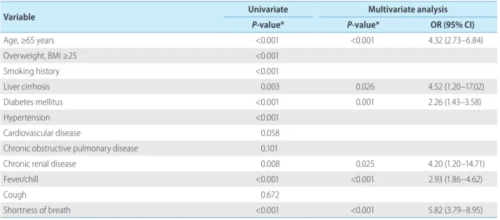
Table 3. Independent risk factors for severe COVID-19
Variable Univariate Multivariate analysis
P-value* P-value* OR (95% CI)
Age, ≥65 years <0.001 <0.001 4.32 (2.73–6.84)
Overweight, BMI ≥25 <0.001
Smoking history <0.001
Liver cirrhosis 0.003 0.026 4.52 (1.20–17.02)
Diabetes mellitus <0.001 0.001 2.26 (1.43–3.58)
Hypertension <0.001
Cardiovascular disease 0.058
Chronic obstructive pulmonary disease 0.101
Chronic renal disease 0.008 0.025 4.20 (1.20–14.71)
Fever/chill <0.001 <0.001 2.93 (1.86–4.62)
Cough 0.672
Shortness of breath <0.001 <0.001 5.82 (3.79–8.95)
COVID-19, coronavirus disease 2019; OR, odds radio; CI, confidence interval; BMI, body mass index.
*Calculated by logistic regression analysis.
P= 0.003) (Fig. 1).
Independent predictors of severe pneumonia in COVID-19
In multivariate analysis, higher age (OR, 4.32; 95% CI, 2.73–
6.84; P<0.001), presence of liver cirrhosis (OR, 4.52; 95% CI, 1.20–17.02; P=0.026), diabetes (OR, 2.26; 95% CI, 1.43–3.58;
P=0.001), chronic renal disease (OR, 4.20; 95% CI, 1.20–14.71;
P =0.025), fever and chill (OR, 2.93; 95% CI, 1.86–4.62;
P<0.001), and shortness of breath (OR, 5.82; 95% CI, 3.79–8.95;
P<0.001) were identified as independent predictors of severe CO- VID-19 (Table 3).
Independent predictors of mortality in COVID-19
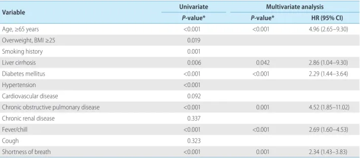
Multivariate analysis showed that higher age (HR, 4.96; 95%
CI, 2.65–9.30; P<0.001), presence of liver cirrhosis (HR, 2.86;
95% CI, 1.04–9.30; P =0.042), diabetes (HR, 2.29; 95% CI, 1.44–3.64; P<0.001), COPD (HR, 4.52; 95% CI, 1.85–11.02;
P= 0.001), fever and chill (HR, 2.69; 95% CI, 1.60–4.53;
P<0.001), and shortness of breath (HR, 2.34; 95% CI, 1.43–3.83;
P=0.001) were all significantly associated with mortality in COV- ID-19 (Table 4).
Clinical outcomes and risk factors for mortality in COVID-19 patients with liver cirrhosis
We determined the clinical outcomes and risk factors for mor- tality in COVID-19 patients with liver cirrhosis. Fourteen COVID-19 patients with cirrhosis (Child- Pugh class A and B in nine and five patients, respectively) were included and analyzed (Table 5). The most common etiology of cirrhosis was chronic hepatitis B (n=5) and alcohol (n=5), followed by chronic hepatitis C (n=2), autoim- mune hepatitis (n=1), and unknown etiology (n=1). Two patients had a history of HCC treatment (radiofrequency ablation), and there was no evidence of recurrence. Seven patients (50%) had decompensated liver cirrhosis. The initial MELD score was 8 (IQR, 7–12). CLIF-C ACLF score and CLIF-OF score were 92 (IQR, 49–
106) and 6 (IQR, 6–6.25), respectively. ACLF was diagnosed in one patient (grade I) on admission. Hepatic flare occurred in three of 14 patients (21.4%), all of whom survived. The most common symptoms were fever (n=8, 57.1%), followed by shortness of breath (n=6, 42.9%). Ten patients (71.4%) received antiviral ther- apy with lopinavir/ritonavir and seven (50%) received hydroxy- chloroquine (Table 6). Four patients (28.6%) experienced septic shock and five patients (35.7%) had ARDS. Among these pa- tients, four (28.6%) died after a median of 23 days (16–32) from COVID-19 diagnosis.
We compared non-survivors (n=4) with survivors (n=10) among COVID-19 patients with liver cirrhosis. There were no significant
Table 4. Independent risk factors for death
Variable Univariate Multivariate analysis
P-value* P-value* HR (95% CI)
Age, ≥65 years <0.001 <0.001 4.96 (2.65–9.30)
Overweight, BMI ≥25 0.019
Smoking history 0.001
Liver cirrhosis 0.006 0.042 2.86 (1.04–9.30)
Diabetes mellitus <0.001 <0.001 2.29 (1.44–3.64)
Hypertension <0.001
Cardiovascular disease 0.092
Chronic obstructive pulmonary disease <0.001 0.001 4.52 (1.85–11.02)
Chronic renal disease 0.337
Fever/chill <0.001 <0.001 2.69 (1.60–4.53)
Cough 0.323
Shortness of breath <0.001 0.001 2.34 (1.43–3.83)
HR, hazard ratio; CI, confidence interval; BMI, body mass index.
*Calculated by Cox proportional hazards regression test.
Table 5. Baseline characteristics of COVID-19 patients with liver cirrhosis (n=14) All (n=14)
Non-survivor (n=4, 28.6%)
Survivor
(n=10, 71.4%) P-value*
Demographic variable
Age (years) 66 (60–81) 74 (62–87) 64 (61–81) 0.478
Age, ≥65 years 7/14 (50.0) 3/4 (75.0) 4/10 (40.0) 0.559
Male gender 10/14 (71.4) 4/4 (100.0) 6/10 (60.0) 0.400
Height (cm) 166 (158–174) 168 (161–175) 166 (157–170) 0.857
Body weight (kg) 65 (56–73) 72 (70–74) 60 (55–67) 0.286
Overweight, BMI ≥25 2/8 (25.0) 1/2 (50.0) 1/6 (16.7) 1.000
Obese, BMI ≥30 1/8 (12.5) 0/2 (0.0) 1/6 (16.7) 1.000
Smoking history
Former or current smokers 6/13 (46.2) 3/3 (100.0) 3/10 (30.0) 0.141
Etiology of cirrhosis 0.301
Chronic hepatitis B 5/14 (35.7) 1/4 (25.0) 4/10 (40.0) –
Alcoholic liver disease 5/14 (35.7) 1/4 (25.0) 0/10 (0.0) –
Chronic hepatitis C 2/14 (14.3) 2/4 (50.0) 4/10 (40.0) –
Autoimmune hepatitis 1/14 (7.1) 0/4 (0.0) 1/10 (10.0) –
Others 1/14 (7.1) 0/4 (0.0) 1/10 (10.0) –
Initial stage of cirrhosis
Decompensated 7/14 (50.0) 1/4 (25.0) 6/10 (60.0) 0.554
Child-Pugh class 0.580
A 9/14 (64.3) 2/4 (50.0) 7/10 (70.0) –
B 5/14 (35.7) 2/4 (50.0) 3/10 (30.0) –
MELD score 8 (7–12) 9 (7–13) 8 (7–12) 1.000
CLIF-OF score 6.0 (6.0–6.3) 6.0 (6.0–6.8) 6.0 (6.0–6.5) 1.000
CLIF-C ACLF score 92 (52-106) 96 (80-108) 73 (40-106) 0.304
Other comorbidity
Diabetes mellitus 5/14 (35.7) 2/4 (50.0) 3/10 (30.0) 0.930
Hypertension 5/14 (35.7) 1/4 (25.0) 4/10 (40.0) 1.000
Cardiovascular disease 2/14 (14.3) 1/4 (25.0) 1/10 (10.0) 1.000
Chronic obstructive pulmonary disease 0/14 (0.0) 0/4 (0.0) 0/10 (0.0) –
Chronic renal disease 1/14 (7.1) 1/4 (25.0) 0/10 (0.0) 0.623
Symptoms on admission
Fever/chill 8/14 (57.1) 4/4 (100.0) 4/10 (40.0) 0.147
Cough 5/14 (35.7) 2/4 (50.0) 3/10 (30.0) 0.930
Shortness of breath 6/14 (42.9) 2/4 (50.0) 4/10 (40.0) 1.000
Gastrointestinal symptoms (vomiting/diarrhea) 2/14 (14.3) 0/4 (0.0) 2/10 (20.0) 0.904
Myalgia 1/14 (7.1) 1/4 (25.0) 0/10 (0.0) 0.623
Headache 2/14 (14.3) 3/4 (75.0) 1/10 (10.0) 1.000
Time from symptom onset to admission (days) 8 (1–8) 8 (8–8) 2 (0–8) 0.667
Vital signs at presentation
Body temperature (°C) 36.6 (36.2–37.3) 36.8 (36.1–37.9) 36.5 (36.2–37.1) 0.635
differences between non-survivors and survivors in age, comor- bidities, etiology, and stage of cirrhosis. Clinical symptoms, labo- ratory findings, and radiologic findings were not also related to prognosis. ICU admission, septic shock, ARDS, and acute kidney injury were more common in non-survivors than in survivors (P<0.05). Among the four non-survivors, all patients developed septic shock and ARDS, and all died of respiratory failure due to the progression of COVID-19. There was no worsening or new on- set of jaundice, ascites, or cirrhosis-related complications, such as spontaneous bacterial peritonitis, esophageal variceal bleeding,
or hepatic encephalopathy.
DISCUSSION
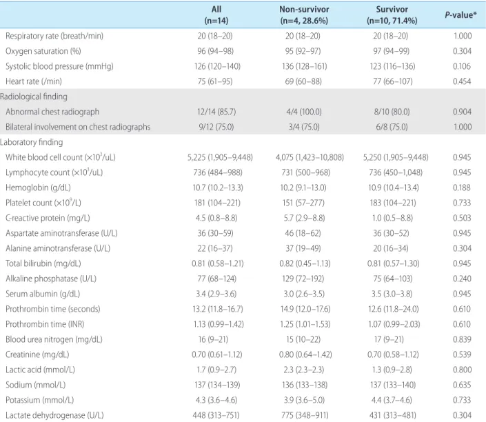
Due to the rapid spread of COVID-19 and the subsequent over- whelmed medical resources in many countries, it is important to evaluate the risk factors affecting the course of COVID-19. Glob- ally, chronic liver disease represents a significant disease bur- den.7,22 Moreover, as the severity of infection is known to be All
(n=14) Non-survivor
(n=4, 28.6%) Survivor
(n=10, 71.4%) P-value*
Respiratory rate (breath/min) 20 (18–20) 20 (18–20) 20 (18–20) 1.000
Oxygen saturation (%) 96 (94–98) 95 (92–97) 97 (94–99) 0.304
Systolic blood pressure (mmHg) 126 (120–140) 136 (128–161) 123 (116–136) 0.106
Heart rate (/min) 75 (61–95) 69 (60–88) 77 (66–107) 0.454
Radiological finding
Abnormal chest radiograph 12/14 (85.7) 4/4 (100.0) 8/10 (80.0) 0.904
Bilateral involvement on chest radiographs 9/12 (75.0) 3/4 (75.0) 6/8 (75.0) 1.000
Laboratory finding
White blood cell count (×103/uL) 5,225 (1,905–9,448) 4,075 (1,423–10,808) 5,250 (1,905–9,448) 0.945
Lymphocyte count (×103/uL) 736 (484–988) 731 (500–968) 736 (450–1,048) 0.945
Hemoglobin (g/dL) 10.7 (10.2–13.3) 10.2 (9.1–13.0) 10.9 (10.4–13.4) 0.188
Platelet count (×109/L) 181 (104–221) 151 (57–277) 183 (104–221) 0.733
C-reactive protein (mg/L) 4.5 (0.8–8.8) 5.7 (2.9–8.8) 1.0 (0.5–8.8) 0.503
Aspartate aminotransferase (U/L) 36 (30–59) 46 (18–62) 36 (30–52) 0.945
Alanine aminotransferase (U/L) 22 (16–37) 37 (19–49) 20 (16–34) 0.304
Total bilirubin (mg/dL) 0.81 (0.58–1.21) 0.82 (0.45–1.13) 0.81 (0.57–1.30) 0.945
Alkaline phosphatase (U/L) 77 (68–124) 129 (72–192) 75 (64–103) 0.240
Serum albumin (g/dL) 3.4 (2.9–3.6) 3.0 (2.6–3.5) 3.5 (3.0–3.8) 0.945
Prothrombin time (seconds) 13.2 (11.8–16.7) 14.9 (12.0–17.6) 12.6 (11.8–24.0) 0.610
Prothrombin time (INR) 1.13 (0.99–1.42) 1.25 (1.01–1.53) 1.07 (0.99–2.03) 0.610
Blood urea nitrogen (mg/dL) 16 (9–21) 15 (10–22) 17 (9–21) 0.839
Creatinine (mg/dL) 0.70 (0.61–1.12) 0.80 (0.64–1.42) 0.70 (0.58–1.12) 0.539
Lactic acid (mmol/L) 1.7 (0.9–2.7) 2.3 (2.3–2.3) 1.3 (0.9–2.8) 0.800
Sodium (mmol/L) 137 (134–139) 136 (133–138) 137 (133–140) 0.635
Potassium (mmol/L) 4.3 (3.6–4.6) 3.9 (3.6–5.0) 4.4 (3.7–4.6) 0.733
Lactate dehydrogenase (U/L) 448 (313–751) 775 (348–911) 431 (313–481) 0.304
Values are expressed as median (interquartile range) or numbers (%).
COVID-19, coronavirus disease 2019; BMI, body mass index; MELD, model for end-stage liver disease; ACLF, acute-on-chronic liver failure; INR, internatinal normalized ratio.
*Calculated by Student’s t test (or the Mann-Whitney U test, if appropriate) and chi-squared test (or Fisher’s exact test, if appropriate).
Table 5. Continued
greater in cirrhotic patients than in the general population, stud- ies on the implication of liver-related comorbidity on the outcomes of COVID-19 should be conducted.8 However, the impact of liver disease in SAR-CoV-2 infection remains unclear. In the present study, we assessed the impact of liver-related comorbidity on the clinical outcomes of COVID-19 patients.
In this study, a total of 1,005 patients were included and ana- lyzed. Liver cirrhosis was more common in patients with severe pneumonia than in those with non-severe pneumonia. When comparing patients with liver-related comorbidity to those with- out underlying liver disease, no significant differences were found in respiratory symptoms and clinical outcomes. Patients with liver cirrhosis needed more oxygen therapy and had a higher risk of admission to the ICU, septic shock, ARDS, acute kidney injury, and death. Multivariate analysis revealed that the presence of liv- er cirrhosis was significantly associated with disease severity and
mortality in COVID-19, along with old age, diabetes, fever, and shortness of breath. Among COVID-19 patients with cirrhosis, there were no significant differences in the stage of cirrhosis, clin- ical symptoms, and laboratory findings between non-survivors and survivors.
This study has several strengths and provides some important findings. First, this study demonstrated the impact of liver disease on the clinical outcomes of COVID-19 in a large number of pa- tients. There were no significant differences in clinical outcomes between COVID-19 patients with underlying liver disease and those without coexisting liver-related comorbidity. However, liver cirrhosis was associated with worsening clinical outcomes, dis- ease severity, and mortality of COVID-19, even after adjusting for other risk factors. Such result suggests that liver cirrhosis is an im- portant risk factor of COVID-19. Second, after the outbreak of COVID-19 in South Korea, mild COVID-19 patients were relocated Table 6. Treatments and clinical outcomes of COVID-19 patients with liver cirrhosis (n=14)
All (n=14)
Non-survivor (n=4, 28.6%)
Survivor
(n=10, 71.4%) P-value*
Treatments
Antiviral therapy 10/14 (71.4) 4/4 (100.0) 6/10 (60.0) 0.400
Lopinavir/ritonavir 10/14 (71.4) 4/4 (100.0) 6/10 (60.0) 0.400
Darunavir/cobicistat 0/14 (0.0) 0/4 (0.0) 0/10 (0.0) –
Hydroxychloroquine 7/14 (50.0) 1/4 (25.0) 6/10 (60.0) 0.554
Systemic glucocorticoid 5/14 (35.7) 3/4 (75.0) 2/10 (20.0) 0.186
Immunoglobulin 2/14 (14.3) 1/4 (25.0) 1/10 (10.0) 1.000
Oxygen support 8/14 (57.1) 4/4 (100.0) 4/10 (40.0) 0.085
High-flow nasal cannula+invasive mechanical ventilation 6/14 (42.9) 4/4 (100.0) 2/10 (20.0) 0.033
High-flow nasal cannula 3/14 (21.4) 1/4 (25.0) 2/10 (20.0) 1.000
Invasive mechanical ventilation 3/14 (21.4) 3/4 (75.0) 0/10 (0.0) 0.018
Invasive mechanical ventilation and ECMO 0/14 (0.0) 0/4 (0.0) 0/10 (0.0) –
Complication
ICU admission 5/14 (35.7) 4/4 (100.0) 1/10 (10.0) 0.011
Septic shock 4/14 (28.6) 4/4 (100.0) 0/10 (0.0) 0.002
Acute respiratory distress syndrome 5/14 (35.7) 4/4 (100.0) 1/10 (10.0) 0.011
Acute kidney injury 3/14 (21.4) 3/4 (75.0) 0/10 (0.0) 0.018
Continuous renal-replacement therapy 1/14 (7.1) 1/4 (25.0) 0/10 (0.0) 0.623
Secondary infection 1/14 (7.1) 0/4 (0.0) 1/10 (10.0) 1.000
Clinical outcome
Hospital stay (days) 23 (16–32) 23 (17–30) 23 (14–34) 0.945
Valuees are expressed as median (interquartile range) or numbers (%).
COVID-19, coronavirus disease 2019; ECMO, extracorporeal membrane oxygenation; ICU, intensive care unit.
*Calculated by Student’s t test (or the Mann-Whitney U test, if appropriate) and chi-squared test (or Fisher’s exact test, if appropriate).
to “life treatment centers,” which were temporary residence for isolation. As their symptoms progressed, the patients were re- ferred to the hospital.12,14 Since then, the number of confirmed pa- tients has gradually decreased. Therefore, we can evaluate the mortality rate and treatment outcomes of COVID-19 in a relatively stable situation with little depletion of medical resources. By June 3, 2020, the case fatality rates of COVID-19 in South Korea was 2.4%.13 Third, most of the patients in our study cohort experi- enced discharge or death, and only 6% were hospitalized when the analyses were performed. Therefore, analysis on the risk fac- tors of mortality could be conducted accurately.
Liver injury in COVID-19 patients might be caused by not only viral infection of liver cells, but also severe inflammatory response, such as cytokine storm, hypoxia, and drug-induced liver injury caused by antiviral agents, including lopinavir/ritonavir, remdesi- vir, and chlorquine.23,24 In COVID-19 patients with liver cirrhosis, poor outcomes might be associated with their systemic immuno- compromised status. Cirrhosis is associated with dysfunctions of the innate and adaptive immunity. Deterioration of liver function causes a reduction in the number and dysfunction of neutrophils, monocytes, and innate immunity proteins. Both B and T lympho- cytes involved in acquired immunity are also reduced in number and show functional impairment.25,26 Moreover, a previous report showed that multiple factors exacerbate alveolar epithelial injury and increase vascular permeability in cirrhotic patients with ARDS.
Systemic inflammation also promotes decompensation, organ fail- ures and ACLF in patients with cirrhosis and ARDS.27,28
In our study, 14 patients had liver cirrhosis. Among them, seven patients had decompensated liver cirrhosis. Four patients had his- tory of esophageal variceal bleeding, one had history of both eso- phageal variceal bleeding and hepatic encephalopathy, and the other two patients had ascites. During hospitalization, there was no worsening of ascites or jaundice, and ascites were well-con- trolled by taking diuretics. New-onset cirrhosis-related complica- tions, such as spontaneous bacterial peritonitis, esophageal variceal bleeding, and hepatic encephalopathy, did not occur.
Moreover, the presence of decompensation, stage of cirrhosis, and laboratory findings did not affect the prognosis of COVID-19 patients with liver cirrhosis. Among the non-survivors, all patients died from respiratory failure. Unlike our study, recent studies have shown that lower lymphocyte and platelet counts, as well as higher serum direct bilirubin levels were related to poor prognosis in COVID-19 patients with liver cirrhosis, and 29% of cirrhotic pa- tients died of end-stage-liver disease.10,11 This inconsistency of re- sults might be due to the small number of cirrhotic patients in our
study, and further studies with a large number of patients are needed to resolve this issue.
Of a total of 47 patients with chronic liver disease, 24 patients (2.4%) had chronic hepatitis B infection. Among them, 19 pa- tients had chronic hepatitis and five patients had liver cirrhosis. In patients with chronic hepatitis B infection, viral replication status and medication history of nucleos(t)ide analogues could not be confirmed in all patients. However, initial transaminase elevation over 60 U/L was observed only in one patient, and the patient had taken tenofovir disoproxil fumarate. After three days, transaminase levels decreased to under 50 U/L. Based on this re- sult, there was no acute exacerbation of chronic hepatitis B during hospitalization in COVID-19 patients.
A recent study showed that NAFLD identified with HSI more than 36 points and/or abdominal ultrasound was associated with COVID-19 progression.29 In our study, NAFLD, defined by HSI, was not related to clinical outcomes such as severe COVID-19 and mortality (P=0.244 and P=0.631, respectively). However, being overweight (BMI ≥25 kg/m2) was associated with disease severity and death in COVID-19 patients, and further studies are needed to validate this result.
This study showed a mortality rate of 7.7%. By June 3, 2020, the actual case fatality rate of COVID-19 in South Korea was 2.4%
according to the Korea Centers for Disease Control and Preven- tion.13 This difference occurred as patients with mild disease were not included in our study. Since patients with mild disease con- firmed in outpatient settings who were treated in life treatment centers or at home had only brief medical information and limited laboratory testing, we excluded them from this study. Our study cohort included patients who were admitted to tertiary hospitals;
therefore, it may represent the more severe patients of COVID-19.
This study had some limitations. First, some cases had incom- plete records of clinical symptoms, laboratory testing, viral repli- cation status, and medication history due to the retrospective na- ture of the study. Second, as described earlier in discussion, patients with mild disease were not included in this study; there- fore, our study cohort may represent the more severe patients of COVID-19. To investigate the effect of liver-related comorbidity on clinical outcomes, further studies including mild patients as well as patients with severe disease will be required. However, since the prevalence of liver disease in our study was similar to that of the entire population, our findings are thought to be meaningful.
Amid the explosive outbreak of COVID-19 worldwide, research on the risk factors of this infectious disease is important. This study suggests that liver cirrhosis is an important risk factor of
COVID-19. Stronger personal protection for COVID-19 is recom- mended, and more attention is needed in treating patients with advanced liver disease.
Authors’ contribution
Conception or design of the work: W.J. Chung, S.Y. Park; Data collection; Y.R. Lee, M.K. Kang, J.E. Song, H.J. Kim, B.S. Kim; Data analysis and interpretation: Y.R. Lee, W.J. Chung, B.S. Kim, S.Y.
Park; Drafting the article: Y.R. Lee, Y.O. Kweon, W.Y. Tak, S.Y.
Jang; Critical revision of the article; J.G. Park, C. Lee, J.S. Hwang, B.K. Jang, J.I. Suh; Final approval of the version to be published:
W.J. Chung, B.S. Kim, S.Y. Park
Conflicts of Interest
The authors have no conflicts to disclose.
SUPPLEMENTARY MATERIAL
Supplementary material is available at Clinical and Molecular Hepatology website (http://www.e-cmh.org).
REFERENCES
1. World Health Organization (WHO). Coronavirus disease (COVID-19) Situation Report – 134. WHO web site, <https://www.who.int/docs/
default-source/coronaviruse/situation-reports/20200602-covid- 19-sitrep-134.pdf?sfvrsn=cc95e5d5_2>. Accessed 3 Jun 2020.
2. Chen N, Zhou M, Dong X, Qu J, Gong F, Han Y, et al. Epidemio- logical and clinical characteristics of 99 cases of 2019 novel coro- navirus pneumonia in Wuhan, China: a descriptive study. Lancet 2020;395:507-513.
3. Guan WJ, Ni ZY, Hu Y, Liang WH, Ou CQ, He JX, et al. Clinical characteristics of coronavirus disease 2019 in China. N Engl J Med 2020;382:1708-1720.
4. Wang D, Hu B, Hu C, Zhu F, Liu X, Zhang J, et al. Clinical charac- teristics of 138 hospitalized patients with 2019 novel Coronavirus- infected pneumonia in Wuhan, China. JAMA 2020;323:1061-1069.
5. Wang B, Li R, Lu Z, Huang Y. Does comorbidity increase the risk of patients with COVID-19: evidence from meta-analysis. Aging (Albany NY) 2020;12:6049-6057.
6. Zhang C, Shi L, Wang FS. Liver injury in COVID-19: management and challenges. Lancet Gastroenterol Hepatol 2020;5:428-430.
7. Asrani SK, Devarbhavi H, Eaton J, Kamath PS. Burden of liver dis- eases in the world. J Hepatol 2019;70:151-171.
8. Fernández J, Gustot T. Management of bacterial infections in cirrho- sis. J Hepatol 2012;56 Suppl 1:S1-S12.
9. Fernández J, Acevedo J, Castro M, Garcia O, de Lope CR, Roca D, et al. Prevalence and risk factors of infections by multiresistant bacte- ria in cirrhosis: a prospective study. Hepatology 2012;55:1551-1561.
10. Iavarone M, D’Ambrosio R, Soria A, Triolo M, Pugliese N, Del Poggio P, et al. High rates of 30-day mortality in patients with cirrhosis and COVID-19. J Hepatol. 2020 Jun 9. doi: 10.1016/j.jhep.2020.06.001.
11. Qi X, Liu Y, Wang J, Fallowfield JA, Wang J, Li X, et al. Clinical course and risk factors for mortality of COVID-19 patients with pre- existing cirrhosis: a multicentre cohort study. Gut. 2020 May 20.
doi: 10.1136/gutjnl-2020-321666.
12. Korean Society of Infectious Diseases and Korea Centers for Disease Control and Prevention. Analysis on 54 mortality cases of coronavi- rus disease 2019 in the Republic of Korea from January 19 to March 10, 2020. J Korean Med Sci 2020;35:e132.
13. Korea Centers for Disease Control and Prevention (KCDC). The update of COVID-19 in Korea as of June 3. KCDC web site, <http://
ncov.mohw.go.kr/en/>. Accessed 3 Jun 2020.
14. Korean Society of Infectious Diseases; Korean Society of Pediatric Infectious Diseases; Korean Society of Epidemiology; Korean Society for Antimicrobial Therapy; Korean Society for Healthcare-associated Infection Control and Prevention; Korea Centers for Disease Control and Prevention. Report on the epidemiological features of coronavi- rus disease 2019 (COVID-19) outbreak in the Republic of Korea from January 19 to March 2, 2020. J Korean Med Sci 2020;35:e112.
15. World Health Organization (WHO). Laboratory testing for corona- virus disease 2019 (COVID-19) in suspected human cases: interim guidance, 2 March 2020. WHO web site, <https://apps.who.int/iris/
handle/10665/331329>. Accessed 3 Jun 2020.
16. World Health Organization (WHO). Laboratory testing of human suspected cases of novel coronavirus (nCoV) infection: interim guid- ance, 10 January 2020. WHO web site, <https://apps.who.int/iris/
handle/10665/330374>. Accessed 3 Jun 2020.
17. World Health Organization (WHO). Clinical management of severe acute respiratory infection (SARI) when COVID-19 disease is sus- pected: interim guidance, 13 March 2020. WHO web site, <https://
apps.who.int/iris/handle/10665/331446>. Accessed 3 Jun 2020.
18. Kidney disease improving global outcomes (KDIGO). KDIGO clinical practice guideline for acute kidney injury. KDIGO web site, <https://
kdigo.org/wp-content/uploads/2016/10/KDIGO-2012-AKI-Guide- line-English.pdf>. Accessed 3 Jun 2020.
19. Yeom SK, Lee CH, Cha SH, Park CM. Prediction of liver cirrhosis, us- ing diagnostic imaging tools. World J Hepatol 2015;7:2069-2079.
20. Lee JH, Kim D, Kim HJ, Lee CH, Yang JI, Kim W, et al. Hepatic steato- sis index: a simple screening tool reflecting nonalcoholic fatty liver disease. Dig Liver Dis 2010;42:503-508.
21. European Association for the Study of the Liver. EASL clinical prac-
tice guidelines for the management of patients with decompensated cirrhosis. J Hepatol 2018;69:406-460.
22. Chung W. The cost of liver disease in Korea: methodology, data, and evidence. Clin Mol Hepatol 2015;21:14-21.
23. Sun J, Aghemo A, Forner A, Valenti L. COVID-19 and liver disease.
Liver Int 2020;40:1278-1281.
24. Chai X, Hu L, Zhang Y, Han W, Lu Z, Ke A, et al. Specific ACE2 ex- pression in cholangiocytes may cause liver damage after 2019-nCoV infection. bioRxiv. 2020 Feb 4. doi: 10.1101/2020.02.03.931766.
25. Albillos A, Lario M, Álvarez-Mon M. Cirrhosis-associated immune dysfunction: distinctive features and clinical relevance. J Hepatol 2014;61:1385-1396.
26. Bernsmeier C, Pop OT, Singanayagam A, Triantafyllou E, Patel VC, Weston CJ, et al. Patients with acute-on-chronic liver failure have
increased numbers of regulatory immune cells expressing the recep- tor tyrosine kinase MERTK. Gastroenterology 2015;148:603-615.
e14.
27. Bernardi M, Moreau R, Angeli P, Schnabl B, Arroyo V. Mechanisms of decompensation and organ failure in cirrhosis: from peripheral arterial vasodilation to systemic inflammation hypothesis. J Hepatol 2015;63:1272-1284.
28. Gacouin A, Locufier M, Uhel F, Letheulle J, Bouju P, Fillatre P, et al.
Liver cirrhosis is independently associated with 90-day mortality in ARDS patients. Shock 2016;45:16-21.
29. Ji D, Qin E, Xu J, Zhang D, Cheng G, Wang Y, et al. Implication of non-alcoholic fatty liver diseases (NAFLD) in patients with CO- VID-19: a preliminary analysis. J Hepatol 2020;73:451-453.