Copyright © 2021 Korean Stroke Society
This is an Open Access article distributed under the terms of the Creative Commons Attribution Non-Commercial License (http://creativecommons.org/licenses/by-nc/4.0/) which permits unrestricted non-commercial use, distribution, and reproduction in any medium, provided the original work is properly cited.
Original Article
Prediction of Early Recanalization after Intravenous Thrombolysis in Patients with Large-Vessel Occlusion
Young Dae Kim,
aHyo Suk Nam,
aJoonsang Yoo,
b,cHyungjong Park,
a,bSung-Il Sohn,
bJeong-Ho Hong,
bByung Moon Kim,
dDong Joon Kim,
dOh Young Bang,
eWoo-Keun Seo,
eJong-Won Chung,
eKyung-Yul Lee,
fYo Han Jung,
f,gHye Sun Lee,
hSeong Hwan Ahn,
iDong Hoon Shin,
jHye-Yeon Choi,
kHan-Jin Cho,
lJang-Hyun Baek,
m,nGyu Sik Kim,
oKwon-Duk Seo,
o,pSeo Hyun Kim,
qTae-Jin Song,
r,sJinkwon Kim,
c,tSang Won Han,
uJoong Hyun Park,
uSung Ik Lee,
pJoonNyung Heo,
aJin Kyo Choi,
a,dJi Hoe Heo,
aon behalf of the Thrombus Imaging Study Group and the SECRET Study Group
aDepartment of Neurology, Yonsei University College of Medicine, Seoul, Korea
bDepartment of Neurology, Brain Research Institute, Keimyung University School of Medicine, Daegu, Korea
cDepartment of Neurology, Yongin Severance Hospital, Yonsei University College of Medicine, Yongin, Korea
dDepartment of Radiology, Yonsei University College of Medicine, Seoul, Korea
eDepartment of Neurology, Samsung Medical Center, Sungkyunkwan University School of Medicine, Seoul, Korea
fDepartment of Neurology, Gangnam Severance Hospital, Severance Institute for Vascular and Metabolic Research, Yonsei University College of Medicine, Seoul, Korea
gDepartment of Neurology, Changwon Fatima Hospital, Changwon, Korea
hDepartment of Research Affairs, Biostatistics Collaboration Unit, Yonsei University College of Medicine, Seoul, Korea
iDepartment of Neurology, Chosun University College of Medicine, Gwangju, Korea
jDepartment of Neurology, Gachon University Gil Medical Center, Incheon, Korea
kDepartment of Neurology, Kyung Hee University Hospital at Gangdong, Kyung Hee University School of Medicine, Seoul, Korea
lDepartment of Neurology, Pusan National University School of Medicine, Busan, Korea
mDepartment of Neurology, National Medical Center, Seoul, Korea
nDepartment of Neurology, Kangbuk Samsung Hospital, Sungkyunkwan University School of Medicine, Seoul, Korea
oDepartment of Neurology, National Health Insurance Service Ilsan Hospital, Goyang, Korea
pDepartment of Neurology, Wonkwang University Sanbon Hospital, Wonkwang University School of Medicine, Sanbon, Korea
qDepartment of Neurology, Yonsei University Wonju College of Medicine, Wonju, Korea
rDepartment of Neurology, Ewha Womans University Mokdong Hospital, Ewha Womans University School of Medicine, Seoul, Korea
sDepartment of Neurology, Ewha Womans University Seoul Hospital, Ewha Womans University School of Medicine, Seoul, Korea
tDepartment of Neurology, CHA Bundang Medical Center, CHA University, Seongnam, Korea
uDepartment of Neurology, Inje University Sanggye Paik Hospital, Inje University College of Medicine, Seoul, Korea
Background and Purpose We aimed to develop a model predicting early recanalization after in- travenous tissue plasminogen activator (t-PA) treatment in large-vessel occlusion.
Methods Using data from two different multicenter prospective cohorts, we determined the fac- tors associated with early recanalization immediately after t-PA in stroke patients with large-ves- sel occlusion, and developed and validated a prediction model for early recanalization. Clot volume was semiautomatically measured on thin-section computed tomography using software, and the degree of collaterals was determined using the Tan score. Follow-up angiographic studies were performed immediately after t-PA treatment to assess early recanalization.
Results Early recanalization, assessed 61.0±44.7 minutes after t-PA bolus, was achieved in 15.5%
(15/97) in the derivation cohort and in 10.5% (8/76) in the validation cohort. Clot volume (odds ratio [OR], 0.979; 95% confidence interval [CI], 0.961 to 0.997; P=0.020) and good collaterals (OR,
Correspondence: Ji Hoe Heo Department of Neurology, Yonsei University College of Medicine, 50-1 Yonsei-ro, Seodaemun-gu, Seoul 03722, Korea
Tel: +82-2-2228-1605 Fax: +82-2-393-0705 E-mail: [email protected]
https://orcid.org/0000-0001-9898-3321 Received: August 27, 2020
Revised: January 17, 2021 Accepted: February 10, 2021
Introduction
Successful recanalization in acute stroke patients with large-ves- sel occlusion (LVO) is a main target of reperfusion therapy. To achieve early and complete successful recanalization, early treatment with either intravenous tissue plasminogen activator (IV t-PA) or endovascular treatment (EVT) is needed.1,2
Even after the successful introduction of EVT, bridging thera- py with IV t-PA is recommended before EVT.2 However, IV t-PA treatment has low successful recanalization rates in patients with LVO.3-5 In addition, bridging therapy with IV t-PA before EVT may delay the initiation of EVT or increase the risk of bleeding complications, which may lead to unfavorable out- comes.6 In contrast, early recanalization that can be achieved with IV t-PA may be beneficial for outcomes.7 Although bridg- ing therapy with IV t-PA is currently a debated issue,8 a specific group of patients may benefit from IV t-PA before EVT and se- lection of good responders would be helpful for improving pa- tient outcomes.
A large clot burden determined either quantitatively or qual- itatively is strongly associated with successful recanalization after IV t-PA treatment.5,9-13 In our previous study, almost all patients with thrombus measuring >200 mm3 did not achieve early recanalization after IV t-PA; these patients were suggest- ed to be the primary candidates for direct EVT.5 However, many patients had thrombus measuring <200 mm3, and approxi- mately 70% of them still did not achieve early recanalization.
Furthermore, predicting patients who might achieve early re- canalization after IV t-PA would be helpful for deciding IV t-PA treatment in those with LVO. This suggests the necessity of an improved model that predicts not only nonrecanalization but also successful recanalization.
While a clot burden is associated with early recanalization, the collateral status may also have a role.14 The presence of
good collaterals is an indicator of a favorable outcome in pa- tients undergoing recanalization treatment.15 Although aiding in keeping the penumbra area durable until achieving recanali- zation is the main reason, an enhanced thrombolytic effect by delivering more t-PA to the clot via backflow may also contrib- ute to the favorable outcome.14,16 Therefore, we aimed to deter- mine the predictors of early recanalization after IV t-PA treat- ment, including both clot volume and the degree of collaterals.
We also aimed to develop and validate a simple prediction model for early recanalization using data from two different prospective cohorts.
Methods
Study population
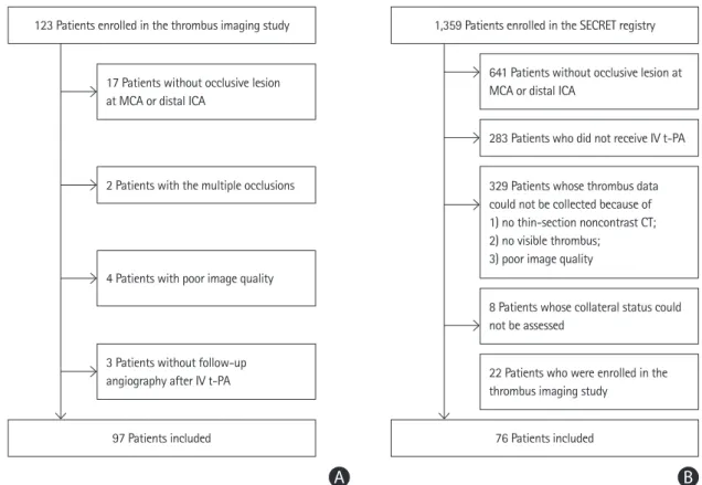
We used data from two different multicenter stroke registries for developing a prediction model. For the derivation cohort, we used data from the Thrombus Imaging Study registry, which was a multicenter prospective study designed to provide clini- cal evidence for thrombus imaging for outcome prediction in reperfusion therapy.5 The inclusion criteria were treatment with IV t-PA within 4.5 hours of symptom onset and visible thrombi at the occlusion site of the distal internal carotid artery (ICA) and/or middle cerebral artery (MCA) on thin-section noncon- trast computed tomography (NCCT). Patients were consecu- tively enrolled between February 2016 and August 2017 from nine hospitals.5 In this analysis, we included 97 patients with visible thrombus at the distal ICA or MCA (Figure 1).
For the validation cohort, we used data from the Selection Criteria in Endovascular Thrombectomy and Thrombolytic Ther- apy (SECRET) registry (Clinicaltrials.gov: NCT02964052).17 The SECRET registry is a nationwide multicenter registry for explor- ing the selection criteria for patients who would benefit from reperfusion therapies. This study included 1,026 patients who 6.129; 95% CI, 1.592 to 23.594; P=0.008) were significant factors associated with early recanali-
zation. The area under the curve (AUC) of the model including clot volume was 0.819 (95% CI, 0.720 to 0.917) and 0.842 (95% CI, 0.746 to 0.938) in the derivation and validation cohorts, re- spectively. The AUC improved when good collaterals were added (derivation cohort: AUC, 0.876;
95% CI, 0.802 to 0.950; P=0.164; validation cohort: AUC, 0.949; 95% CI, 0.886 to 1.000; P=0.036).
The integrated discrimination improvement also showed significantly improved prediction (0.097;
95% CI, 0.009 to 0.185; P=0.032).
Conclusions The model using clot volume and collaterals predicted early recanalization after intra- venous t-PA and had a high performance. This model may aid in determining the recanalization treatment strategy in stroke patients with large-vessel occlusion.
Keywords Ischemia; Stroke; Thrombosis; Thrombolysis; Reperfusion
had been registered retrospectively from 15 hospitals between January 2012 and December 2015 and 333 patients who had been registered prospectively from 13 hospitals between No- vember 2016 and December 2017. There were 98 patients with visible thrombi in the distal ICA and/or MCA on initial thin-sec- tion NCCT and those with imaging data available before and after IV t-PA. However, there was an overlap of the study peri- od between the Thrombus Imaging Study and the SECRET study. Therefore, we excluded 22 patients (anonymized) who were enrolled during the overlapped period. Finally, 76 patients were included for the validation (Figure 1). This study was ap- proved by the Institutional Review Board of each study hospi- tal, and written informed consent was obtained from all pa- tients enrolled prospectively.
Imaging protocol and reperfusion therapy
The patients included in this study underwent thin-section (1 or 1.25 mm) NCCT and computed tomography angiography (CTA) before IV t-PA. All patients were treated with IV t-PA (Actilyse, Boehringer-Ingelheim, Ingelheim, Germany) at a standard dose of 0.9 mg/kg (10% as a bolus followed by 90%
as an infusion for 60 minutes) within 4.5 hours of stroke onset.
The inclusion and exclusion criteria for IV t-PA treatment were based on the guidelines, which were not different among the
study hospitals. Further EVT was sequentially performed if the patients did not show clinical improvement or did not achieve arterial recanalization after IV t-PA treatment. Follow-up CTA or magnetic resonance angiography (MRA) was immediately performed after the end of IV t-PA if the patients were not treated with EVT.
Assessment of imaging findings
The volume and density of thrombus were semiautomatically measured on baseline thin-section NCCT using three-dimen- sional software (Xelis, Infinitt, Seoul, Korea). The detailed methods have been previously described.5,18,19 Briefly, when a hyperdense artery (pixels between 50 and 100 Hounsfield units) was identified on thin-section NCCT, this thrombus area was automatically shown as a red area. When clicking any por- tion within the thrombus area and subsequently the “Dilate”
icon, every slice of thrombus was merged with automatic pixel dilation and region growing. Thereafter, the volume and density of the thrombus were shown on a screen within 1 minute by automatic calculation. Simultaneously, CTA maximum-intensi- ty projection images and thin-section NCCT images could be synchronized on a screen, which could enable the easy identi- fication of thrombus.
The presence of early recanalization was assessed on CTA or
1,359 Patients enrolled in the SECRET registry
76 Patients included
641 Patients without occlusive lesion at MCA or distal ICA
283 Patients who did not receive IV t-PA
22 Patients who were enrolled in the thrombus imaging study
8 Patients whose collateral status could not be assessed
329 Patients whose thrombus data could not be collected because of 1) no thin-section noncontrast CT;
2) no visible thrombus;
3) poor image quality
B
123 Patients enrolled in the thrombus imaging study
97 Patients included
17 Patients without occlusive lesion at MCA or distal ICA
2 Patients with the multiple occlusions
4 Patients with poor image quality
3 Patients without follow-up angiography after IV t-PA
A
Figure 1. Patient selection: (A) derivation cohort, (B) validation cohort. MCA, middle cerebral artery; ICA, internal carotid artery; IV t-PA, intravenous tissue plasminogen activator; SECRET, Selection Criteria in Endovascular Thrombectomy and Thrombolytic Therapy; CT, computed tomography.
MRA images taken immediately at the end of IV t-PA or on digital subtraction angiography images taken during EVT. Suc- cessful recanalization was defined as a modified Thrombolysis in Cerebral Infarction (TICI) grade of 2b or 3 on digital sub- traction angiography20 or an arterial occlusive lesion score of 3 on CTA or MRA. We also determined the degree of collater- als using the Tan collateral score11,16 based on the single-phase CTA axial image performed before IV t-PA treatment, and the presence of good collaterals was defined as a Tan score of 3.
We also collected the Alberta Stroke Program Early CT score.
Two stroke neurologists (Y.D.K. and J.Y.) measured the throm- bus volume and density and determined the Tan score, Alberta Stroke Program Early CT score, and TICI grade. Entire imaging analyses were independently performed without knowledge of any clinical information. In the case of discrepancies, a con- sensus was reached for the derivation cohort and adjudicated by the imaging adjudication committee for the validation co- hort.
Clinical variables
We collected data on demographics, risk factors, and premor- bid disability (dependent status defined as a modified Rankin Scale score of 3–5). Time parameters such as the intervals from stroke onset to initial brain imaging, from onset to IV t-PA, and from the initiation of IV t-PA to follow-up angiographic studies were determined. Data on the total dose of t-PA for each pa- tient were collected. The stroke severity was assessed using the National Institutes of Health Stroke Scale.
Statistical analyses
Data are presented as frequency (percentage) for categorical variables and mean±standard deviation or median (interquartile range) for continuous variables. Differences between the groups were assessed using Student’s t-test or the Mann-Whitney U-test for continuous variables and the chi-square test or Fish- er’s exact test for categorical variables, as appropriate.
Univariable and multivariable logistic regression analyses were performed to determine the independent factors for early successful recanalization after IV t-PA. The collinearity of com- binations of variables in the derivation cohort was evaluated using variation inflation factors (<2 being considered nonsig- nificant). Odds ratios (ORs) with 95% confidence intervals (CIs) for each variable included in the model were finally calculated.
On the basis of the results of multivariable logistic regression analysis, we developed a prediction model and a nomogram.
We compared the predictive ability between the model in- cluding clot volume only and the model in which the presence of good collaterals was added to clot volume by computing the
area under the curve (AUC) using the Delong method. The inte- grated discrimination improvement (IDI) index was computed to investigate whether adding the presence of good collaterals to clot volume would improve the predictive power. We also plotted decision curves to assess the net benefit of nomo- gram-assisted decisions.
We examined the calibration (calibration plots and Hos- mer-Lemeshow test) and discrimination (AUC). To assess the external validity of model performance, we applied our predic- tion model to the validation cohort. We examined the perfor- mance of the final model both in the derivation and external validation cohorts in terms of discrimination using AUC and in terms of calibration by plotting the agreement between the predicted and observed probabilities across the quartiles of scores. In addition, we determined the optimal cutoff value of the prediction value based on nomogram, which was verified by calculating the Youden index.
Statistical analyses were performed using R software pack- age version 3.6.2 (http://www.R-project.org). All statistical tests were two-tailed; P<0.05 was considered statistically signifi- cant, whereas 0.05≤P<0.2 was considered a trend toward sig- nificance to increase the sensitivity for detecting potential se- lection bias.
Results
Baseline characteristics
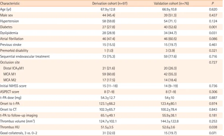
Follow-up angiographic studies were performed at a mean of 61.0±44.7 minutes after the bolus injection of t-PA (65.1±49.1 minutes in the derivation cohort and 55.9±38.1 minutes in the validation cohort, P=0.181). The baseline characteristics includ- ing clot volume and the frequency of good collaterals were not different between the derivation and validation cohorts, except diabetes and dyslipidemia, which had lower frequencies in the derivation cohort (Table 1).
Determinants for successful recanalization
The occluded artery was recanalized immediately after IV t-PA in 15.5% (15/97) of the derivation cohort and in 10.5% (8/76) of the validation cohort. The factors associated with early reca- nalization in the derivation cohort were smaller clot volumes, good collaterals, and lower National Institutes of Health Stroke Scale scores on univariable analysis. Multivariable logistic re- gression analysis showed that clot volume (OR, 0.979 per 1 mm3; 95% CI, 0.961 to 0.997; P=0.02) and good collaterals (OR, 6.129; 95% CI, 1.592 to 23.594; P=0.008) were indepen- dent and significant predictors of early recanalization (Table 2).
These findings were still observed when we divided the study
Table 1. Baseline characteristics of the derivation and external validation cohorts
Characteristic Derivation cohort (n=97) Validation cohort (n=76) P
Age (yr) 67.9±12.8 66.9±10.8 0.620
Male sex 44 (45.4) 39 (51.3) 0.437
Hypertension 58 (59.8) 54 (71.1) 0.124
Diabetes 27 (27.8) 40 (52.6) 0.001
Dyslipidemia 28 (28.9) 34 (44.7) 0.031
Atrial fibrillation 46 (47.4) 46 (60.5) 0.086
Previous stroke 15 (15.5) 15 (19.7) 0.461
Premorbid disability 1 (1.0) 3 (3.9) 0.321
Sequential endovascular treatment 73 (75.3) 59 (77.6) 0.716
Occlusion site 0.727
Distal ICA±M1 21 (21.6) 20 (26.3)
MCA M1 59 (60.8) 42 (55.3)
MCA M2 17 (17.5) 14 (18.4)
Initial NIHSS score 15 (11–18) 14 (9–19) 0.736
ASPECT score 8 (7–9) 8 (7–9) 0.306
t-PA dose (mg) 54.3±12.7 54±10 0.887
Onset to t-PA 123.1±66.2 123.4±80.1 0.974
Onset to CT 102.3±65.7 100.2±78.4 0.843
t-PA to follow-up imaging 65.1±49.1 55.9±38.1 0.181
Thrombus volume (mm3) 124.7±102.1 144.3±122.8 0.253
Thrombus HU 51.5±3.5 52.6±3.6 0.038
Good collaterals, 3 vs. 0–2 31 (32.0) 15 (19.7) 0.071
Values are presented as mean±standard deviation, number (%), or median (interquartile range).
ICA, internal carotid artery; MCA, middle cerebral artery; NIHSS, National Institutes of Health Stroke Scale; ASPECT, Alberta Stroke Program Early Computed Tomography; t-PA, tissue plasminogen activator; CT, computed tomography; HU, Hounsfield unit.
Table 2. Univariable and multivariable analyses for early recanalization
Variable Univariable analysis Multivariable analysis
Odds ratio (95% CI) P Odds ratio (95% CI) P
Age 0.968 (0.929–1.008) 0.117
Male sex 0.496 (0.162–1.524) 0.221
Hypertension 3.130 (0.821–11.934) 0.095
Diabetes 1.364 (0.419–4.435) 0.606
Dyslipidemia 0.570 (0.148–2.198) 0.414
Atrial fibrillation 0.700 (0.228–2.146) 0.533
Previous stroke 2.347 (0.634–8.688) 0.201
Premorbid disability 0.000 (0.000–NA) 1.000
Initial NIHSS score 0.882 (0.797–0.976) 0.015 0.963 (0.852–1.089) 0.551
ASPECT score 1.458 (0.933–2.279) 0.098
t-PA dose (mg) 1.010 (0.965–1.056) 0.670
Onset to t-PA 0.999 (0.991–1.008) 0.886
Onset to CT 1.000 (0.992–1.009) 0.928
t-PA to follow-up imaging 1.004 (0.995–1.013) 0.394
Clot volume (mm3) 0.976 (0.961–0.992) 0.004 0.979 (0.961–0.997) 0.020
Clot HU 0.873 (0.744–1.023) 0.094
Good collateral, 3 vs. 0–2 8.525 (2.441–29.769) 0.001 6.129 (1.592–23.594) 0.008
CI, confidence interval; NA, not applicable; NIHSS, National Institutes of Health Stroke Scale; ASPECT, Alberta Stroke Program Early Computed Tomography;
t-PA, tissue plasminogen activator; CT, computed tomography; HU, Hounsfield unit.
population into two groups based on the timing (≥45 or ≤75 minutes) of follow-up angiographic studies after IV t-PA (Sup- plementary Table 1).
As the clot volume was a strong factor associated with early recanalization, we determined whether adding good collaterals to the prediction model would improve the model performance.
When the presence of good collaterals was added to clot vol- ume, the AUC improved from 0.819 (95% CI, 0.720 to 0.917) to 0.876 (95% CI, 0.802 to 0.950) in the derivation cohort (P=0.164) (Figure 2A) and from 0.842 (95% CI, 0.746 to 0.938) to 0.949 (95% CI, 0.886 to 1.000) in the validation cohort (P=0.036). The decision curves for the probability of successful recanalization in the derivation cohort showed better perfor- mance when the presence of good collaterals was added to clot volume (Figure 2B). The computation of IDI also showed significantly improved prediction (IDI, 0.097; 95% CI, 0.009 to 0.185; P=0.032). The Hosmer-Lemeshow tests were not signifi- cant in the derivation cohort (P=0.970) and the validation co- hort (P=0.935), which means a good fit. Figure 3 demonstrates the predicted versus observed probability based on the quartile of the scores, which showed good correlation.
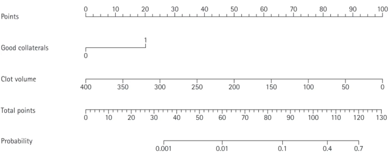
Accordingly, we developed a model predicting early recanali- zation after IV t-PA treatment using both clot volume and good collaterals. For easy use of the prediction model, we also developed a nomogram predicting early recanalization by as- signing a graphic score to each of clot volume and good collat- erals, with a point range from 0 to 20. The scores were summed to generate a total score, which was finally converted into an individual probability of early successful recanalization after IV
t-PA (Figure 4). Higher total points based on the sum of the assigned number of points for good collaterals and clot volume were associated with the chance of early recanalization (Sup- plementary Figure 1). The cut off value of >0.1107 of the pre- diction model value for predicting early recanalization, which was based on the Youden index, had good sensitivity (100%) and specificity (68.3%).
Discussion
In the present study, early recanalization after IV t-PA was sig- nificantly associated with clot volume and collateral status (thus, the model was named “2C”). The prediction model using both clot volume and good collaterals could reliably predict early recanalization after IV t-PA treatment among patients with LVO. This 2C model was externally validated using another population enrolled in a nationwide multicenter registry. We also developed a nomogram using these two variables for rapid and easy use in clinical practice, which can help identify pa- tients who would be expected to achieve early recanalization after IV t-PA treatment.
With EVT becoming the standard treatment, bridging therapy with IV t-PA before EVT in patients with LVO is a debated is- sue.8 This is because, in patients with LVO, recanalization is achieved immediately after t-PA in only about 10% to 15%4,5,13,14,19,21 and t-PA may increase the risk of bleeding and distal embolization of partially lysed clots. It may also delay the begin.
Recently, the Direct Intraarterial Thrombectomy in Order to
A
Sensitivity
1-Specificity 1.0 -
0.75 -
0.5 -
0.25 -
0.0 -
0.2 0.25 0.5 0.7 1.0
Model using only clot volume Model using both clot volume and good collateral
B
Net benefit
Threshold probability (%) 0.2 -
0.15 -
0.1 -
0.05 -
0.0 -
-0.05 -
0 20 40 60 80 100
All None
Model using only clot volume Model using both clot volume and good collateral
Figure 2. Comparison of receiver operating characteristic (ROC) curves between two models (A) and decision curve analysis (B). (A) ROC curves of the deriva- tion cohorts and area under the curve (AUC) values (model using only clot volume, 0.819; model using both clot volume and good collaterals, 0.876). (B) Deci- sion curve analysis showing that the model using both clot volume and good collaterals was the preferred model.
A
Sensitivity
1-Specificity 1.0 -
0.8 -
0.6 -
0.4 -
0.0 - 0.2 -
0.0 0.2 0.4 0.6 0.8 1.0
B
(%)
Quartile of predicted probability 50 -
40 -
30 -
20 -
0.0 - 10 -
Q1 Q2 Q3 Q4
Predicted outcome Observed outcome
Figure 3. Assessment of discrimination and calibration in the derivation cohort (A, B) and the validation cohort (C, D). (A) Receiver operating characteristic (ROC) curves of the derivation cohort and area under the curve (AUC) values (AUC, 0.876; 95% confidence interval [CI], 0.802 to 0.950). (B) Calibration plot per quartile of the scores in the derivation cohort. (C) ROC curves of the validation cohort and AUC values (AUC, 0.949; 95% CI, 0.886 to 1.000). (D) Calibra- tion plot per quartile of the scores in the validation cohort.
C
Sensitivity
1-Specificity 1.0 -
0.8 -
0.6 -
0.4 -
0.0 - 0.2 -
0.0 0.2 0.4 0.6 0.8 1.0
D
(%)
Quartile of predicted probability 40 -
30 -
20 -
0.0 - 10 -
Q1 Q2 Q3 Q4
Predicted outcome Observed outcome
Figure 4. Nomogram for predicting successful recanalization within 1 hour after intravenous tissue plasminogen activator.
Points 0 10 20 30 40 50 60 70 80 90 100
Good collaterals 1
0
Clot volume
400 350 300 250 200 150 100 50 0
Total points
0 10 20 30 40 50 60 70 80 90 100 110 120 130
Probability
0.001 0.01 0.1 0.4 0.7
Revascularize Acute Ischemic Stroke Patients with Large Vessel Occlusion Efficiently in Chinese Tertiary Hospitals: a Multi- center Randomized Clinical Trial (DIRECT-MT) demonstrated that EVT alone was noninferior to combined IV t-PA and EVT with respect to functional outcomes, but it was associated with lower rates of successful recanalization than the com- bined therapy.22 Although similar trials (NCT03192332, NCT03494920, and ISRCTN80619088) are ongoing,23,24 it is questionable whether one strategy (either direct EVT or com- bined intravenous thrombolysis and EVT) should be the only treatment option for all patients with LVO. A certain group of patients would benefit from IV t-PA before EVT, whereas direct EVT would be better in another group of patients.8 In this con- text, prediction of early recanalization after t-PA would be helpful for deciding the strategy between direct EVT and com- bined intravenous thrombolysis and EVT. For example, IV t-PA before EVT may be considered when the probability of early re- canalization by IV t-PA is high based on the prediction model.
When the probability is very low, direct EVT may be considered as IV t-PA may increase the risk of bleeding and lead to delay the initiation of EVT.
In t-PA treatment, clot volume is an important factor associ- ated with early recanalization. This study showed that good col- laterals also enhance the chance for early successful recanaliza- tion. In a previous study based on perfusion magnetic resonance imaging, good collaterals were associated with recanalization within 3 hours after t-PA treatment.14 Good collaterals can lead to a bidirectional action of t-PA (forward and back flow). This may be able to enhance the proteolytic action of t-PA by in- creasing the surface area contacting the t-PA.14,16,25
Although there have been several prognostic models or scor- ing systems for estimating safety or efficacy of thrombolytic therapy with IV t-PA for acute ischemic stroke, little is known on the model for predicting the successful recanalization by IV t-PA within 1 hour. In this study, recanalization was assessed about 60 minutes after IV t-PA injection, immediately after the end of IV t-PA infusion when making a decision regarding fur- ther EVT is necessary. Therefore, prediction of recanalization at this time point is important for determining the treatment strategy. We found that only two variables (clot volume and good collaterals) were sufficient for reliably predicting recanal- ization with high performance at this time point. The nomo- gram that we developed may also be helpful for making a rapid decision.
This study had some limitations. Although collaterals are easily determined on CTA, measurement of clot volume re- quires a software program. Therefore, the wide use of this pre- diction model is limited because it depends on the availability
of the software. We only included patients with occlusion of the MCA or distal ICA. Thereby, the predictive ability of our model for occlusion at other cerebral arterial segments is un- known. In addition, although we tried to show the 2C model in this study could be useful for predicting the early recanaliza- tion, the rate of early recanalization was low. This might lead the relatively low statistical power. So, we thought further analysis including larger amount of data would be necessary.
Conclusions
We showed that clot volume and good collaterals were associ- ated with early recanalization after IV t-PA treatment, and the prediction model using clot volume and good collaterals showed high performance in the derivation and validation co- horts. Our model may aid in the decision making with respect to the recanalization strategy.
Supplementary materials
Supplementary materials related to this article can be found online at https://doi.org/10.5853/jos.2020.03622.
Disclosure
The authors have no financial conflicts of interest.
Acknowledgments
This work was supported by a grant from the Basic Science Re- search Program through the National Research Foundation of Korea (NRF) funded by the Ministry of Education (NRF- 2018R1A2A3074996); a faculty research grant of Yonsei Uni- versity College of Medicine (6-2020-0202); and the “Dongwha”
Faculty Research Assistance Program of Yonsei University Col- lege of Medicine (6-2019-0191).
References
1. Goyal M, Menon BK, van Zwam WH, Dippel DW, Mitchell PJ, Demchuk AM, et al. Endovascular thrombectomy after large-vessel ischaemic stroke: a meta-analysis of individual patient data from five randomised trials. Lancet 2016;387:
1723-1731.
2. Powers WJ, Rabinstein AA, Ackerson T, Adeoye OM, Bam- bakidis NC, Becker K, et al. Guidelines for the early manage- ment of patients with acute ischemic stroke: 2019 update to the 2018 guidelines for the early management of acute isch-
emic stroke. A guideline for healthcare professionals from the American Heart Association/American Stroke Associa- tion. Stroke 2019;50:e344-e418.
3. Lee KY, Han SW, Kim SH, Nam HS, Ahn SW, Kim DJ, et al.
Early recanalization after intravenous administration of re- combinant tissue plasminogen activator as assessed by pre- and post-thrombolytic angiography in acute ischemic stroke patients. Stroke 2007;38:192-193.
4. Campbell BCV, Mitchell PJ, Churilov L, Yassi N, Kleinig TJ, Dowling RJ, et al. Tenecteplase versus alteplase before thrombectomy for ischemic stroke. N Engl J Med 2018;378:1 573-1582.
5. Yoo J, Baek JH, Park H, Song D, Kim K, Hwang IG, et al. Throm- bus volume as a predictor of nonrecanalization after intrave- nous thrombolysis in acute stroke. Stroke 2018;49:2108-2115.
6. Fischer U, Kaesmacher J, Mendes Pereira V, Chapot R, Sid- diqui AH, Froehler MT, et al. Direct mechanical thrombecto- my versus combined intravenous and mechanical thrombec- tomy in large-artery anterior circulation stroke: a topical re- view. Stroke 2017;48:2912-2918.
7. Tsivgoulis G, Saqqur M, Sharma VK, Brunser A, Eggers J, Mi- kulik R, et al. Timing of recanalization and functional recov- ery in acute ischemic stroke. J Stroke 2020;22:130-140.
8. Albers GW. Thrombolysis before thrombectomy: to be or DI- RECT-MT? N Engl J Med 2020;382:2045-2046.
9. Zhu G, Michel P, Jovin T, Patrie JT, Xin W, Eskandari A, et al.
Prediction of recanalization in acute stroke patients receiv- ing intravenous and endovascular revascularization therapy.
Int J Stroke 2015;10:28-36.
10. Rohan V, Baxa J, Tupy R, Cerna L, Sevcik P, Friesl M, et al.
Length of occlusion predicts recanalization and outcome af- ter intravenous thrombolysis in middle cerebral artery stroke.
Stroke 2014;45:2010-2017.
11. Tan IY, Demchuk AM, Hopyan J, Zhang L, Gladstone D, Wong K, et al. CT angiography clot burden score and collateral score: correlation with clinical and radiologic outcomes in acute middle cerebral artery infarct. AJNR Am J Neuroradiol 2009;30:525-531.
12. Heo JH, Kim K, Yoo J, Kim YD, Nam HS, Kim EY. Computed tomography-based thrombus imaging for the prediction of recanalization after reperfusion therapy in stroke. J Stroke 2017;19:40-49.
13. Behrens L, Möhlenbruch M, Stampfl S, Ringleb PA, Hametner C, Kellert L, et al. Effect of thrombus size on recanalization by bridging intravenous thrombolysis. Eur J Neurol 2014;21:
1406-1410.
14. Seners P, Roca P, Legrand L, Turc G, Cottier JP, Cho TH, et al.
Better collaterals are independently associated with post-thrombolysis recanalization before thrombectomy.
Stroke 2019;50:867-872.
15. Derraz I, Bourcier R, Soudant M, Soize S, Hassen WB, Hossu G, et al. Does clot burden score on baseline T2*-MRI impact clinical outcome in acute ischemic stroke treated with me- chanical thrombectomy? J Stroke 2019;21:91-100.
16. Bang OY, Goyal M, Liebeskind DS. Collateral circulation in ischemic stroke: assessment tools and therapeutic strategies.
Stroke 2015;46:3302-3309.
17. Kim YD, Heo JH, Yoo J, Park H, Kim BM, Bang OY, et al. Im- proving the clinical outcome in stroke patients receiving thrombolytic or endovascular treatment in Korea: from the SECRET Study. J Clin Med 2020;9:717.
18. Kim EY, Lee SK, Kim DJ, Suh SH, Kim J, Heo JH, et al. Detec- tion of thrombus in acute ischemic stroke: value of thin-sec- tion noncontrast-computed tomography. Stroke 2005;36:
2745-2747.
19. Kim YD, Nam HS, Kim SH, Kim EY, Song D, Kwon I, et al.
Time-dependent thrombus resolution after tissue-type plas- minogen activator in patients with stroke and mice. Stroke 2015;46:1877-1882.
20. Higashida RT, Furlan AJ, Roberts H, Tomsick T, Connors B, Barr J, et al. Trial design and reporting standards for intra-ar- terial cerebral thrombolysis for acute ischemic stroke. Stroke 2003;34:e109-e137.
21. Seners P, Caroff J, Chausson N, Turc G, Denier C, Piotin M, et al. Recanalization before thrombectomy in tenecteplase vs.
alteplase-treated drip-and-ship patients. J Stroke 2019;21:
105-107.
22. Yang P, Zhang Y, Zhang L, Zhang Y, Treurniet KM, Chen W, et al. Endovascular thrombectomy with or without intravenous alteplase in acute stroke. N Engl J Med 2020;382:1981-1993.
23. Katsanos AH, Malhotra K, Goyal N, Arthur A, Schellinger PD, Köhrmann M, et al. Intravenous thrombolysis prior to me- chanical thrombectomy in large vessel occlusions. Ann Neu- rol 2019;86:395-406.
24. Suzuki K, Kimura K, Takeuchi M, Morimoto M, Kanazawa R, Kamiya Y, et al. The randomized study of endovascular ther- apy with versus without intravenous tissue plasminogen ac- tivator in acute stroke with ICA and M1 occlusion (SKIP study). Int J Stroke 2019;14:752-755.
25. Zhang S, Zhang X, Yan S, Lai Y, Han Q, Sun J, et al. The veloc- ity of collateral filling predicts recanalization in acute isch- emic stroke after intravenous thrombolysis. Sci Rep 2016;6:
27880.
Supplementary Table 1. Multivariable analyses for early recanalization and the area under curves of models by the timing of follow-up angiographic studies
Variable ≥45 minutes ≤75 minutes
OR (95% CI) P OR (95% CI) P
Clot volume (mm3) 0.980 (0.963–0.997) 0.024 0.972 (0.946–0.999) 0.040
Good collaterals 3.972 (0.840–18.796) 0.082 10.714 (1.790–64.139) 0.009
AUC of model 0.862 0.908
OR, odds ratio; CI, confidence interval; AUC, area under the curve.
1 0
400 350 300 250 200 150 100 50 0
0 10 20 30 40 50 60 70 80 90 100 110 120 130
0.001 0.01 0.1 0.4 0.7
Points 10 20 30 40 50 60 70 80 90 100
Good collaterals
Clot volume
Total points
Probability
0
Supplementary Figure 1. Nomogram for predicting early successful recanalization after intravenous tissue plasminogen activator. When using nomogram, higher total points based on the sum of the assigned number of points for good collaterals and clot volume were associated with the chance of early recanali- zation. Firstly, points based on good collateral and clot volume should be calculated. Then, the summation of these points indicates the probability of the early recanalization. For example, a patient with good collaterals and clot volume of 50 mm3 would have a total of 108 points (20 points for good collaterals [red solid line] and 88 points for clot volume [red dashed line]). The predicted early successful recanalization (blue solid line) is 44.4% for this patient.