약학회지 제 31권 제 5 호 308〜 314 (1987) Yakhak Hoeji Vol. 3 1,No. 5
고혈압 동물에서 혈압변동과 적혈구변형능의 상관성
고 광 호 • 이 명 걸 • 김 낙 두 • 조 윤 성 . 권 석 윤 . 윤재 순 * 서 울대 학교 약학대 학,*이 화여 자대 학교 약학대 학
(Received June 9,1987)
Relationship between Blood Pressure Changes and Erythrocyte Deformability in Hypertensive Rats
Kwang Ho Ko, Myung Gull Lee, Nak Doo Kim, Yun Sung Chough, Seok Yoon Kwon and Jae Soon Yun*
College o f Pharmacy, Seoul National University, Seoul 151 and
^College o f Pharmacy, Ewha Womans University, Seoul 120, Korea
Abstract—In cardiovascular disease the flow adaptation of erythrocytes can be affected by reduced shear stresses and metabolic influences on red cell fluidity as a consequence of tissue hypoxia. In addition there are indications that risk factors of cardiovascular diseases are able to decrease the intrinsic red cell deformability. Erythrocyte deformability was studied by the filtration technique of Reid et al. to investigate the relationship between blood pressure chances and erythr
ocyte deformability. In this experiment normotensive rats, spontaneously and DOCA-salt treated hypertensive rats were used. Erythrocyte deformability was significantly reduced by blood pressure elevation in hypertensive rats but was not fully recovered by normalization of blood pressure after antihypertensive drug treatment. Therefore other factors than blood pressure may be involved in erythrocyte deformability reduction during blood pressure elevation.
적 혈구는 정지 상태 에서는 직 경 7. 5 micron 정 도의 biconcave disk 상태를 유지하고 있으나 모 세 혈관내 혈류에 서 는 혈관벽 으로부터 간접 적 으 로 힘을 받아 그 형태가 변화하게 된다. 이와 같이 적혈구가 외부로부터 힘을 받아 형태가 변 화되 는 성 질을 적 혈구의 변형 능 (erythrocyte de
formability) 이라 하며, 일반적으로 모세혈관을 비롯한 미소순환(microcirculation) 영역에서 혈 액의 유동성을 결정하는 데 중요한 의의를 가지 고 있다. 1
적 혈구의 변형능이 갖는 생 리적 인 역 할이 보 고되 었고,특히 Dormandy 등에 의 해 말초순환장 해와의 관련성이 알려졌다. 6,7) 아울러 흡연, 동 맥 경 화, 당뇨병 , 비 만, 고뇨산혈 증 (hyperurice
mia), 고지방혈중 (hyperlipidemia) 등 심장혈관계 의 위험인자들에 대한 적혈구변형능의 관련성도
보고되 었다. 8,9>
고혈압의 경우 혈관의 긴장도가 증가하게 되 고 혈관의 긴 장도 증가는 적 혈구의 변형능에 악 영 향을 미칠 가능성 이 보고되 어 있으나 적혈구 변형능이 혈관의 긴장도와는 상관성 이 없을 것 이라는 주장도 있어서 고혈압현상과 적혈구 변 형능의 상관관계 에 대 해서 는 아직 분명 하자 않 은 점이 많이 있다. 11,121
본 연구에서는 혈압변동의 척도로서 선천성 및 후천성 고혈압을 사용하여 혈압변동에 의한 혈관긴장도의 변화가 적혈구의 변형능에 미치는 영향을 알아보고자, 정상혈압,선천성 및 후천 성 고혈압상태 인 흰쥐를 사용하여 각 경우의 적 혈구의 변형능을 측정 하고 혈 압강하제 등 혈 압변 동을 나타내는 약물투여시에 혈압변동과 적혈구 의 변형능이 갖는 상관성 을 알아보고자 하였 다.
308
고혈압 동물에서 혈암변동과 처혈구변형능의 상관에 대한 연구
실 험 방 법
실험동물 및 시약一실험동물은 선천성 고혈압 쥐 (SHR: spontaneously hypertensive rat) 와 정 상 혈압을 유지 하는 쥐 (W K Y rat) 를 대 상으로 하 였으며,어느 경우나 생후 4〜 5주에 이유시킨 뒤 충분한 물과 사료로 사육시켰 다. 시 약은 투 여 약물로서 sodium chloride, pentoxifylline, clo
nidine, deoxycorticosterone acetate (D O CA ) 를 사 용하였으며, 항응고제로서는 Na2E D T A ,혈장단 백으로는 bovine serum albumin을 사용하였다.
기타 pH 를 조절하기 위한 시약들로서는 Tris,
K H 2P 0 4, K 2H P 0 4, NaOH 등율 사용하였다.
고혈압 유발一선천성 고혈압쥐 (S H R )의 경우 는 생후 7주에서부터 자연적으로 나타나는 혈압 상승을 기준으로 하였으며,후천성 고혈압쥐의 경 우는 정 상쥐 (W K Y rat) 에 게 cotton oil에 용해 시킨 deoxycorticosterone acetate를 3일에 한번씩
12.5m g/kg씩 피하주사로 투 여하 고 물대신 1%
소금물을 주어 유발시킨 고혈압 을 기준으로 하 였다.
혈압측정一실험동물을 혈압 측정전에 온도조 절장치가 부착된 housing holder (Narco, Biosy- stem) 에 40oC 에서 10분간 고정시켜 꼬리정맥이 완전히 확장되게 한 후 Pfeffer13) 등의 indirect tail-cuff법에 의해 혈압을 측 정 하 였 다 . 그 방법 으로서 는 Programmed Electrosphygmomanometer PE-300 (Narco, Biosystem)에 연결된 occlusion cuff를 통하여 일정압력 을 걸어 준 후 Korotkotf Sounds microphone으로 측정 된 혈 압을 physio- graph (Narco Trace T M -80 ) 에 기록 하였으며,
이때 처음 맥박이 나타나는 점을 수축기혈압으 로 하였다. 혈압은 각 동물당 5회 반복한 평균 치를 채택하였다.
적혈구변형능의 측정ᅳ적혈구의 변형능 은 Reid5) 등에 의한 “A simple method for uring erythrocyte deformability” 의 방법 을 사용
측정 meas-
lm l SY R IN G E
A
0
25ml G RIFFIN PIPETTE FILLER
WATER
W M ER RESERVOIR
A Schematic representation of the filtration apparatus.
Vol' 31,No. 5,1987
S10 고광호 • 이 명 걸 • 김 낙부 • 조윤성 • 권석 윤 • 은재 순
T
Fig. 1—Blood pressure of each group at 7,10 and 14 weeks after birth. Each bar indicates Mean 土 SD from at least 5 animals. *,**, ***
indicate significant differences from normal Wistar. (*p<0. 05," * * P < 0 . 01, ***p<0. 001)
J. Pharm. Soc. Korea 연령증가에 따른 혈압변화ᅳ정상쥐의 경우 연
령중가에 따라 혈압상승은 거의 나타나지 않았 다. 그러나 선천성 및 후천성 고혈압쥐에서는 연령증가에 따라 유의성 있게 혈압상승이 관찰 되었다 . 한편 선천성 및 후천성 고혈압쥐 사이 의 혈압은 거의 차이가 없었다(Fig. 1) .
연령증가에 따른 적혈구의 변형능 변화一정상 쥐 의 적 혈구 변형능은 연령 증가에 따라 다소 감 소되는 경 향을 나타내었으며,이와 같은 경 향은
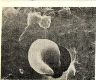
Scanning electron microscopy: red cell (8ᄍ diameter) passing through a filter (5// diameter)
하였다 . 혈액의 여과시에 사용한 nucleopore filter (pore diameter 5jwm, pore density 4 XlO 5/
cm2, thickness 13jwm, effective filtrative area 0.8cm 2) 와 Filter holder는 General Electric회사 (Pleasantown California 94566,U SA ) 제 품을 사 용하였다.
유의 성 검정 一One tailed student t-test에 의 하 여 실험치 사이의 유의 성 검정 을 실시 하였다.
그리고 모든 실험치들은 평균치±표준편차로 표 시하였다.
실험디자인一혈압측정과 적혈구 변형능 측정 은 생후 7주,10 주, 14 주에서 선천성 및 후천성
고혈압쥐에서 실시하였으며, 약물투여 전후의
혈압측정과 적혈구 변형능 측정도 7주, 10 주,
14 주에서 실시하였다.
실 험 결 고^
선천성 및 우천성 고혈압쥐에서도 관찰되었다.
약물을 투여받지 않은 선천성 고혈압쥐와 후천 성 고혈압쥐의 적혈구 변형능은 모두 정상쥐에 비해 유의성 있게 저하되었는데 특히 선천성 고 혈압쥐에서 현저하였다(Fig. 2).
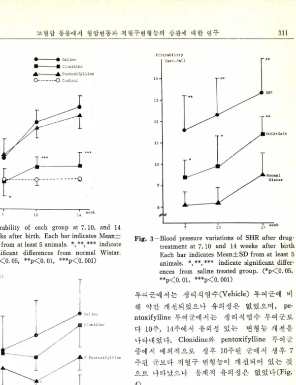
선천성 고혈압쥐에서 약물 투여 후의 혈압변 화ᅳ선천성 고혈압쥐에 생리식염수, clonidine, pentoxifylline 등의 약물을 2주 동안 투여한 후 혈압변화를 관찰하였는데 생 리 식 염 수는 vehicle로 투여 하였 다. Clonidine 투여 에 의 해 서 는 7주,• 10 주, 14 주에서 모두 유의성 있는 혈압강하를 나 타내 었으며, 적 혈구 변형 능 개 선제 인 pentoxi
fylline 에 의해서는 유의성 있는 혈압강하를 나타 내지 못하였다(Fig. 3).
선천성 고혈압쥐에서 약물투여 후의 적혈구 변 형능 변화ᅳ선천성 고혈압쥐에 생리식염수,clo
nidine, pentoxifylline 등의 약물을 2 주 동안 투 여한 후 적혈구 변형능을 관찰하였다. Clonidine
(mmHg)
210
00
90
) S H R
|D O C A + S a l t
L N o r m a l W i s t a r
i
I
고 혈 압 동물에서 혈 압 변 동 과 적 혈 구 변 형 능 의 상관에 대 한 연구 S11
Fig. 2—Filtrability of each group at 7,10,and 14 weeks after birth. Each bar indicates Mean 士 SD from at least 5 animals. *, **, *** indicate significant differences from normal Wistar.
(*p<0. 05, **p<0. 01, ,ScH!*p<0. 001)
Fig. 4—Filtrability variations of SHR after drug- treatment at 7,10 and 14 weeks after birth Each bar indicates Mean士 SD from at least 5 animals. *, *** indicate significant diffe
rences from salinetreated group. (*p<0. 05,
**|)<0.01,***p<0.001)
Fig. 3—Blood pressure variations of SHR after drug- treatment at 7,10 and 14 weeks after birth Each bar indicates Mean士 SD from at least 5 animals. *,**, *** indicate significant differ
ences from saline treated group. (* p < 0 .05,
**p<0. 01, ***p<0. 001)
투여군에서는 생리식염수(Vehicle) 투여군에 비 해 약간 개선되었으나 유의성은 없었으며, Pe
ntoxifylline 투여군에서는 생 리 식 염 수 투여군보 다 10 주, 14 주에 서 유의 성 있는 변형능 개 선을 나타내었다. Clonidine와 pentoxifylline 투여군 중에서 예의적으로 생후 10 주된 군에서 생후 7 주된 군보다 적 혈구 변형능이 개 선되 어 있는 것 으로 나타났으나 통계적 유의성은 없 었 다 (Fig.
4).
후천성 고혈압쥐에서 약물투여 후의 혈압 변 화ᅳ후천성 고혈압쥐에서도 생 리 식 염 수 ,cloni
dine, pentoxifylline 등의 약물을 2주간 투여한 후 혈압변화를 관찰하였다. Clonidine을 투여한 경우에는 7주, 10 주, 14 주에서 모두 유의성 있 는 혈 압강하를 나타내 었으며,적 혈구 변형능 개 선제 인 pentoxifylline을 투여 한 경 우에 는 유의 성 있는 혈압강하를 나타내지 못 했 다 (Fig. 5).
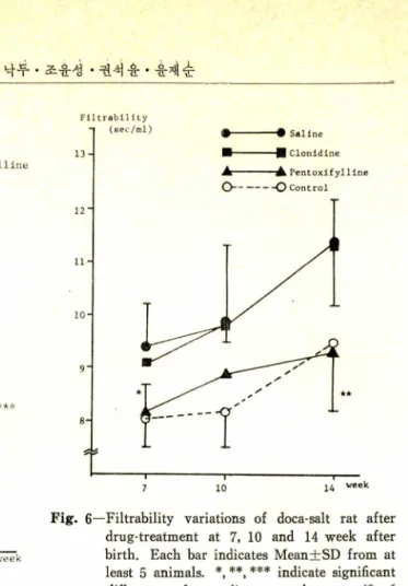
후천성 고혈압쥐에서 약물투여후의 적혈구 변 형능 변 화 一 후천성 고혈압쥐에 생 리식 염수 ,
* P e n to x ify l】
312 고 광 호 • 이 명 설 • ^ 낙 羊 • 조윤^ • 권^ 去 • 奋예 ^
B .P . (m m H g)
2 2 0.
210 2 0 0'
190' 180-1 170- 160 150.
A--- 麵
P e n t o x i f y l l i n e
o---o C o n t r o l
Fig. 5—Blood pressure variations of doca-salt rats after drug-treatment at 7,10 and 14 weeks after birth. Each bar indicates Mean士SD from at least 5 animals. *,**, *** indicate significant differences from saline treated gr
oup. (*p<0. 05, pCO.Ol, p<0. 001) clonidine, pentoxifylline 등의 약물을 2주간 투
여 한 후의 적 혈구 변형 능은 혈압강하제 인 cloni
dine 투여군에서는 생리식염수 투여군에 비해 약 간 개선되었으나 유의성은 없었다. 적혈구 변형 능 개선제인 pentoxifylline을 투여 한 군에 서 는 생리식염수 투여군보다 적혈구 변형능이 많이 개 선되 었으나 7주, 14주에 서만 유의 성 이 있었고 10주에서는 유의성이 없었다(Fig. 6).
고 찰
본 실험의 결과는 혈압이 상승됨에 따라 선천 성 고혈 압쥐 (SH R) 나 후천성 고혈 압쥐 (DOCA- Salt rat) 모두에서 적 혈구의 변형능이 저 하됨 을 나타내고 있다. 이것은 혈압상승과 적혈구의 변 형능 저하와의 상관성 이 있을 수 있다는 것을
F i ltrabilIty
Fig. 6—Filtrability variations of doca-salt rat after drug-treatment at 7, 10 and 14 week after birth. Each bar indicates Mean士SD from at least 5 animals. *, **, *** indicate significant differences from saline treated group. (*p<
0.05,**p<0. 01, ***p<0. 001)
시사하고 있으며, 이러한 결과는 Lagrue11) 등의 인체에 대한 실험 결과와도 일치한다. 동일한 동물들에서 상승된 혈압이 clonidine을 투여하여 유의 성 있게 저 하되 었을 때 적 혈구 변형능은 유 의성 있는 증가를 나타내지 않았다. 이 사실은 고혈압에서 정상 혈압 상태로 변화하는 현상이 적 혈구 변형능의 개 선과 상관성 이 없음을 시 사 한다고 해석할 수도 있다. 그리고 동물에서 나 타나는 고혈압 현상이 적 혈구 변형능의 감소와 연관성을 나타낸 사실과 약물투여에 의해 고혈 암이 정 상혈압으로 회 복될 때 적 혈구 변형능의 감소가 정상으로 회복되지 않은 사실은 상호 모 순된 해 석 을 가능하게 한다. 이 러 한 모순은 다 음의 이유들에 기인할 가능성이 있다. 적혈구 변형능은 한가지 특정 요인에 의 해 결정되 는 것 이 아니라 여러가지 요인들의 평형에 의해 정해 진 다는 사실 이 다. 5) 즉 적 혈구의 표면적 /용적 비 율, 내용물의 유동성,세포내 대사,헤모글로빈 농도 등의 내 인성 요인과 혈액의 pH, 삼투압,
J. Pharm. Soc. Korea
고 혈 압 동물에서 혈 압 변 동 과 적 혈 구 변 형 능 의 상관에 .대한 연구 313 산소분압 및 세 포내 유리 지방산의 농도등의 외
인성 요인등이 적 혈구의 변형능에 영 향을 미 치 며, 이 요인들의 영향은 가역적인 것으로 알려 져 있다. 14,15) 또 한가지 는 적 혈구 변형능 측정 방법의 다양성이다. 본 실험에서는 Reid 등이 고 안한 whole blood filtration 방법을 사용하였는 바 이때 혈액의 여과시간을 결정하는 요인은 적 혈구 변형능이 주된 것이 나 그외 에 fitrinogen, polymorphonuclears, cholesterol 등이 부수적 인 영향을 미칠 가능성도 있다. 6) 이 문제점은 low hematocrit, washed red cell 사용 및 자체 혈장 액에의 현탁 등의 조건을 검색하여 그 관련성 여부를 결정해야 하므로 후속실험이 추가되어야 할 필요성이 있다. 이상의 해석상의 어려움에도 불구하고 본 실험의 결과에서 추측할 수 있는 것은 첫째,적혈구의 변형능 저하는 고혈압 유 발요인일 가능성이 있거나 또는 고혈압 현상에 의해 결과적으로 발생한 현상일 수 있다. 둘째,
고혈압에서 정상혈압 상태로의 변화현상이 적혈 구 변형능과 상관성 이 없다는 점 을 완전히 증명 할 수는 없으나 적어도 clonidine에 의한 고혈압 치료 효과는 적혈구 변형능의 증가에 기 인하는 것은 아님을 나타낸다. 따라서 선천성 및 후천 성 고혈압 동물의 경우 혈압상승시에 나타나는 적혈구 변형능의 저하는 혈압상승 이외의 다른 요인이 개재되어 있다고 생각해 볼 수 있음을 의미한다 하겠다.
결 론
선천성 및 후천성 고혈압쥐에서 고혈압 상태 와 정 상혈 압 상태 를 유도하여 적 혈구 변형능을 측정하여 다음의 결과를 얻었다.
1) 선천성 및 후천성 고혈압쥐에서 혈압상승 에 따라 적 혈구의 변형능은 저 하되 었으며, 이 러 한 상관성은 선천성 고혈압쥐에서 후천성 고혈 압쥐보다 유의성 있었다.
2) 선천성 고혈압쥐에서 혈압강하제인 Cloni
dine 을 투여한 경우에 혈압은 유의 성 있게 감소 되 었으나 적 혈구 변형능의 개 선은 나타나지 않 았다.
3) 후천성 고혈압쥐에서도 혈압강하제인 C lo
nidine 을 투여하여 혈압은 유의성 있게 감소되 었 으나 적 혈구 변형능 개 선은 나타나지 않 았 다 .
4) 위의 사실들은 고혈압 동물의 경우 혈 압 상 승에 따르는 적혈구 변형능의 저하에는 혈 압 이 외의 다른 요인이 개재할 가능성을 시 사 한 다 .
본 연구는 부분적으로 한국과학재단 및 서울 대학교 약학대 학 부설 종합약학연구소의 연구비 지원에 의해 수행되었다.
문 헌
1) Bagge, U.’ Braenemark, P., Karlson, R. and Skalak, R .: Three dimensional observations of red blood cell deformation in capillaries. Blood Cells 6, 231 (1980).
2) Faehraeus, R. and Lindquist, T.: The viscosity of blood in narrow capillary tubes. Am. J. Physiol.
96, 562 (1981).
3) Gaehtgens, P.: Flow of blood through narrow capillaries; Rheological mechanisms determining capillary hematocrit and apparent viscosity. Bior- heology 17,183 (1980).
4) Schmid-Schonbein, H. and Wells, R .: Fluid drop
like transmission of erythrocytes under shear.
Science 165,288 (1969).
5) Reid, H.L., Barnes, A.J., Lock, P.J., Dormandy, J. A. and Dormandy, T .L .: A simple method for measuring erythrocyte deformability. J. Clin.
Path. 29,855 (1976).
6) Dormandy, J.A., Hoare, E.,Colley, J., Arrows- mith D.E. and Dormandy, T .L .: Clinical, haemo- dynamic, rheological, and biochemical findings in 126 patients with intermittent claudication. Brit.
Med. J. iv, 576 (1973).
7) Dormandy, J.A., Hoare, E., Dhattab, A.H., Arro- wsmith, D.E. and Dormandy, T .L .: Prognostic significance of rheological and biochemical findings in patients with intermittent claudication, Brit.
Med. J. iv, 581 (1973).
8) Ehrly, A .M .: Rhe이ᄋgically induced impairment of the vascular microcirculation: A new patho
physiological CQncept of intermittent claudication.
314 고 광 호• 이명걸 • 김낙 두 • 조윤성 . 권석윤• 윤재순 In: IXth Int. Congr. Angiology. Florence 1,410
(1974).
9) Ehrly, A.M. and Kohler H .J.: Altered deforma- bility of erythrocytes from patients with chronic occlusive arterial disease. Vasa 5,319 (1976).
10) Reid, H.L., Dormandy, J.A., Barnes, A.J., Lock, P.J. and Dormandy, T .L.: Impaired red cell deformability in peripheral vascular desease, Lancet i, 666 (1976).
11) Lagrue, G., Marcel, G.A., Faucher, G.,Gallais, M. and Branellec, A.: Deformabilite erythrocyta- ire: Influence du tabagisme et des facteurs de risque vasculaire. Nouv. Presse Med. 8, 4079 (1979).
12) C. Isles, G.D.O. Lowe, M.M. Drummond, C.D.
rheology in malignant phase hypertension. Sc and.
J. Clin. Lab. Invest. 41, Suppl. 156 (1981).
13) Pfeffer, J.M., Pfeffer, M.A. and Frohlich, E.D.:
Validity of an indirect tail-cuff method for deter
mining systolic arterial pressure in unanesthetized normotensive and spontaneously hypertensive rats.
J. Lab. Clin. Med. 78,957, (1971).
14) Schubotz, R. and Muhlfellner, O.: The effect of pentoxifylline on erythrocyte deformability and on phosphatide fatty acid distribution in the erythr
ocyte membrane. Curr. Med. Res. Opin. 4, 609 (1977).
15) Stefanovich, V.: Effect of pentoxifylline on ery
throcyte adenine nucleotide levels in rats. Int. Res, Forbes, A,C.,Kennedy, and A.F. Lever: Blood Commun. Sci, 26,1882 1975.